-
Články
- Vzdělávání
- Časopisy
Top články
Nové číslo
- Témata
- Kongresy
- Videa
- Podcasty
Nové podcasty
Reklama- Kariéra
Doporučené pozice
Reklama- Praxe
Polymorphisms in the Mitochondrial Ribosome Recycling Factor Compromise Cell Respiratory Function and Increase Atorvastatin Toxicity
Mitochondrial translation, essential for synthesis of the electron transport chain complexes in the mitochondria, is governed by nuclear encoded genes. Polymorphisms within these genes are increasingly being implicated in disease and may also trigger adverse drug reactions. Statins, a class of HMG-CoA reductase inhibitors used to treat hypercholesterolemia, are among the most widely prescribed drugs in the world. However, a significant proportion of users suffer side effects of varying severity that commonly affect skeletal muscle. The mitochondria are one of the molecular targets of statins, and these drugs have been known to uncover otherwise silent mitochondrial mutations. Based on yeast genetic studies, we identify the mitochondrial translation factor MEF2 as a mediator of atorvastatin toxicity. The human ortholog of MEF2 is the Elongation Factor Gene (EF-G) 2, which has previously been shown to play a specific role in mitochondrial ribosome recycling. Using small interfering RNA (siRNA) silencing of expression in human cell lines, we demonstrate that the EF-G2mt gene is required for cell growth on galactose medium, signifying an essential role for this gene in aerobic respiration. Furthermore, EF-G2mt silenced cell lines have increased susceptibility to cell death in the presence of atorvastatin. Using yeast as a model, conserved amino acid variants, which arise from non-synonymous single nucleotide polymorphisms (SNPs) in the EF-G2mt gene, were generated in the yeast MEF2 gene. Although these mutations do not produce an obvious growth phenotype, three mutations reveal an atorvastatin-sensitive phenotype and further analysis uncovers a decreased respiratory capacity. These findings constitute the first reported phenotype associated with SNPs in the EF-G2mt gene and implicate the human EF-G2mt gene as a pharmacogenetic candidate gene for statin toxicity in humans.
Published in the journal: . PLoS Genet 8(6): e32767. doi:10.1371/journal.pgen.1002755
Category: Research Article
doi: https://doi.org/10.1371/journal.pgen.1002755Summary
Mitochondrial translation, essential for synthesis of the electron transport chain complexes in the mitochondria, is governed by nuclear encoded genes. Polymorphisms within these genes are increasingly being implicated in disease and may also trigger adverse drug reactions. Statins, a class of HMG-CoA reductase inhibitors used to treat hypercholesterolemia, are among the most widely prescribed drugs in the world. However, a significant proportion of users suffer side effects of varying severity that commonly affect skeletal muscle. The mitochondria are one of the molecular targets of statins, and these drugs have been known to uncover otherwise silent mitochondrial mutations. Based on yeast genetic studies, we identify the mitochondrial translation factor MEF2 as a mediator of atorvastatin toxicity. The human ortholog of MEF2 is the Elongation Factor Gene (EF-G) 2, which has previously been shown to play a specific role in mitochondrial ribosome recycling. Using small interfering RNA (siRNA) silencing of expression in human cell lines, we demonstrate that the EF-G2mt gene is required for cell growth on galactose medium, signifying an essential role for this gene in aerobic respiration. Furthermore, EF-G2mt silenced cell lines have increased susceptibility to cell death in the presence of atorvastatin. Using yeast as a model, conserved amino acid variants, which arise from non-synonymous single nucleotide polymorphisms (SNPs) in the EF-G2mt gene, were generated in the yeast MEF2 gene. Although these mutations do not produce an obvious growth phenotype, three mutations reveal an atorvastatin-sensitive phenotype and further analysis uncovers a decreased respiratory capacity. These findings constitute the first reported phenotype associated with SNPs in the EF-G2mt gene and implicate the human EF-G2mt gene as a pharmacogenetic candidate gene for statin toxicity in humans.
Introduction
The primary function of the mitochondria is the aerobic production of ATP, a process that is reliant on a series of protein complexes that comprise the electron transport chain. Several components of the electron transport chain are encoded in the mitochondrial genome, the translation of which is governed largely by nuclear encoded genes. Increasingly, mutations within these genes are being implicated with respiratory deficiency, an underlying factor in a number of diseases, including myopathies and liver failure [1], [2], [3], [4]. For example, pathogenic mutations in the human mitochondrial elongation factor genes, EF-G1mt and EF-Tu(mt), have been implicated with severe lactic acidosis and encephalopathy [1], [2], [3]. Recently a mutation in a novel gene, believed to be a member of the class of mitochondrial peptide release factors, was identified in patients exhibiting symptoms of Leigh syndrome [4].
In addition to disease, there is also emerging evidence that respiratory deficiencies are responsible for adverse drug reactions. Consequently, treatment with certain drugs have uncovered otherwise silent mitochondrial mutations [5], [6]. The group of cholesterol-lowering drugs, statins, are one example. The primary target of statins is 3-hydroxy-3-methylglutaryl-coenzyme A (HMG-CoA) reductase, the rate limiting enzyme of the sterol synthesis pathway, but increasingly, studies are reporting signs of statin-induced mitochondrial dysfunction [7], [8]. This is believed to be a factor in the myopathic side-effects of statins. Approximately 0.1 to 0.5 percent of statin users experience severe myopathic symptoms (defined as serum creatine kinase levels more than 10 times the upper limit of normal) and many more suffer milder musculoskeletal pain [9], [10]. Frequently such patients present symptoms that are similar to those of patients with mitochondrial myopathies [11]. To date, there have been several case studies reporting the presence of a subclinical MELAS (mitochondrial encephalopathy, lactic acidosis and stroke-like episodes) mutation within the mitochondrial DNA (mtDNA) of patients who have developed severe myopathic symptoms following statin medication [12], [13], [14]. It is expected that existing weakness in mitochondrial function can be exacerbated upon exposure to statin, leading to the uncovering of previously asymptomatic mutations in mitochondrial genes.
The yeast Saccharomyces cerevisiae has been the model of choice for studies of mitochondrial function. In addition to mitochondrial similarities with human cells, the ability of yeast to survive in the absence of mtDNA, the simplicity with which both nuclear and mtDNA can be manipulated and the extensive number of tools and resources available specifically for yeast research has greatly contributed to an understanding of potentially pathogenic mutations [15], [16], [17]. Statins were first isolated as secondary metabolites from fungi, the presumption being that the strong antifungal properties of statins provide an ecological advantage for the producer over other fungi, similar to that of antibiotics. We and others have demonstrated that upon exposure to statin, yeast, as well as having reduced cell viability, also display evidence of mitochondrial dysfunction [18], [19], [20].
In this study, we identify a nuclear gene encoding a mitochondrial translation factor as a modulator of atorvastatin toxicity in yeast (MEF2) and human cell lines (EF-G2mt). The eukaryotic mitochondrial protein synthesis system consists of four phases; initiation, elongation, termination and ribosome recycling, each carefully orchestrated by a series of nuclear encoded proteins [21], [22]. The human EF-G2mt gene, originally named a mitochondrial elongation factor based on sequence homology with bacterial EF-G, has since been shown to function as a ribosome recycling factor [23], [24]. EF-G2mt is believed to interact with the already known ribosome recycling factor (RRF1) to promote dissociation of the ribosomal subunits following termination of translation [23]. In bacteria, the dual role of translocation and ribosome recycling are shared by a single EF-G protein [25]. Eukaryotic cells harbour two EF-G proteins in their mitochondria and it appears that these have distinct functions, the EF-G1mt protein for translocation and the EF-G2mt protein for ribosome recycling [23], [26]. The human EF-G2mt protein is conserved across the majority of eukaryotic species [23]. With its yeast Mef2p counterpart, the human EF-G2mt protein shares greater than 32 percent homology and four of the five protein domains. We use the atorvastatin-sensitive phenotype of the yeast MEF2 gene to uncover naturally occurring human variants of EF-G2mt that have respiratory deficient phenotypes. These findings have ramifications for patient drug response and possibly also for disease.
Results
Identification of the yeast MEF2 gene as a cell mediator of atorvastatin toxicity
In light of the emerging evidence that mitochondria are important in dictating statin toxicity, which in turn can reveal underlying respiratory defects that have important health implications, experiments were designed to discover mitochondrial lesions that affect statin sensitivity. Two published fitness profiling experiments in yeast have observed statin sensitivity in hundreds of heterozygous deletion mutants following statin exposure for 20 generations of growth [27], [28]. Using the Gene Ontology term finder available on the Saccharomyces genome database website (www.yeastgenome.org), we discovered approximately 14–17% of genes conferring statin-sensitivity are associated with the mitochondria. One of the most sensitive of these mitochondrial associated genes was the MEF2 gene, encoding a mitochondrial translation factor believed to have a role in ribosome recycling [23], [27].
The growth of deletion mutants in a competition style assay is a very sensitive method of detecting differences in growth rate. However, these assays are prone to a higher incidence of false positive and non-replicable results [29]. To confirm the MEF2 phenotype, a number of yeast deletion mutants that ranked as the most statin sensitive in the fitness profiling experiments were compared [27], [28]. Both heterozygous and haploid mutants were tested and cell viability was assessed after five days exposure to 110 µM atorvastatin. The concentration of 110 µM atorvastatin is approximate to those concentrations used in the original genome-wide fitness profiling screens (62.5 µM and 125 µM atorvastatin) [27]. Furthermore, during a previous investigation of the effects of different atorvastatin concentrations in yeast, we have shown that 110 µM atorvastatin does not inhibit cell growth but is sufficient to cause a significant decrease in intracellular ergosterol (approximately 85%), accompanied by loss of cell viability after prolonged exposure (5 days) [18]. Of the heterozygous mutants tested, only the hmg1Δ/HMG 1 strain was confirmed to be sensitive to atorvastatin (Figure 1A). However, of the haploid mutants tested, three displayed a statin hypersensitive phenotype (Figure 1B). The mef2Δ mutant emerged as the strain which exhibited the greatest loss of cell viability in the presence of atorvastatin, with an almost 20-fold reduction in cell viability compared with the wild-type. The hmg1Δ strain displayed a 5-fold reduction in cell viability and disruption of the HTZ1 gene, encoding a histone protein, resulted in a 2-fold loss of cell viability (Figure 1B).
Fig. 1. Viability of yeast heterozygous and haploid deletion mutants in atorvastatin after 5 days. 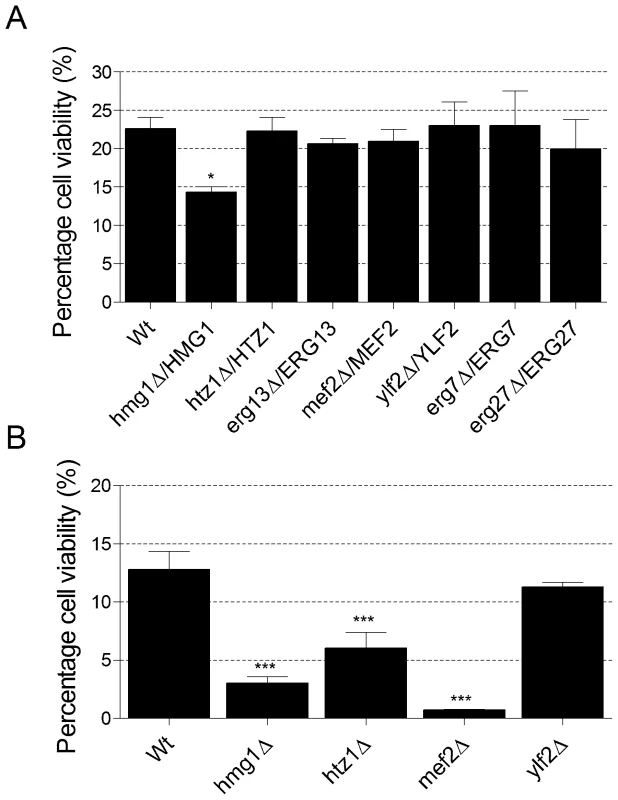
(A) Percentage cell viability of heterozygous deletion mutants in 110 µM atorvastatin relative to viability in the solvent control. (B) Percentage cell viability of haploid deletion mutants in 110 µM atorvastatin relative to viability in the solvent control. Data represent mean ± SEM (n = 3). A one-way ANOVA, followed by a Dunnett's multiple comparison test, was used to compare the mean percentage viability of the mutant strains to that of the wild-type. *P<0.05, ***P<0.001. Yeast mutants defective in mitochondrial translation undergo rapid loss of mtDNA and we have previously shown this to be the case for the mef2Δ mutant [30]. Consequently, mef2Δ is ρ0 (completely devoid of mtDNA). To determine whether atorvastatin-sensitivity is the consequence of abrogation of the MEF2 gene or simply from the absence of mtDNA, mef2Δ statin sensitivity was compared with that of ethidium bromide generated cytoplasmic ρ0 mutants. In order to ensure that there were no secondary site mutations in the mef2Δ deletion mutant that originated from the S. cerevisiae gene deletion collection, new mef2Δ haploid strains were created. Results show that although the ρ0 strains were more sensitive to atorvastatin than the respiratory positive parent, they did not display the same degree of sensitivity as mef2Δ (Table 1). Therefore, mutation of the MEF2 gene is a critical determinant of statin sensitivity through mtDNA dependent and independent functions.
Tab. 1. Percentage cell viability relative to solvent control after 5 days exposure to 110 µM atorvastatin. 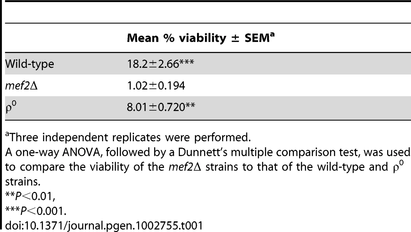
Three independent replicates were performed. Silencing of the human EF-G2mt gene compromises OXPHOS and exacerbates atorvastatin toxicity in human cell lines
The human EF-G2mt gene, ortholog of the yeast MEF2 gene, encodes a recently characterised mitochondrial ribosome recycling factor [23], but to date, no functional analysis has been performed for this gene. To determine whether the EF-G2mt gene is essential for human cell function and to ascertain whether depletion influences statin toxicity, an siRNA pool comprising of four individual EF-G2mt targeted siRNAs was used to silence EF-G2mt expression in the human rhabdomyosarcoma (RD) cell line. The RD cell line has previously been established as a skeletal muscle model for mitochondrial disorders and has also been used in studies of statin toxicity [31], [32], [33]. At 72 hours post-transfection, greater than 80 percent silencing was consistently achieved and cells remained viable. Cells were then re-transfected at this time point to enable continued depletion of EF-G2mt activity. This strategy had previously uncovered an essential role for cell viability for the first discovered ribosomal recycling factor gene, RRF1 [34]. However, at 72 hours post-re-transfection (six days after the initial transfection), RD cells remained viable even though EF-G2mt mRNA concentration had decreased by 99.9 percent. Additionally, there was no decrease in mtDNA levels upon EF-G2mt silencing as analysed using quantitative PCR (Figure S1) [35].
Although gross inhibition of mitochondrial translation in human cell lines results in loss of cell viability [34], a more subtle mitochondrial phenotype may be masked by the phenomenon of the Crabtree effect whereby many human cell lines, when grown in the presence of glucose, derive their energy almost solely by fermentative means [36]. To circumvent this effect and force cells to rely on mitochondrial respiration as their primary energy source, galactose was used to replace glucose as the carbon source [37]. Silencing of EF-G2mt led to a marked decline in the growth of RD cells at four days post-transfection, which was maintained for a period of seven days (Figure 2). This growth defect on galactose medium signals an impairment in oxidative phosphorylation (OXPHOS).
Fig. 2. Growth of siRNA transfected RD cells in glucose and galactose medium. 
At 24 hours post-transfection, cells were seeded into wells of a 96-well plate in DMEM medium containing 10% fetal bovine serum and either 4.5 g/L glucose (left panel) or 4.5 g/L galactose (right panel). Cell proliferation was determined daily using a luminescent cell viability assay. An untransfected cell line and the rho0 cell line were included as controls. Cell proliferation is shown as a percentage of the maximum cell growth (100%) of the untransfected control. Error bars represent the mean ± SEM for three independent measurements. Based on the findings in yeast, it was predicted that decreased EF-G2mt activity would also enhance the effects of atorvastatin toxicity. EF-G2mt silenced RD cells were subjected to various concentrations of atorvastatin in medium containing either galactose or glucose as the carbon source for a period of 48 hours. In glucose medium, there was no difference in statin sensitivity between the EF-G2mt silenced cells and those transfected with the non-targeting control. However, based on IC50 values (defined here as a 50% loss of viability at 48 hours) in galactose medium, EF-G2mt silenced cells were over 20 percent more sensitive to atorvastatin than cells transfected with the non-targeting siRNA pool (Table 2). These results confirm a role for the human EF-G2mt gene in cell resistance to atorvastatin in a human skeletal muscle cell line. Notably, a similar increase in sensitivity (17%) was observed using the human hepatic HepG2 cell line (a model for statin-induced liver toxicity), although it should be noted that HepG2 cells are approximately 10 times more statin resistant than RD cells and this elevation in atorvastatin sensitivity was not statistically significant in these experiments (Table 2).
Tab. 2. IC50 values for EF-G2mt silenced cells exposed to atorvastatin. 
IC50 values of EF-G2mt silenced cells were compared to their respective non-targeting siRNA transfected cell lines using the extra sum-of-squares F-test. EF-G2mt protein variants, when created in yeast Mef2p, increase atorvastatin toxicity
A global alignment of the amino acid sequence of the human EF-G2mt protein (Isoform I, AAH15712.1) with the yeast Mef2 protein (CAA59392) reveals 32.1% amino acid sequence identity (Figure 3 and Figure S2). At the commencement of this study, there were nine published non-synonymous Single Nucleotide Polymorphisms (SNPs) in the human EF-G2mt gene, of which five were either conserved or semi-conserved in the yeast MEF2 gene. Three of these variants, EF-G2mtI627T, EF-G2mtE594G and EF-G2mtK334R, are considered rare, with a heterozygosity frequency below one percent. One of the variants, EF-G2mtR744G, has a heterozygosity frequency of three percent and the EF-G2mtR774Q allele has a heterozygosity frequency greater than 20 percent. These five SNPs were selected for functional analysis using the yeast MEF2 gene as a model.
Fig. 3. EF-G2mt protein variants. 
Alignment of the protein amino acid sequence of the human EF-G2mt protein with the yeast Mef2 protein. Dark shaded areas represent conserved amino acid residues and grey shaded areas represent semi-conserved residues. EF-G2mt SNPs that are semiconserved in yeast MEF2 are shown in italics and fully conserved SNPs are depicted in bold. The five alleles selected for functional characterisation are outlined. The five EF-G2mt protein domains are represented below the alignment. Global alignment of protein sequences was performed using Lalign and the BioEdit sequence alignment editor was used to generate the graphical representation. For each of the five selected human EF-G2mt variants, single nucleotide base pair substitutions were constructed directly into the chromosomal copy of the yeast MEF2 gene to replace the codon specific to the wild-type amino acid residue with a codon that corresponds to the amino acid present in the human EF-G2mt protein variants. The Mef2p variants constructed were mef2K769Q, mef2R740G, mef2I616T, mef2D578G and mef2K308R which correspond to EF-G2mtR774Q, EF-G2mtR744G, EF-G2mtI627T, EF-G2mtE594G and EF-G2mtK334R respectively. To assess for respiratory competence, mef2 mutants were grown on medium containing the non-fermentable carbon source glycerol. After 72 hours, all five mutants were proficient in the production of colonies on both glucose and glycerol medium. The number and size of colonies produced by the mutant strains on glycerol medium was equal to that of the wild-type, indicating that all mutants are respiratory competent (Figure 4A). Moreover, based on measurements of cell growth in both glucose and glycerol liquid medium, there were no growth defects exhibited by any of the mef2 mutants (Table S1). Mitochondrial DNA stability was measured periodically for up to 32 generations. The frequency of cells which spontaneously lose mtDNA amongst populations of each mef2 mutant remained equal to that of the wild-type (approximately 2 to 3%), verifying that mtDNA is stable over successive generations.
Fig. 4. Functional characterisation of EF-G2mt equivalent SNPs in the yeast MEF2 gene. 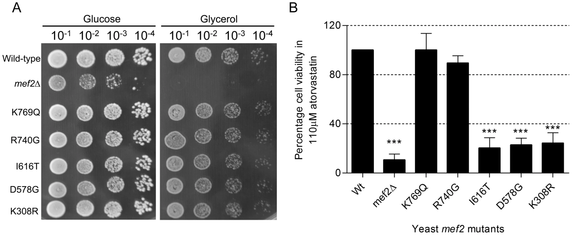
(A) Growth of wild-type, mef2 deletion strain and mef2 mutants in yeast medium containing either glucose as the carbon source or the non-fermentable carbon source glycerol. Cells from exponentially growing cultures were serially diluted and 5 µl of each dilution spotted onto each plate. Growth was assessed after 72 hours incubation at 30°C. (B) Percentage cell viability of yeast mef2 mutants in 110 µM atorvastatin relative to viability of the wild-type strain following exposure to atorvastatin for 5 days. Data represent mean ± SEM (n = 3). A one-way ANOVA followed by a Dunnett's multiple comparison test was used to compare the mean percentage viability of the mef2 variants to that of the wild-type. ***P<0.001. The five mef2 mutants were then assayed for atorvastatin sensitivity. Following five days of exposure to 110 µM atorvastatin, three of the mef2 mutants exhibited a statin hypersensitive phenotype. Viability of the mef2I616T, mef2D578G and mef2K308R mutants was reduced to 20.3, 22.8 and 24.2 percent respectively (Figure 4B). The two other mef2 mutants, mef2K769Q and mef2R740G, did not exhibit a statin sensitive phenotype. The unmasking of a phenotype for the mef2K308R, mef2D578G and mef2I616T variants by atorvastatin is a strong indicator that these mutations have an effect on Mef2p function. A similar effect conferred by these alleles in the human EF-G2mt protein could have vital consequences for statin users.
Atorvastatin-sensitive mef2 mutants also have reduced respiration rates
Although no obvious growth phenotype was observed, the statin-sensitive phenotype of three of these mutants indicates a subtle defect in mitochondrial function. Staining of mef2 mutant cells with the nucleic acid staining dye 4′,6-diamidino-2-phenylindol (DAPI) confirmed the presence of mtDNA nucleoids in cells of all five mutants and quantification of mtDNA copy number using quantitative PCR (qPCR) [38] showed that mtDNA levels were the same as that of the wild-type (Figure S3). It therefore appears that the EF-G2mt equivalent mutations do not destabilise Mef2p function so as to compromise mtDNA stability.
To investigate the possibility of a respiratory phenotype, oxygen consumption for the three statin-sensitive mef2 mutant cultures was measured (in the absence of atorvastatin) using a non-invasive oxoluminescent device [35]. All three mutants, mef2K308R, mef2D578G and mef2I616T, exhibited a significantly reduced respiration capacity, approximately one third lower than that of the wild-type (Figure 5). The two mef2 mutants that did not display an atorvastatin-sensitive phenotype had respiration rates much closer to that of the wild-type. Together, these results demonstrate a respiratory phenotype conferred by at least three of the EF-G2mt equivalent mef2 variants and this correlates with the inability of these mef2 mutants to tolerate atorvastatin toxicity.
Fig. 5. Oxygen consumption rates of mef2 mutant cultures. 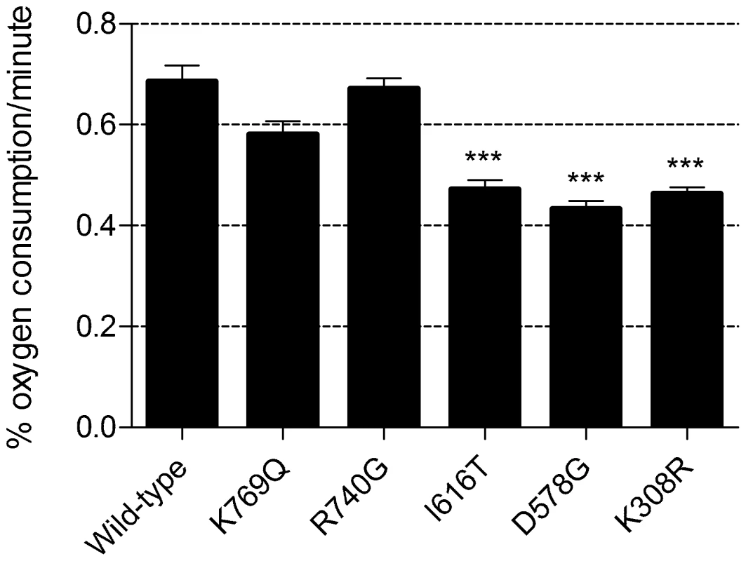
Figure depicts the rate of oxygen consumption per minute per 30 mL of yeast culture at an OD600 of 0.2. Data represent mean ± SEM (n = 3). A one-way ANOVA followed by a Dunnett's multiple comparison test was used to compare the mean oxygen consumption rate of mef2 variants to that of the wild-type. ***P<0.001. Interestingly, the partially respiratory deficient mef2 mutants exhibit a greater statin sensitivity than the ethidium bromide generated ρ0 strains, which are completely devoid of respiratory function (Table 1). This, in support of the mef2Δ results, indicates the role of a non-respiratory function of MEF2 in the statin response. In addition to a lack of mtDNA, we have previously shown, by staining cells with MitoTracker Red CMXRos, that the mef2Δ strain has a reduced mitochondrial membrane potential (ΔΨ) and that mitochondria appear fewer, with a tendency to aggregate [30]. The MitoTracker Red CMXRos probe enters the mitochondrial matrix dependent on ΔΨ. This same method was used to test ΔΨ and mitochondrial morphology in the mef2 mutants. Cells were visualised using a laser scanning confocal microscope and, in contrast to the mef2Δ strain, all five mef2 mutants displayed mitochondria that stain brightly and are arranged in a tubular network, typical of mitochondria in wild-type yeast. Staining of the three partially respiratory deficient mutants is shown in Figure 6. These results indicate that the mef2 mutations do not disrupt the function of Mef2p in maintaining ΔΨ and so does not explain the enhanced statin sensitivity of these mutants. It is known that statin toxicity can cause loss of ΔΨ [39]. Therefore one possibility is that atorvastatin acts synergistically with a mutated Mef2p to exacerbate loss of ΔΨ and compromise cell viability. However, other mechanisms, such as modulation of the mitochondrial retrograde response, cannot be discounted [40].
Fig. 6. Visualisation of mitochondrial membrane potential. 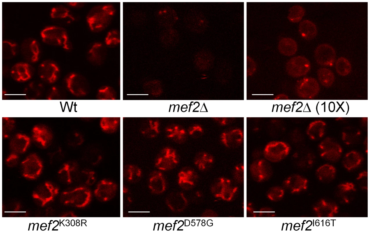
Cells were stained with MitoTracker Red CMXRos and observed using a laser scanning confocal microscope. To better visualise mitochondrial structure within the mef2 deletant, cells were stained with a 10× concentration of MitoTracker Red. Scale bar represents 5 µm. Protein homology modelling of the human EF-G2mt protein predicts function of EF-G2mt protein variants
The crystal structure of the human EF-G2mt protein has not yet been elucidated. Therefore, to gain insight into the molecular effects of the five chosen amino acid variations, a computational model of the EF-G2mt protein was constructed using the SWISS-MODEL server [41]. The model is based on the experimentally determined crystal structure of the Thermus. thermophilus EF-G protein [42] which shares 39 percent identity with the human EF-G2mt protein and four of the five EF-G2mt protein domains. As the N-terminal and C-terminal regions of the EF-G2mt protein share particularly low homology with the template, the initial 65 and final 10 amino acid residues could not be accurately modelled. For this reason, the location of amino acid variant EF-G2mtR774Q, positioned very close to the C-terminus, was omitted (Figure 7A). Stereochemical quality of the model was assessed by generating a Ramachandran plot using PROCHECK. Eighty seven percent of residues fall within the most favoured regions of the plot, indicating ideal stereochemistry.
Fig. 7. In silico model of the human EF-G2mt protein. 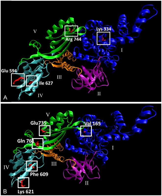
(A) Model depicting four of the five amino acid variants that were functionally characterised in this study. (B) Five newly discovered variants that have yet to be functionally characterised. Helices are shown as ribbons, beta-sheets are depicted as flat broad arrows and loops and coils appear as thin tubes. The five protein domains of the EF-G2mt protein are distinguished by different colours. Domain I, the GTP binding domain is shown in blue, domain II is purple, domain III is orange, domain IV is cyan and domain V is green. Model was visualised using the Visual Molecular Dynamics program (VMD), version 1.8.7 (University of Illinois). Using the in silico model, we can make some hypothetical predictions about the function of the protein variants. The human EF-G2mtK334R variant, which corresponds to the respiratory compromised yeast mef2K308R variant, is located on a coil close to the surface of the GTP-binding domain (domain I) that is highly conserved in EF-G2mt homologs from all major eukaryotic species. The presence of GTP is essential for ribosome dissociation at the termination of translation, and subsequent hydrolysis of GTP is then required in order to release the EF-G2mt protein from the ribosome [23]. The EF-G2mtE594G and EF-G2mtI627T variants, equivalent to the respiratory compromised mef2D578G and mef2I616T mutants respectively, are both located in domain IV and occur close to the EF-G2mt protein surface. The EF-G2mtE594G variant is expected to diminish the largely negative electrostatic surface potential of this domain, thereby interfering in the protein's interaction with both the mitochondrial ribosome and Rrf1 [23], [25]. The EF-G2mtI627T variant is located on an alpha helix whose structural integrity is disturbed by the threonine hydroxyl group. The final two EF-G2mt protein variants, in which the corresponding mef2 mutants did not exhibit a phenotype, are located in domain V, the C-terminal region that accelerates (but is not essential for) the ribosome recycling action of the EF-G2mt protein [23].
In the last year, data from the 1000 Genomes project has expanded the number of known polymorphisms in the human genome and a further 11 non-synonymous SNPs have been discovered within the human EF-G2mt gene (dbSNP, National Centre for Biotechnology Information (NCBI)) [43]. Of these, one is fully conserved in the S. cerevisiae Mef2 protein and another five are semi-conserved (Figure 3 and Figure S2). The location of five of these variants is shown on the EF-G2mt protein model in Figure 7B. Although experimental confirmation is essential, based on the findings above it is hypothesised that the domain IV (EF-G2mtK621N and EF-G2mtF609Y) and domain I (EF-G2mtV165G) variants will affect protein activity. Therefore, these naturally occurring EF-G2mt variants may have respiratory deficient consequences.
Discussion
By exploiting the genetic tractability of yeast, complemented by siRNA silencing studies in human cell lines, we have identified a mitochondrial translation factor as a mediator of atorvastatin toxicity and also made fundamental discoveries about the function of human variants within the EF-G2mt gene. The human EF-G2mt gene, ortholog of the yeast MEF2 gene, was originally identified as a mitochondrial elongation factor gene. However, a recently published comprehensive analysis of the EF-G2mt protein has shown that it functions as a ribosome recycling factor, interacting with the first discovered ribosome recycling factor protein, Rrf1, to dissociate the ribosomal subunits at the termination of translation [23]. In yeast, deletion of the MEF2 gene results in loss of mtDNA [30], a circumstance which would be lethal in higher eukaryotic cells without the supplementation of uridine and pyruvate [44]. Nevertheless, siRNA silencing experiments show that in contrast to its RRF1 counterpart, knockdown of human EF-G2mt expression does not compromise cell viability in glucose medium. Furthermore, silencing of EF-G2mt expression does not deplete cellular mtDNA content. It is possible that some compensatory mechanism enables ribosome recycling to continue to a sufficient degree to maintain cell viability in fermentative cell lines.
When galactose is used as the carbon source instead of glucose, the ATP produced via glycolysis is insufficient for cell energy requirements; therefore, there is a greater reliance on the production of ATP through the oxidative metabolism of glutamine. This more closely resembles the metabolic activity of cells in a human physiological system [37], [45]. By using galactose to force reliance on the mitochondria for cell energy production, it was shown that reduced EF-G2mt activity does indeed compromise cell proliferation in respiring cells. It is consequently expected that the EF-G2mt gene is essential for cell function in a human system. Furthermore, in support of the notion that abnormalities in mitochondrial function sensitise cells to statin toxicity, the growth defect of EF-G2mt silenced cells in galactose medium was even further exacerbated upon exposure to atorvastatin.
For cells in which mitochondrial function is challenged, exposure to mitochondrial toxicants, such as statins, places additional stress on mitochondrial function and this has the potential to trigger pathogenicity. Indeed we have shown that ρ0 strains, lacking respiratory capacity, are more sensitive to atorvastatin than those with a functioning mitochondrial genome in both yeast and human cells. Studies have shown that statins exert their mitochondrial toxicity effects by inhibiting function of the electron transport chain but there is also evidence of non-respiratory mitochondrial consequences [46], [47], [48]. These include a loss of mitochondrial membrane potential, aberrant mitochondrial morphology and apoptosis [39]. These effects, in combination with the absence of respiratory function may explain the hypersensitivity of ρ0 cells to statin.
The hypersensitivity of respiratory deficient cells to statins may have clinical ramifications for patients that have variations within mitochondrial functioning genes. Statins have been known to aggravate clinically silent disease associated mutations resulting in myopathies. In fact, mutations (which in many cases were asymptomatic) for three common myopathic diseases; carnitine palmitoyltransferase II deficiency, McArdle disease and myoadenylate deaminase deficiency (AMPD deficiency), are thought to be the underlying determinants responsible for statin-induced myopathy in up to 10 percent of patients showing adverse effects [5]. There are also reports of the statin-induced triggering of MELAS syndrome in patients whose MELAS mutations were clinically silent [13], [14]. As MELAS syndrome arises from mutations in mtDNA, it strongly implicates mitochondrial dysfunction in susceptibility to statin toxicity. This is further supported by the identification of two commonly occurring SNPs in the human COQ2 gene that are associated with an increased risk of statin intolerance [49]. The COQ2 gene is required for the synthesis of coenzyme Q10, an essential component of the mitochondrial electron transport chain [49].
Despite the presence of a number of SNPs within the EF-G2mt gene, the function of these variants for cell fitness had never been investigated. In this study, five EF-G2mt equivalent SNPs were selected for functional analysis in the yeast MEF2 gene. Unlike the mef2Δ deletant, all mef2 mutants grew proficiently on both glucose and the non-fermentable carbon source glycerol. Furthermore, the mutations had no effect on mtDNA stability, ΔΨ or mitochondrial morphology. However, exposure of the mutants to toxic concentrations of atorvastatin has uncovered a phenotype for three of the mef2 mutants; mef2K308R, mef2D578G and mef2I616T, equivalent to the EF-G2mtK334R, EF-G2mtE594G and EF-G2mtI627T alleles respectively. In silico protein homology modelling reveals these three mutations are located in either the GTP binding domain (domain I) of the EF-G2mt protein or an external domain (domain IV) necessary for ribosomal interaction.
Based on the observations that statins exacerbate clinically silent disease associated mutations, it was predicted that the three mutations compromise mitochondrial function in a subtle yet potentially significant way. The subsequent observation that oxygen consumption was significantly reduced in the statin-sensitive mef2 mutants confirmed this hypothesis and demonstrates a sub-optimal mitochondrial function for the three EF-G2mt equivalent mef2 mutants. This sub-optimal mitochondrial function is expected to contribute to the atorvastatin-sensitive phenotype of the three mef2 mutants. However, comparison of the statin sensitive phenotype of the mef2K308R, mef2D578G, mef2I616T and mef2Δ mutants with cytoplasmic ρ0 strains indicates that statin sensitivity is not fully explained by the reduced respiratory capacity of these mutants and further studies are required to completely elucidate the precise mechanism.
This study constitutes the first report of a phenotype associated with EF-G2mt, demonstrating an essential role for aerobic respiration in human cell lines and an importance for cell tolerance to atorvastatin. Atorvastatin constituted the focus of this study and is the highest selling and also one of the more potent of the statins [50]. Although the various statins have been shown to differ in their cellular toxicity effects, all of them have been implicated with mitochondrial dysfunction [48], [50], [51], [52], [53]. It would therefore be expected that a mitochondrial mediator of atorvastatin toxicity may also mediate cell response to the other statins. In support of this, preliminary experiments confirm that the atorvastatin sensitive mef2 mutants also exhibit sensitivity to lovastatin.
With an estimated 38 million people around the world undertaking statin treatment [9], the identification of novel biomarkers for statin toxicity has the potential to personalise therapy for millions of individuals. To date, only a handful of genes have been associated with statin toxicity [9], but the finding of the EF-G2mt gene as a potential pharmacogenetic candidate has strengthened the association between existing mitochondrial dysfunction and statin hypersensitivity. Importantly, the discovery of naturally occurring human polymorphisms within the EF-G2mt gene that affect respiratory function indicates that these variants, either alone or in combination with other polymorphisms, have significant pathogenic consequences. This opens avenues for further clinical investigations into a possible association between EF-G2mt variants and disease.
Materials and Methods
Chemicals
Atorvastatin calcium was purchased from 7 Chemicals (India). Stock solutions were prepared by dissolving atorvastatin in methanol at a concentration of 20 mg/mL and solutions were stored at −20°C. 5-fluoroorotic acid and geneticin (G418) were purchased from Sigma.
Yeast strains and media
The haploid wild-type S. cerevisiae strains used were of the background BY4741 (MATa his3Δ1 leu2Δ0 met15Δ0 ura3Δ0) and BY4742 (MATα his3Δ1 leu2Δ0 lys2Δ0 ura3Δ0). The diploid strain was BY4743 (MATa/α his3Δ1/his3Δ1 leu2Δ0/leu2Δ0 LYS2/lys2Δ0 MET15/met15Δ0 ura3Δ0/ura3Δ0) [54]. The mef2Δ/MEF2, mef2Δ and the cytoplasmic ρ0 strains were previously described [30]. Prior to experiments, yeast strains were cultured in liquid YEPD (1% yeast extract, 2% peptone, 2% dextrose) medium for 24 hours, subcultured and then grown to exponential phase in Synthetic Complete (SC) medium (0.67% Difco yeast nitrogen base without amino acids, 2% dextrose, 0.79 g L−1 amino acid supplement (Sunrise Science Products, Australia)). Cells were incubated at 30°C with shaking.
Statin viability assay in yeast
Triplicate exponential phase cultures grown in SC medium were diluted to an optical density (OD595 nm) of 0.2 and 1.25 mL of this culture was added to 3.75 mL SC medium with the appropriate concentration of atorvastatin. After a maximum of five days growth, cells were diluted and plated onto solid YEPD (YEPD plus 2% agar) medium and viability counts were performed 48 hours later.
Cell culture
HepG2 cells and RD Cells are from the American Type Culture Collection (ATCC). Cells were cultured at 37°C and 5% CO2 in Dulbecco's Modified Eagle's Medium (DMEM) medium (Gibco) containing 4.5 g/L glucose and 10% fetal bovine serum (FBS) (Bovogen Biologicals, Australia). For experiments in which galactose was used as the carbon source, cells were grown in glucose free DMEM with 10% fetal bovine serum and 4.5 g/L galactose. To generate the ρ0 cell lines, used as respiratory deficient controls, HepG2 and RD cells were cultured for eight weeks in DMEM medium containing 100 ng/mL ethidium bromide and supplemented with 10% FBS, 50 µg/mL uridine and 100 µg/mL sodium pyruvate [31], [44]. Following ethidium bromide treatment, ρ0 cells were maintained in medium supplemented with uridine and sodium pyruvate. Depletion of mtDNA was confirmed using qPCR.
Cell transfection with siRNA
The ON-TARGETplus SMARTpool, comprising of four siRNAs targeting the EF-G2MT transcript (NM_170691) was purchased from Dharmacon (cat. # L-017534-01-0005, Dharmacon, Thermo Fisher Scientific, Lafayette, CO.). The corresponding ON-TARGETplus non-targeting SMARTpool (cat. #D-001810-10-05) and DharmaFECT transfection agents were also purchased from Dharmacon. DharmaFECT agent 2 (0.2 µL/100 µL) and DharmaFECT agent 4 (0.4 µL/100 µL) were used to transfect RD cells and HepG2 cells respectively. All siRNAs were used at a final concentration of 25 nM. To transfect cells, equal volumes of the siRNA and transfection agent were mixed, allowed to incubate for 30 minutes, and 100 µL of the solution was added to 400 µL DMEM medium containing 10% FBS. This solution was added to the attached cells which had been washed in PBS. Gene expression silencing was assessed at 72 hours post-transfection by qPCR. For experiments in which cells were re-transfected, the above procedure was performed again at 72 hours subsequent to the initial transfection.
Cell proliferation assay
At 24 hours post-transfection, approximately 1×10⁴ cells/well of each culture were seeded into eight wells (one for each day of the eight-day proliferation assay) of a 96-well plate and allowed to attach overnight. Each day, the number of viable cells in one of the eight wells was assessed using the CellTiter-Glo assay (Promega) as described above. Cell medium was changed every two days.
IC50 assays
Approximately 5×10⁴ cells/well were seeded into wells of a 96-well plate. For siRNA transfected cells, seeding occurred at 24 hours post-transfection. Following overnight incubation to enable attachment, media was changed to DMEM containing 10% FBS plus the appropriate atorvastatin concentration. Atorvastatin concentrations ranged from 0 to 128 µM for RD cells and 0 to 1024 µM for HepG2 cells. Cell survival after 48 hours in the presence of atorvastatin was estimated using the CellTiter-Glo luminescent cell viability assay (Promega), which measures intracellular ATP concentration. The assay was performed according to the manufacturer's instructions and luminescence was quantified using a Tecan Genios microplate reader. IC50 values were calculated from dose-response curves that were generated using least-squares linear regression.
Construction of SNP mutations in yeast MEF2 gene
Codon modifications used to alter yeast Mef2p amino acid residues to those of the corresponding EF-G2mt variants are A2305C, A2219G, T1848C, A1734G and A924G for the yeast mef2K769Q, mef2R740G, mef2I616T, mef2D578G and mef2K308R respectively. The GenBank accession number for the MEF2 sequence used was NC_001142.9. The single base pair substitutions were created in the yeast MEF2 gene according to the double-strand break mediated delitto perfetto method [55]. The pGSKU plasmid containing the CORE-I-SceI cassette was kindly provided by Francesca Storici (Georgia Tech, Atlanta, GA). The CORE-I-SceI cassette was PCR amplified with chimeric primers (Table S2) that contain 50 bp homologous to the site of insertion and 20 bp for amplification of the cassette. Cassette amplification was performed in 50 µL PCR reactions (25 µL 2× Phusion Flash PCR Master mix (Finnzymes), 0.5 µM each primer and approximately 1–10 ng purified pGSKU plasmid) using a PTC-200 thermal cycler (MJ Research) (Initial denaturation, 98°C for 10 seconds; denature, 98°C for 1 second; anneal/extend, 72°C for 75 seconds; repeat denature and anneal/extend for 30 cycles; final extension, 72°C for 1 min). Cells were transformed with 10 µL of the concentrated PCR product as previously described [55]. Transformants were selected by plating onto synthetic medium lacking uracil (0.67% Difco yeast nitrogen base without amino acids, 2% dextrose, 0.79 g L−1 uracil dropout amino acid supplement (Sunrise Science Products, Australia) and 2% agar) and after 24 hours, G418 resistance was checked by replica plating onto solid YEPD containing 200 µg/mL G418 (Sigma). Colony PCR was performed to confirm accurate integration of the cassette. Cells containing the integrated CORE-I-SceI cassette were then transformed with 0.5 nM of each strand of a pair of complementary 80 bp oligonucleotides (Table S2) containing the desired substitution and possessing 40 bp on either side of the oligo which is targeted to the regions adjacent to the integrated marker (GeneWorks, Adelaide). To induce the I-SceI mediated double strand break at the recombination site, prior to transformation cells were cultured in 50 mL of synthetic complete medium containing 2% galactose instead of glucose and incubated at 30°C with shaking for a period of six to eight hours. Following transformation, cells were plated onto solid YEPD medium and after 24 hours, replica plated onto synthetic complete medium containing 1 g/L 5-fluoroorotic acid and 60 mg/L uracil to check for loss of the URA3 marker. Loss of the CORE-I-SceI cassette was confirmed using colony PCR (primers listed in Table S2) and the resulting PCR product was sequenced to ensure the desired mutation was present. Due to complete disruption of the MEF2 gene during insertion of the cassette, all resulting mef2 mutants were lacking mtDNA. Therefore, to reintroduce mtDNA, mutant strains were mated with the wild-type of opposite mating type (the BY4741 strain (MATa)). Diploid strains were then sporulated and dissected, resulting in 2∶2 segregation of wild-type and mef2 mutant colonies, all of which were ρ+. Sequencing was used to identify the colonies which possessed the desired mutation.
Measurements of oxygen utilisation
Cell respiration was measured by monitoring dissolved oxygen levels in 30 mL of exponential phase yeast cells at an optical density of 0.2 in YEPD medium. Cells were cultured in 50 mL Erlenmeyer flasks sealed with a rubber bung to minimise the exchange of oxygen with the external environment. Each flask was equipped with a PreSens PSt3 oxygen sensitive spot (NomaCorc) and the percentage of dissolved oxygen in the medium was measured using an oxoluminescent device, the NOMASense oxygen analyser system (NomaCorc) [35], every 15 minutes until percent oxygen reached below 10% for the wild-type strain.
Visualisation of mitochondrial membrane potential (ΔΨ)
MitoTracker Red CMXRos (Molecular Probes) was added directly to a 500 µL volume of exponential phase culture in YEPD to a final concentration of 250 nM (1×) or 2500 nM (10×). Cells were incubated at 30°C for 30 minutes, washed with fresh medium and resuspended in YEPD medium. Cells were mounted onto a glass slide and viewed immediately using a laser scanning confocal microscope (Zeiss LSM 710, Carl Zeiss Microimaging, Germany) controlled using the ZEN 2010 software (Carl Zeiss Microimaging, Germany). The excitation line used was 543 nm and the laser power was set at 2%. Cells were viewed using 630× optical magnification and 3× digital magnification. All samples were analysed using the same settings.
Protein homology modelling
The protein model of the human EF-G2mt protein was based on the crystal structure of the Thermus thermophilus EF-G protein (Protein Data Bank 2bm0) [42]. This shares 39% residue identity. Selection of the T. thermophilus template and homology modelling was carried out using the SWISS-MODEL server in ‘project mode’ to enable alignment inspection prior to modelling [41]. The completed model was then submitted to the SWISS-MODEL suite of quality check programs which tests for model quality and stereochemistry using algorithms such as ANOLEA [56] and PROCHECK [57]. The model was visualised using the Visual Molecular Dynamics program (VMD), version 1.8.7 (University of Illinois) and this software was also used for the assignment of protein secondary structure and the assessment of electrostatic potential.
Bioinformatics
All sequences, both yeast and human, were obtained from the Ensembl database. There are three known human EF-G2mt protein isoforms but isoform I (AAH15712.1) was used throughout this study. Human EF-G2mt SNPs were identified using the dbSNP database available on the National Center for Biotechnology Information (NCBI) website (www.ncbi.nlm.nih.gov/SNP/index.html). The Lalign program available on the Swiss EMBnet server (www.ch.embnet.org/software/lalign_form.html) was used to generate global alignments of protein sequences, based on the BLOSUM50 matrix [58]. Graphical representations were constructed using the BioEdit version 7.0.5 sequence alignment editor.
Supporting Information
Zdroje
1. CoenenMJAntonickaHUgaldeCSasarmanFRossiR 2004 Mutant mitochondrial elongation factor G1 and combined oxidative phosphorylation deficiency. N Engl J Med 351 2080 2086
2. AntonickaHSasarmanFKennawayNGShoubridgeEA 2006 The molecular basis for tissue specificity of the oxidative phosphorylation deficiencies in patients with mutations in the mitochondrial translation factor EFG1. Hum Mol Genet 15 1835 1846
3. ValenteLTirantiVMarsanoRMMalfattiEFernandez-VizarraE 2007 Infantile encephalopathy and defective mitochondrial DNA translation in patients with mutations of mitochondrial elongation factors EFG1 and EFTu. Am J Hum Genet 80 44 58
4. AntonickaHOstergaardESasarmanFWeraarpachaiWWibrandF 2010 Mutations in C12orf65 in patients with encephalomyopathy and a mitochondrial translation defect. Am J Hum Genet 87 115 122
5. VladutiuGSimmonsZIsacksonPTarnopolskyMPeltierW 2006 Genetic risk factors associated with lipid-lowering drug-induced myopathies. Muscle Nerve 32 153 162
6. BaruffiniELodiT 2010 Construction and validation of a yeast model system for studying in vivo the susceptibility to nucleoside analogues of DNA polymerase gamma allelic variants. Mitochondrion 10 183 187
7. De PinieuxGChariotPAmmi-SaidMLouarnFLejoncJ 1996 Lipid-lowering drugs and mitochondrial function: effects of HMG-CoA reductase inhibitors on serum ubiquinone and blood lactate/pyruvate ratio. Br J Clin Pharmacol 42 333 337
8. WagnerBKKitamiTGilbertTJPeckDRamanathanA 2008 Large-scale chemical dissection of mitochondrial function. Nat Biotechnol 26 343 351
9. VladutiuG 2008 Genetic predisposition to statin myopathy. Curr Opin Rheumatol 20 648 655
10. BuettnerCDavisRBLeveilleSGMittlemanMAMukamalKJ 2008 Prevalence of musculoskeletal pain and statin use. J Gen Intern Med 23 1182 1186
11. PhillipsPSHaasRHBannykhSHathawaySGrayNL 2002 Statin-associated myopathy with normal creatine kinase levels. Ann Intern Med 137 581 585
12. TaySDiMauroSPangALaiPYapH 2008 Myotoxicity of lipid-lowering agents in a teenager with MELAS mutation. Pediatr Neurol 39 426 428
13. ChariotPAbadiaRAgnusDDananCGherardiRK 1993 Simvastatin-induced rhabdomyolysis followed by a MELAS syndrome. Am J Med 94 109 110
14. ThomasJELeeNThompsonP 2007 Statins provoking MELAS syndrome. A case report. Eur Neurol 57 232 235
15. RinaldiTDallabonaCFerreroIFrontaliLBolotin-FukuharaM 2010 Mitochondrial diseases and the role of the yeast models. FEMS Yeast Res 10 1006 1022
16. BarrientosA 2003 Yeast models of human mitochondrial diseases. IUBMB Life 55 83 95
17. SchwimmerCRakMLefebvre-LegendreLDuvezin-CaubetSPlaneG 2006 Yeast models of human mitochondrial diseases: from molecular mechanisms to drug screening. Biotechnol J 1 270 281
18. CallegariSMcKinnonRAAndrewsSde Barros LopesMA 2010 Atorvastatin-induced cell toxicity in yeast is linked to disruption of protein isoprenylation. FEMS Yeast Res 10 188 198
19. MacreadieIGJohnsonGSchlosserTMacreadiePI 2006 Growth inhibition of Candida species and Aspergillus fumigatus. FEMS Microbiol Lett 262 9 13
20. FowlerDMCooperSJStephanyJJHendonNNelsonS 2011 Suppression of statin effectiveness by copper and zinc in yeast and human cells. Mol Biosyst 7 533 544
21. TowpikJ 2005 Regulation of mitochondrial translation in yeast. Cell Mol Biol Lett 10 571 594
22. SmitsPSmeitinkJvan den HeuvelL 2010 Mitochondrial translation and beyond: Processes implicated in combined oxidative phosphorylation deficiencies. J Biomed Biotechnol 2010 1 24
23. TsuboiMMoritaHNozakiYAkamaKUedaT 2009 Ef-G2mt is an exclusive recycling factor in mammalian mitochondrial protein synthesis. Mol Cell 35 502 510
24. HammarsundMWilsonWCorcoranMMerupMEinhornS 2001 Identification and characterisation of two novel human mitochondrial elongation factor genes, hEFG2 and hEFG1, phylogenetically conserved through evolution. Hum Genet 109 542 550
25. GaoNZavialovAVEhrenbergMFrankJ 2007 Specific interaction between EF-G and RRF and its implication for GTP-dependent ribosome splitting into subunits. J Mol Biol 374 1345 1358
26. ChristianBHaqueESpremulliL 2009 Ribosome shifting or splitting: it is all up to the EF-G. Mol Cell 35 400 402
27. GiaeverGFlahertyPKummJProctorMNislowC 2004 Chemogenomic profiling: identifying the functional interactions of small molecules in yeast. Proc Natl Acad Sci U S A 101 793 798
28. LumPYArmourCDStepaniantsSBCavetGWolfMK 2004 Discovering modes of action for therapeutic compounds using a genome-wide screen of yeast heterozygotes. Cell 116 121 137
29. HillenmeyerMEEricsonEDavisRWNislowCKollerD 2010 Systematic analysis of genome-wide fitness data in yeast reveals novel gene function and drug action. Genome Biol 11 1 17
30. CallegariSMcKinnonRAAndrewsSde Barros LopesMA 2011 The MEF2 gene is essential for yeast longevity, with a dual role in cell respiration and maintenance of mitochondrial membrane potential. FEBS Lett 585 1140 1146
31. VerganiLPrescottARHoltIJ 2000 Rhabdomyosarcoma ρ0 cells: isolation and characterization of a mitochondrial DNA depleted cell line with ‘muscle-like’ properties. Neuromuscul Disord 10 454 459
32. NishimotoTTozawaRAmanoYWadaTImuraY 2003 Comparing myotoxic effects of squalene synthase inhibitor T-91485, and 3-hydroxy-3-methylglutaryl coenzyme A (HMG-CoA) reductase inhibitors in human myocytes. Biochem Pharmacol 66 2133 2139
33. ArakiMMotojimaK 2008 Hydrophobic statins induce autophagy in cultured human rhabdomyosarcoma cells. Biochem Biophys Res Commun 367 462 467
34. RorbachJRichterRWesselsHJWydroMPekalskiM 2008 The human mitochondrial ribosome recycling factor is essential for cell viability. Nucleic Acids Res 36 5787 5799
35. WittmannCKimHMJohnGHeinzleE 2003 Characterization and application of an optical sensor for quantification of dissolved O2 in shake-flasks. Biotechnol Lett 25 377 380
36. Rodríguez-EnríquezSJuárezORodríguez-ZavalaJSMoreno-SánchezR 2001 Multisite control of the Crabtree effect in ascites hepatoma cells. Eur J Biochem 268 2512 2519
37. MarroquinLDHynesJDykensJAJamiesonJDWillY 2007 Circumventing the crabtree effect: replacing media glucose with galactose increases susceptibility of HepG2 cells to mitochondrial toxicants. Toxicol Sci 97 539 547
38. TaylorSDZhangHEatonJSRodehefferMSLebedevaMA 2005 The conserved Mec1/Rad53 nuclear checkpoint pathway regulates mitochondrial DNA copy number in Saccharomyces cerevisiae. Mol Biol Cell 16 3010 3018
39. KaufmannPTorokMZahnoAWaldhauserKMBrechtK 2006 Toxicity of statins on rat skeletal muscle mitochondria. Cell Mol Life Sci 63 2415 2425
40. LiuZButowRA 2006 Mitochondrial retrograde signaling. Annu Rev Genet 40 159 185
41. BordoliLKieferFArnoldKBenkertPBatteyJ 2009 Protein structure homology modeling using SWISS-MODEL workspace. Nat Protoc 4 1 13
42. HanssonSSinghRGudkovATLiljasALoganDT 2005 Crystal structure of a mutant elongation factor G trapped with a GTP analogue. FEBS Lett 579 4492 4497
43. KaiserJ 2008 DNA sequencing: a plan to capture human diversity in 1000 genomes. Science 319 395
44. KingMPAttardiG 1989 Human cells lacking mtDNA: repopulation with exogenous mitochondria by complementation. Science 246 500 503
45. ReitzerLJWiceBMKennellD 1979 Evidence that glutamine, not sugar, is the major energy source for cultured HeLa cells. J Biol Chem 254 2669 2676
46. SirventPBordenaveSVermaelenMRoelsBVassortG 2005 Simvastatin induces impairement in skeletal muscle while heart is protected. Biochem Biophys Res Commun 338 1426 1434
47. SirventPMercierJVassortGLacampagneA 2005 Simvastatin triggers mitochondria-induced Ca2+ signaling alteration in skeletal muscle. Biochem Biophys Res Commun 329 1067 1075
48. SirventPFabreOBordenaveSHillaire-BuysDRaynaud De MauvergerE 2012 Muscle mitochondrial metabolism and calcium signaling impairment in patients treated with statins. Toxicol Appl Pharmacol 259 263 268
49. OhJBanMRMiskieBAPollexRLHegeleRA 2007 Genetic determinants of statin intolerance. Lipids Health Dis 6 1 5
50. ChongPSeegerJDFranklinC 2001 Clinically relevant differences between the statins: implications for therapeutic selection. Am J Med 111 390 400
51. SchickBALaaksonenRFrohlichJJLehtimakiTHumphriesKH 2007 Decreased skeletal muscle mitochondrial DNA in patients treated with high-dose simvastatin. Clin Pharmacol Ther 81 650 653
52. BergmanMSalmanHDjaldettiMAlexandrovaSPunskyI 2003 Ultrastructure of mouse striated muscle fibers following pravastatin administration. J Muscle Res Cell Motil 24 417 420
53. WestwoodFRScottRCMarsdenAMBigleyARandallK 2008 Rosuvastatin: Characterization of induced myopathy in the rat. Toxicol Pathol 36 345 352
54. WinzelerEAShoemakerDDAstromoffALiangHAndersonK 1999 Functional characterization of the S. cerevisiae genome by gene deletion and parallel analysis. Science 285 901 906
55. StoriciFDurhamCLGordeninDAResnickMA 2003 Chromosomal site-specific double-strand breaks are efficiently targeted for repair by oligonucleotides in yeast. Proc Natl Acad Sci U S A 100 14994 14999
56. MeloFFeytmansE 1998 Assessing protein structures with a non-local atomic interaction energy. J Mol Biol 277 1141 1152
57. LaskowskiRAMacArthurMWMossDThorntonJM 1993 PROCHECK: a program to check the stereochemical quality of protein structures. J Appl Crystallogr 26 283 291
58. HuangXMillerW 1991 A time-efficient, linear-space local similarity algorithm. Adv Appl Math 12 337 357
Štítky
Genetika Reprodukční medicína
Článek Uracil-Containing DNA in : Stability, Stage-Specific Accumulation, and Developmental InvolvementČlánek Fuzzy Tandem Repeats Containing p53 Response Elements May Define Species-Specific p53 Target GenesČlánek Preferential Genome Targeting of the CBP Co-Activator by Rel and Smad Proteins in Early EmbryosČlánek Protective Coupling of Mitochondrial Function and Protein Synthesis via the eIF2α Kinase GCN-2Článek Cohesin Proteins Promote Ribosomal RNA Production and Protein Translation in Yeast and Human CellsČlánek TERRA Promotes Telomere Shortening through Exonuclease 1–Mediated Resection of Chromosome EndsČlánek Attenuation of Notch and Hedgehog Signaling Is Required for Fate Specification in the Spinal CordČlánek Genome-Wide Functional Profiling Identifies Genes and Processes Important for Zinc-Limited Growth ofČlánek MicroRNA93 Regulates Proliferation and Differentiation of Normal and Malignant Breast Stem Cells
Článek vyšel v časopisePLOS Genetics
Nejčtenější tento týden
2012 Číslo 6- Růst a vývoj dětí narozených pomocí IVF
- Délka menstruačního cyklu jako marker ženské plodnosti
- Vztah užívání alkoholu a mužské fertility
- Intrauterinní inseminace a její úspěšnost
- Šanci na úspěšný průběh těhotenství snižují nevhodné hladiny progesteronu vznikající při umělém oplodnění
-
Všechny články tohoto čísla
- Rumors of Its Disassembly Have Been Greatly Exaggerated: The Secret Life of the Synaptonemal Complex at the Centromeres
- Mimetic Butterflies Introgress to Impress
- Tipping the Balance in the Powerhouse of the Cell to “Protect” Colorectal Cancer
- Selection-Driven Gene Loss in Bacteria
- Decreased Mitochondrial DNA Mutagenesis in Human Colorectal Cancer
- Parallel Evolution of Auditory Genes for Echolocation in Bats and Toothed Whales
- Diverse CRISPRs Evolving in Human Microbiomes
- The Rad4 ATR-Activation Domain Functions in G1/S Phase in a Chromatin-Dependent Manner
- Stretching the Rules: Monocentric Chromosomes with Multiple Centromere Domains
- Uracil-Containing DNA in : Stability, Stage-Specific Accumulation, and Developmental Involvement
- Fuzzy Tandem Repeats Containing p53 Response Elements May Define Species-Specific p53 Target Genes
- Adaptive Introgression across Species Boundaries in Butterflies
- G Protein Activation without a GEF in the Plant Kingdom
- Synaptonemal Complex Components Persist at Centromeres and Are Required for Homologous Centromere Pairing in Mouse Spermatocytes
- An Engineering Approach to Extending Lifespan in
- Incompatibility and Competitive Exclusion of Genomic Segments between Sibling Species
- Effects of Histone H3 Depletion on Nucleosome Occupancy and Position in
- Patterns of Evolutionary Conservation of Essential Genes Correlate with Their Compensability
- Interplay between Synaptonemal Complex, Homologous Recombination, and Centromeres during Mammalian Meiosis
- Preferential Genome Targeting of the CBP Co-Activator by Rel and Smad Proteins in Early Embryos
- A Mouse Model of Acrodermatitis Enteropathica: Loss of Intestine Zinc Transporter ZIP4 (Slc39a4) Disrupts the Stem Cell Niche and Intestine Integrity
- Protective Coupling of Mitochondrial Function and Protein Synthesis via the eIF2α Kinase GCN-2
- Geographic Differences in Genetic Susceptibility to IgA Nephropathy: GWAS Replication Study and Geospatial Risk Analysis
- Cohesin Proteins Promote Ribosomal RNA Production and Protein Translation in Yeast and Human Cells
- TERRA Promotes Telomere Shortening through Exonuclease 1–Mediated Resection of Chromosome Ends
- Stimulation of Host Immune Defenses by a Small Molecule Protects from Bacterial Infection
- A Broad Requirement for TLS Polymerases η and κ, and Interacting Sumoylation and Nuclear Pore Proteins, in Lesion Bypass during Embryogenesis
- Genome-Wide Identification of Ampicillin Resistance Determinants in
- The CCR4-NOT Complex Is Implicated in the Viability of Aneuploid Yeasts
- Gustatory Perception and Fat Body Energy Metabolism Are Jointly Affected by Vitellogenin and Juvenile Hormone in Honey Bees
- Genetic Variants on Chromosome 1q41 Influence Ocular Axial Length and High Myopia
- Is a Key Regulator of Pancreaticobiliary Ductal System Development
- The NSL Complex Regulates Housekeeping Genes in
- Attenuation of Notch and Hedgehog Signaling Is Required for Fate Specification in the Spinal Cord
- Dual-Level Regulation of ACC Synthase Activity by MPK3/MPK6 Cascade and Its Downstream WRKY Transcription Factor during Ethylene Induction in Arabidopsis
- Genome-Wide Functional Profiling Identifies Genes and Processes Important for Zinc-Limited Growth of
- Base-Pair Resolution DNA Methylation Sequencing Reveals Profoundly Divergent Epigenetic Landscapes in Acute Myeloid Leukemia
- MicroRNA93 Regulates Proliferation and Differentiation of Normal and Malignant Breast Stem Cells
- Phylogenomic Analysis Reveals Dynamic Evolutionary History of the Drosophila Heterochromatin Protein 1 (HP1) Gene Family
- Found: The Elusive ANTAR Transcription Antiterminator
- The Mutation in Chickens Constitutes a Structural Rearrangement Causing Both Altered Comb Morphology and Defective Sperm Motility
- Control of CpG and Non-CpG DNA Methylation by DNA Methyltransferases
- Polymorphisms in the Mitochondrial Ribosome Recycling Factor Compromise Cell Respiratory Function and Increase Atorvastatin Toxicity
- Gene Expression Profiles in Parkinson Disease Prefrontal Cortex Implicate and Genes under Its Transcriptional Regulation
- Global Regulatory Functions of the Endoribonuclease III in Gene Expression
- Extensive Evolutionary Changes in Regulatory Element Activity during Human Origins Are Associated with Altered Gene Expression and Positive Selection
- The Regulatory Network of Natural Competence and Transformation of
- Brain Expression Genome-Wide Association Study (eGWAS) Identifies Human Disease-Associated Variants
- Quantifying the Adaptive Potential of an Antibiotic Resistance Enzyme
- Divergence of the Yeast Transcription Factor Affects Sulfite Resistance
- The Histone Demethylase Jhdm1a Regulates Hepatic Gluconeogenesis
- RNA Methylation by the MIS Complex Regulates a Cell Fate Decision in Yeast
- Rare Copy Number Variants Observed in Hereditary Breast Cancer Cases Disrupt Genes in Estrogen Signaling and Tumor Suppression Network
- Genome-Wide Location Analysis Reveals Distinct Transcriptional Circuitry by Paralogous Regulators Foxa1 and Foxa2
- A Mutation Links a Canine Progressive Early-Onset Cerebellar Ataxia to the Endoplasmic Reticulum–Associated Protein Degradation (ERAD) Machinery
- The Mechanism for RNA Recognition by ANTAR Regulators of Gene Expression
- Limits to the Rate of Adaptive Substitution in Sexual Populations
- PLOS Genetics
- Archiv čísel
- Aktuální číslo
- Informace o časopisu
Nejčtenější v tomto čísle- Rumors of Its Disassembly Have Been Greatly Exaggerated: The Secret Life of the Synaptonemal Complex at the Centromeres
- The NSL Complex Regulates Housekeeping Genes in
- Tipping the Balance in the Powerhouse of the Cell to “Protect” Colorectal Cancer
- Interplay between Synaptonemal Complex, Homologous Recombination, and Centromeres during Mammalian Meiosis
Kurzy
Zvyšte si kvalifikaci online z pohodlí domova
Autoři: prof. MUDr. Vladimír Palička, CSc., Dr.h.c., doc. MUDr. Václav Vyskočil, Ph.D., MUDr. Petr Kasalický, CSc., MUDr. Jan Rosa, Ing. Pavel Havlík, Ing. Jan Adam, Hana Hejnová, DiS., Jana Křenková
Autoři: MUDr. Irena Krčmová, CSc.
Autoři: MDDr. Eleonóra Ivančová, PhD., MHA
Autoři: prof. MUDr. Eva Kubala Havrdová, DrSc.
Všechny kurzyPřihlášení#ADS_BOTTOM_SCRIPTS#Zapomenuté hesloZadejte e-mailovou adresu, se kterou jste vytvářel(a) účet, budou Vám na ni zaslány informace k nastavení nového hesla.
- Vzdělávání


