
Histone Methylation Restrains the Expression of Subtype-Specific Genes during Terminal Neuronal Differentiation in
Although epigenetic control of stem cell fate choice is well established, little is known about epigenetic regulation of terminal neuronal differentiation. We found that some differences among the subtypes of Caenorhabditis elegans VC neurons, particularly the expression of the transcription factor gene unc-4, require histone modification, most likely H3K9 methylation. An EGF signal from the vulva alleviated the epigenetic repression of unc-4 in vulval VC neurons but not the more distant nonvulval VC cells, which kept unc-4 silenced. Loss of the H3K9 methyltransferase MET-2 or H3K9me2/3 binding proteins HPL-2 and LIN-61 or a novel chromodomain protein CEC-3 caused ectopic unc-4 expression in all VC neurons. Downstream of the EGF signaling in vulval VC neurons, the transcription factor LIN-11 and histone demethylases removed the suppressive histone marks and derepressed unc-4. Behaviorally, expression of UNC-4 in all the VC neurons caused an imbalance in the egg-laying circuit. Thus, epigenetic mechanisms help establish subtype-specific gene expression, which are needed for optimal activity of a neural circuit.
Published in the journal:
. PLoS Genet 9(12): e32767. doi:10.1371/journal.pgen.1004017
Category:
Research Article
doi:
https://doi.org/10.1371/journal.pgen.1004017
Summary
Although epigenetic control of stem cell fate choice is well established, little is known about epigenetic regulation of terminal neuronal differentiation. We found that some differences among the subtypes of Caenorhabditis elegans VC neurons, particularly the expression of the transcription factor gene unc-4, require histone modification, most likely H3K9 methylation. An EGF signal from the vulva alleviated the epigenetic repression of unc-4 in vulval VC neurons but not the more distant nonvulval VC cells, which kept unc-4 silenced. Loss of the H3K9 methyltransferase MET-2 or H3K9me2/3 binding proteins HPL-2 and LIN-61 or a novel chromodomain protein CEC-3 caused ectopic unc-4 expression in all VC neurons. Downstream of the EGF signaling in vulval VC neurons, the transcription factor LIN-11 and histone demethylases removed the suppressive histone marks and derepressed unc-4. Behaviorally, expression of UNC-4 in all the VC neurons caused an imbalance in the egg-laying circuit. Thus, epigenetic mechanisms help establish subtype-specific gene expression, which are needed for optimal activity of a neural circuit.
Introduction
Epigenetic regulation of gene expression, e.g., through histone modification, is essential to silence key developmental genes, prevent neural differentiation, and maintain the pluripotency of embryonic stem cells (ESCs) [1]. For example, methylation on the lysine 27 of histone 3 (H3K27) suppresses the expression of genes required for the neural lineage and prevents the differentiation of the mammalian ESCs into neural precursor cells (NPCs); ESCs deficient in the Polycomb-group (PcG) proteins, which promote H3K27 trimethylation, show an increased propensity to differentiate [2], [3]. H3K27 trimethylation and histone deacetylaiton are also responsible for silencing neuron-specific genes and inhibiting neurogenesis during the differentiation of NPCs into astroglial cells [4]. Despite the importance of epigenetic control in the cell fate choice of stem cells or neural precursor cells, the involvement of chromatin modification in the terminal differentiation of neurons has not been reported.
Here we show that histone methylation restrains the expression of a functionally important transcription factor (TF) in a specific neuronal subtype in Caenorhabditis elegans. Within the ventral cord of C. elegans the six VC motor neurons help control egg laying [5]. VC neurons can be categorized into two subtypes according to their proximity to the vulva, their morphology, and their gene expression. The vulva VC neurons, VC4 and VC5, flank the vulva, have short processes in the ventral cord, and send branched processes dorsally along the vulval hypodermis on each side of the vulval slit. In contrast, the nonvulval VC neurons, VC1-3 and VC6, which are more distant from the vulva, send less-branched processes to the vulva and have longer processes in the ventral cord. All VC axons extend dorsal branches that innervate vm2 vulval muscles, but only VC1-3 and VC6 innervate ventral body muscles. All VC neurons generate acetylcholine (ACh), but its activity is only known for the vulval VC cells where it acts as a neuromodulator that inhibits the activity of egg-laying-inducing HSN motor neuron [5]. In addition, only the vulval VC cells release serotonin to activate vulval muscle and promote egg laying [5]. Since loss of VC4 and VC5 neurons increases egg laying [6], their overall activity is biased toward inhibition.
We find that the vulval VC neurons, but not the nonvulval VC neurons, express the TF UNC-4 and that this expression is determined by both external signals from the vulva, which trigger unc-4 transcription in the adjacent vulval VC neurons through EGF signaling, and internal histone methylation, which silences unc-4 in the nonvulval VC neurons in the absence of EGF signals. Mutation of the H3K9 methyltransferase MET-2, the human HP1 homolog HPL-2 and the MBT repeats-containing protein LIN-61, which are recruited to H3K9me2/3, and a novel chromodomain protein CEC-3 leads to the loss of subtype-specificity of unc-4 expression; the gene is expressed in all six VC neurons. Epigenetic silencing of unc-4 occurs initially in all six neurons, but is relieved in the vulval VC cells due to the action of EGF signaling and the LIM domain TF LIN-11. Functionally, this release of epigenetic silencing of unc-4 expression in the vulval VC neurons helps balance the choice between egg retention and egg laying.
Results
Only the vulval VC neurons express unc-4
The transiently expressed UNC-4 homeodomain protein plays an important role in the differentiation and synaptic formation of ventral nerve cord motor neurons in C. elegans [7]. To monitor the dynamics of unc-4 expression pattern, we used a 2.5 kb promoter of the unc-4 gene (unc-4p) to drive the expression of a rapidly degraded form of GFP (uIs45; [8]). When compared to the transgene of unc-4 promoter-driven regular GFP, the expression from uIs45 labeled far fewer cells at nearly every developmental stage (Figure S1). uIs45 expression began in DA neurons in 3-fold embryos and lasted until the middle of first larval (L1) stage. The reporter was expressed next in the VA neurons beginning with the most anterior cells during the L2 stage. This expression was lost soon afterward; by the late L2 stage the most posterior VA neurons had expressed and then lost the reporter (Figure S2). Although head neurons SAB, AVF, and I5 constantly expressed the reporter throughout the larval and adult stages, virtually no ventral cord neurons expressed it from the L3 to early L4 stage.
The reporter was expressed in VC4 and VC5 (the vulval VC neurons; Figure 1A) beginning at the same time as anchor cell invasion in early L4 animals. The expression stabilized in the mid-L4 when the hermaphrodite vulva formed (Figure 1A) and lasted throughout adulthood. The reporter was not observed in the other VC neurons at any time. In males, no ventral cord neuron expresses the reporter after L3 stage despite the expression in VA and DA neurons during earlier larval stages.

Genetic screen identified mutants with abnormal unc-4 expression pattern
We screened F2 animals representing 25,000 haploid genomes after EMS mutagenesis for mutants with increased expression of uIs45 in adult ventral cord neurons. Twenty-three mutants had more than the two neurons found in the parent strain (Table S1). Twelve mutants with strong phenotypes were identified by whole genome sequencing (see Methods) and had defects in three genes (pqe-1, cec-3, and ceh-20). The remaining eleven mutants have either weak phenotypes or low penetrance and were not studied further; all complemented null alleles of pqe-1, cec-3, and ceh-20.
Of the twelve mutants we analyzed, the eight pqe-1 mutants and the two cec-3 mutants expressed uIs45 in all six VC neurons, whereas the two ceh-20 mutants prolonged uIs45 expression in adult VA neurons (Figure S3). In this paper we focus on the abnormal activation of unc-4 promoter in the VC neurons and the mechanisms inhibiting unc-4 expression in these neurons.
Mutation of pqe-1 results in ectopic unc-4 promoter expression in all six VC neurons
pqe-1 was originally identified as a modifier of polyglutamine neurotoxicity; mutation of pqe-1 significantly enhanced polyglutamine-induced neurodegeneration of the ASH neurons [9]. All eight alleles in our screen harbored nonsense mutations and caused uIs45 expression in all six adult VC neurons with 100% penetrance (Table S1; Figure 1B). The allele u825 was used in subsequent studies. The extra GFP-expressing cells in these animals were identified as the VC1-3 and VC6 neurons because the labeled cells were in the correct anatomical positions and hermaphrodite-specific. Furthermore, the expression of a RFP version of uIs45 overlapped with the expression of the VC marker vsIs13[lin-11p::pes-10p::GFP] [6] in these extra cells, confirming their identity (Figure S4).
The pqe-1 gene encodes two isoforms: the a isoform has a glutamine/proline-rich domain, whereas the b isoform has an additional C-terminal RNA exonuclease domain ([9]; Figure 1C). Because, ok1983, an allele that deletes a large portion of both isoforms and causes a subsequent frame shift, produced the same ectopic uIs45 expression as our eight pqe-1 alleles, all these alleles are likely to be null alleles. Additionally, since expression of the pqe-1a isoform from the VC-expressed promoter lin-11p::pes-10p, which contains a 500 bp lin-11 enhancer and a pes-10 basal promoter [6], prevented the ectopic unc-4 expression, PQE-1 acts cell-autonomously and does not require the exonuclease domain (Figure 1B). Consistent with cell-autonomous activity, 4.7 kb of DNA upstream of the start codon of pqe-1 drove RFP expression in ventral cord neurons including the VC neurons (Figure S5) and in head and tail neurons. Because lin-11p::pes-10p is also expressed in 2° vulval cells, uterine pi cell progeny, and the spermatheca, we repeated the rescue experiments by expressing pqe-1a(+) from the ida-1 promoter [10], which is expressed in many neurons but only overlaps with the expression pattern of lin-11p::pes-10p in the VC cells. We obtained similar results (data not shown). In this paper we show results of VC cell-specific rescue experiments with the lin-11p::pes-10p promoter, but all of the results were confirmed with ida-1 promoter-driven transgenes. Moreover, to rule out the possible non-cell autonomous interactions among the VC neurons, which synapses on one another [11], we also performed mosaic analyses on pqe-1(u825); uIs45 animals with an extragenic pqe-1(+) array (see Text S1 for the method). In the 15 mosaic animals we examined, all VC1-3 and VC6 cells lacking the rescuing array expressed GFP strongly, suggesting that pqe-1 acts cell autonomously.
Chromodomain proteins and histone methyltransferases also restrict unc-4 VC expression
The nonsense mutation cec-3(u830) produced the same phenotype as the pqe-1 mutations. cec-3 encodes a chromodomain-containing protein. The ok3432 allele, which deletes the start codon of CEC-3 gave the same ectopic expression of uIs45 in all adult VC neurons (Figure 1D and 1E) and failed to complement u830, suggesting that u830 is also a null allele. Moreover, CEC-3 expression in VC neurons restored the normal unc-4 promoter expression pattern, indicating that it acts cell-autonomously in the VC neurons (Figure 1D). As with pqe-1, mosaic analysis confirmed the cec-3 cell autonomy in individual VC cells (data not shown).
Because the CEC-3 protein has a chromodomain, a domain which binds to repressive histone modifications and generally mediates transcriptional suppression [12], we suspected that the abnormal expression of uIs45 in cec-3 mutants resulted from dysregulation of epigenetic silencing. Indeed, mutants defective in the histone H3K9 methyltransferase gene met-2 [13] did express uIs45 in all VC neurons (Figure 2A). Mutations in another histone methyltransferase MET-1, which mainly promotes H3K36 methylation [14] but affects the abundance of H3K9 methylation [13], also resulted in ectopic unc-4 expression. The met-2 animals showed higher penetrance and brighter GFP expression in VC1-3 and VC6 cells than the met-1 mutants (Figure 2A and 2C), consistent with previous reports that MET-2 plays a major role in promoting H3K9 methylation, whereas MET-1 is a minor contributor [13].

MET-2 is the C. elegans homolog of human SETDB1 and Drosophila Eggless [15], which specifically trimethylate H3K9 and contribute to HP1-mediated silencing of euchromatic genes [16]. Andersen et al. [13] found that met-2 mutant embryos had significantly less H3K9 trimethylation. Towbin et al. [17], however, subsequently showed that MET-2 was specific to mono - and di-methylation of H3K9, and another histone methyltransferase (HMT), SET-25, mediated H3K9 trimethylation in the germ line and embryos. We found that set-25 mutants did not show ectopic unc-4 expression (Figure 2C), suggesting H3K9 dimethylation may be mainly responsible for the repression of unc-4 in VC1-3 and VC6. Because we cannot rule out the possibility that MET-2 or some other HMTs promote H3K9 trimethylation in adult VC cells to silence unc-4, we have designated the modification caused in the VC cells as H3K9me2/3.
Loss of MET-1 affects both H3K36 and H3K9 trimethylation in embryos [13] and either or both activities could be important for the effect on unc-4 expression. If the action of MET-1 is direct, the latter activity is likely to be important for the repression of unc-4, since 1) H3K36me3 is an epigenetic mark present in the coding sequence of actively transcribed genes [18]; and 2) mutation of mes-4, another HMT responsible for at least germline and embryo H3K36me3 [19], did not result in ectopic unc-4 expression (data not shown). A more indirect effect of MET-1, however, involving H3K36 trimethylation may also occur.
We also examined mutants defective in other chromodomain-containing proteins, which are thought to be tissue or gene specific [20], and found that mutation of hpl-2 but not others genes in this family led to similar ectopic expression of uIs45 (Figure 2B and 2C). HPL-2 is the C. elegans homolog of human HP1 and is known to be recruited to H3K9me2/3 [21], [22]. Therefore, this result suggests that HPL-2 and perhaps the other chromodomain protein CEC-3 mediate the transcriptional repression of unc-4 through H3K9 methylation in VC1-3 and VC6 neurons. In addition, mutation of genes encoding the components of the Polycomb-like chromatin repressive complex (mes-2, mes-3, and mes-6), which promote H3K27 methylation, did not cause ectopic unc-4 expression (n>50 for each mutant). Similarly, treatment with histone deacetylase inhibitors (valproic acid or Trichostatin A) had no effect on the expression pattern of unc-4 (n>50 in both cases). Therefore, H3K9 methylation may contribute most to the silencing of unc-4.
Mutation of pqe-1, cec-3 and met-2 elevates unc-4 transcription in VC1-3 and VC6 neurons
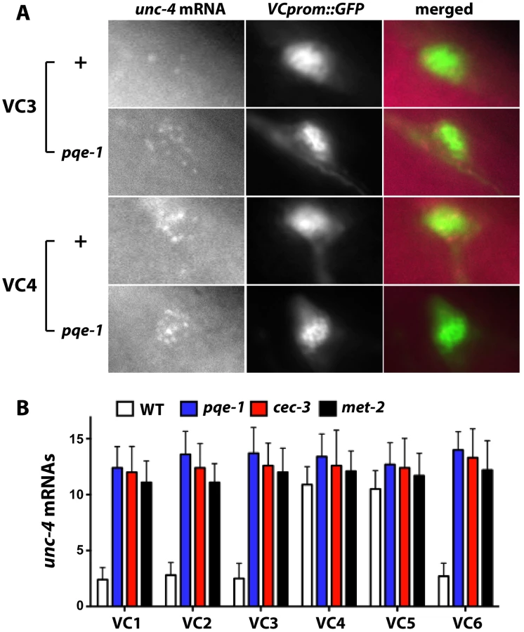
Yamada et al. reported that pqe-1 mutations increased transgene expression but not endogenous gene expression [23]. In contrast, using single molecule fluorescence in situ hybridization, which individually labels at least 80% of the cellular mRNA [24], we found that the level of endogenous unc-4 transcripts increased (Figure 3). VC1-3 and VC6 neurons in wild-type animals contained less than three fluorescently labeled unc-4 mRNA molecules (VC1 : 2.4±0.3, VC2 : 2.8±0.4, VC3 : 2.5±0.4, and VC6 : 2.7±0.4; mean ± SEM, N = 20), whereas VC4 and VC5 had about 11 labeled unc-4 transcripts (VC4 : 10.9±0.5; VC5 : 10.6±0.5). All VC neurons in pqe-1, cec-3 and met-2 mutants had >12 unc-4 transcripts. These results confirmed that unc-4 expression was significantly upregulated in VC1-3 and VC6 neurons in pqe-1, cec-3, and met-2 mutants and that uIs45 truly monitored endogenous unc-4 promoter activity.
Mutation of a subset of synMuv B genes also caused ectopic unc-4 expression
In addition to regulating unc-4 expression in the VC neurons, hpl-2, met-1, and met-2 also repress transcription of lin-3/EGF, which induces vulval development [13], [25]. Because all three genes are synMuv B genes (mutation of any of them, together with a mutation in a synMuv A gene, leads to a synthetic multivulva phenotype [26]), we tested whether other genes regulating vulval development also controlled unc-4 expression. Indeed, mutation of four other synMuv B genes, but no synMuv A genes, caused ectopic expression of unc-4p::MDM2::GFP in the VC neurons (Figure 2C). The four synMuv B genes were efl-1/E2F, which encodes a transcriptional repressor [27]; lin-13, which encodes a zinc-finger protein that forms a complex with HPL-2 and helps localize HPL-2 to certain genomic loci [28]; lin-61, which encodes a protein with four malignant brain tumor repeats that bind to di - and tri-methylated H3K9 [29], [30] and interacts genetically with hpl-2 and met-2 in vulva development [30]; and lin-65, which encodes a large acid-rich protein that lacks obvious similarity to non-nematode proteins [31].
pqe-1 and cec-3 were not, however, synMuv genes. Loss of either pqe-1 or cec-3 in the background of a class A or class B synMuv mutant, such as lin-15A or lin-15B, respectively, did not give a multivulva phenotype (data not shown). Thus, vulval and VC development utilize genetic pathways with overlapping yet divergent regulatory roles.
pqe-1 acts similarly to genes involved in epigenetic silencing
PQE-1 and proteins involved in chromatin modification and remodeling acted similarly in several different situations. cec-3, met-2, and lin-13 mutations, like pqe-1 mutations [23], enhanced transgene expression in AIZ neurons (Figure S6A), suggesting these genes act together to inhibit transcription. Similarly, loss of cec-3, met-1, met-2 and lin-13, like loss of pqe-1 [9], enhanced polyglutamine (polyQ) repeat-induced neurodegeneration in ASH neurons (Figure S6B; see Text S1 for the method). Moreover, Bates et al. found that mutation of several histone deacetylases increased this polyQ-dependent neurodegeneration [32], indicating that histone modification-induced transcriptional suppression was generally protective for polyQ-mediated neuronal cell death. The fact that PQE-1 shares similar functions with HMT and chromodomain protein hints that PQE-1 may also regulate chromatin silencing.
The vulva-inducing signal triggers unc-4 expression in VC neurons
The morphological differentiation of VC neurons requires guidance cues from vulval cells [33]. Only the vulva-flanking VC4 and VC5 neurons branch into the vulval region and innervated vulval muscle in wild-type animals. However, when the vulva is displaced anteriorly to lie between VC3 and VC4 in dig-1 mutants, the axonal branching occurs in the now vulva-flanking VC3 and VC4 neurons, but not in VC5 [33].
unc-4 expression in VC neurons also depended on similar external cues. In dig-1 mutants, VC3 and VC4 neurons flanked the misplaced vulva and expressed unc-4, whereas VC5, which was no longer adjacent to the vulva, did not express unc-4 (Figure 4A). Moreover, the positions of VC neurons were not changed in dig-1 animals, and only VC3 and VC4 underwent morphological differentiation and migrated toward the vulva. These results indicate that the proximity to the vulva determines which VC neurons become the vulval subtype and activate unc-4 transcription.

We also examined five Muv mutants, defective at various points in the pathway that controls vulval induction and development, and found they expressed uIs45 in VC neurons flanking both the vulva and pseudovulvae (Figure 4B and 4C). Thus, the unc-4 expression pattern in VC neurons is regulated by signals from vulval tissue. In addition, VC neurons flanking the pseudovulva, such as VC3 in lin-15AB animals, extended axons to ectopic vulval muscles, mimicking the normal differentiation of vulva-flanking VC4 and VC5 (Figure 4D). Since these morphological changes were not observed in ectopic VC neurons that expressed unc-4 in pqe-1 or cec-3 or other mutant animals (Figure 4D), unc-4 expression was not sufficient to induce these morphological changes. Moreover, both we and Bany et al. [6] found no defects in VC axonal processes in unc-4 mutants, suggesting that unc-4 was not needed for VC morphological differentiation. Other genetic pathways may control the axonal outgrowth of the vulval VC neurons. Therefore, the epigenetic regulation of unc-4 does not determine all the aspects of VC subtype identity.
EGFR signaling induces unc-4 expression in vulval VC neurons
Since EGFR/RAS/MAPK signaling induces vulval development and differentiation [34], we tested whether mutation of genes in this pathway eliminated the expression of unc-4 in VC4 and VC5 neurons. Indeed, animals defective in lin-3/EGF and let-23/EGFR, which are vulvaless, failed to express uIs45 in VC4 and VC5 (Figure 4C and S7A). let-60/RAS and lin-45/RAF mutant, which lack downstream effectors of EGFR, also failed to express unc-4 in vulval VC neurons (Figure 4C and S7B). Importantly, unc-4 expression in VC4 and VC5 neurons was restored in animals with hypomorphic alleles of let-60 and lin-45 by VC-specific expression of the respective wild-type gene, indicating that the EGFR/RAS/RAF signaling cascade functions cell-autonomously in VC neurons (Figure 4C and S7C).
unc-4 expression was also absent in VC neurons in mutants of sem-5/GRB2 (encoding an adaptor protein that links RAS to EGFR; [35]) and mek-2/MAPKK (encoding the downstream kinase of RAS; Figure 4C; [36]). Using the VC marker vsIs13, we have confirmed the presence of the six VC neurons in all the mutants that have diminished unc-4 expression in these cells (data not shown). These results further support the hypothesis that EGFR signaling is essential for inducing unc-4 transcription in the VC4 and VC5 neurons. The expression of unc-4 in the SAB, AVF, and I5 head neurons was not affected by mutations of the EGFR pathway, indicating that other mechanisms maintain the constant expression of unc-4 in these neurons.
We next searched for the origin of the LIN-3/EGF signal that activates unc-4 expression in VC neurons during vulval development. The two known sources of LIN-3 in vulval development are the anchor cell, which secretes LIN-3 at the middle to late L3 stage to induce primary vulval cell fate and pattern the vulval precursor cells [37] and the vulF cells, which secretes LIN-3 to signal the presumptive uv1 cells [38]. The anchor cell is likely not the source of LIN-3 for unc-4 expression in the vulval VC neurons because unc-4 expression occurred much later, after vulval precursor patterning was completed and the anchor cell invasion had started. Laser ablation of vulF cells caused the loss of unc-4 expression in the VC4 and VC5 neurons (Figure 5A), suggesting that vulF cells are responsible for releasing EGF that activates unc-4. Moreover, the vulF cells, which are physically adjacent to the vulval VC neurons, are correctly positioned to activate unc-4 expression in the latter cells with high concentrations of LIN-3/EGF. To further confirm the importance of vulF cells in inducing unc-4, we examined lin-12d mutants, in which all the six vulval precursor cells adopt the 2° vulval cell fate and the 1° lineage progeny vulF cells are not generated [39]. The strong lin-12d allele n137 caused a multivulva phenotype but had no unc-4 expression in any VC cells (N = 35 animals); the weaker lin-12d allele n302 resulted in a vulvaless phenotype and the elimination of unc-4 expression in VC4 and VC5 neurons (95% of 78 animals lacked expression in these cells). These results support the hypothesis that vulF cells are the source of the developmental signal that activates unc-4.

unc-4 expression requires LIN-11
We next wanted to identify the transcription factor (TF) that regulates unc-4 expression in vulval VC cells. Although the ETS-domain-containing TF LIN-1 is a known nuclear target of the EGFR/RAS/MAPK signaling in vulval differentiation [40], loss of lin-1 caused ectopic unc-4 expression in nonvulval VC neurons and a multivulva phenotype instead of diminishing unc-4 transcription (Figure 4C), suggesting that another TF may be involved in activating unc-4 transcription. We screened six TFs (egl-18, lin-11, tag-97, zag-1, vab-15, and hlh-3) known to be expressed in VC neurons and found that mutation of lin-11, which encodes a LIM homeodomain protein [41], eliminated uIs45 expression in VC4 and VC5 (Figure 5B). Expression of lin-11(+) using lin-11p::pes-10p, which is active in VC neurons but transiently expressed in the developing vulval cells, mainly the vulC and vulD cells but not the vulF cells [42], produced a normal unc-4 expression pattern in lin-11 mutants (Figure 5C). We obtained a similar rescue using the ida-1 promoter, which is expressed in many neurons, including the VC neurons, but not vulval cells (data not shown). Thus, the action of lin-11 on unc-4 expression was cell autonomous.
lin-11 expression in VC neurons started at the L2 stage (these cells are generated in the L1 stage) and all of the six VC neurons continued to express lin-11 in subsequent larval and adult stages (Figure 5D). In contrast, unc-4 expression in the vulval VC neurons began in the L4 stage and was absent in the nonvulval VC neurons, which also expressed LIN-11. Therefore, LIN-11 alone was not sufficient to activate unc-4 transcription. Either LIN-11 induction of unc-4 expression requires LIN-11 activation (e.g., through post-translational modification, other coactivators) or changes to downstream genes that allow it to act. Apparently upstream EGFR signaling is needed for these changes.
Epigenetic silencing of unc-4 is independent of the inductive EGFR pathway
Because genes affecting EGFR signaling activate unc-4 expression and epigenetic factors maintain unc-4 silencing in non-vulval VC neurons, we examined the relationship between these two categories of genes. pqe-1 and cec-3 were epistatic to let-60/RAS and lin-45/RAF (Figures 6), since all six VC neurons expressed unc-4 in double mutants. These results suggest that the epigenetic factors that suppress unc-4 expression are independent of the EGFR signaling that induces unc-4 transcription. Importantly, the vulval neurons VC4 and VC5 lacking the epigenetic proteins still expressed unc-4 in the absence of the inductive EGFR signaling, indicating that all six VC neurons expressed unc-4 by default once the silencing mechanism was removed. These results also suggest that the EGFR signaling in vulval VC neurons overrides epigenetic silencing. Moreover, we noticed that the unc-4 reporter expression was clear and strong from early L4 stage in pqe-1, cec-3, or met-2 mutants, whereas expression in vulval VC cells of wild-type animals was not established until the late L4 stage (data not shown). This temporal difference in the onset of unc-4 expression is consistent with the idea that the histone methylation-associated transcriptional repression is established prior to the external EGF signal, which derepresses unc-4 gene by presumably removing the repressive histone modification.

In fact, histone demethylases were also required for the derepression of unc-4 in vulval VC neurons. Among the 13 genes encoding predicted histone demethylases in C. elegans, we found that mutations in jmjd-2, jmjc-1, and spr-5 significantly reduced unc-4 expression in the VC4 and VC5 cells, but not in the unc-4-expressing head neurons (Figure 5E and Table S2). spr-5 encodes the C. elegans homolog of human LSD1. The human enzyme demethylates both H3K4me2 and H3K9me2 [43], [44], but only the H3K4me2 demethylase activity of SPR-5 has been studied in C. elegans [45]. If SPR-5 does demethylate H3K9me2, it may help remove the repressive histone modification on unc-4 gene. jmjd-2 and jmjc-1 encode homologs of human JMJD2a and MINA proteins respectively, both of which are involved in the demethylation of H3K9me3 [46]–[48], but the functions of these C. elegans proteins have not been studied. Thus, the removal of the repressive H3K9me2/3 mark could activate unc-4 expression in vulval VC neurons.
LIN-11 acts downstream of EGFR signaling to induce unc-4 expression
Given the fact that the TF LIN-11 is required to induce unc-4 expression in VC4 and VC5 cells, we expected the doubles of lin-11 with the epigenetic factors should have no unc-4 expression at all. However, to our surprise, lin-11 double mutants with pqe-1, cec-3, met-2, hpl-2, and lin-13 all showed ectopic unc-4 expression in the six VC neurons (Figure 6C). This result suggests that LIN-11 does not directly activate unc-4 transcription. Instead, LIN-11 may be the downstream target of the EGFR signaling that helps remove the repressive chromatin modification of unc-4 gene.
LIN-11 was also needed for the induction of ectopic unc-4 expression in the multivulva mutants, which have excessive EGF signals from the pseudovulvae, since the unc-4 expression in VC neurons near the pseudovulvae was prevented by mutation of lin-11 (Figure 6B). Thus, LIN-11 is required to alleviate the epigenetic silencing of unc-4 in VC neurons in response to the differentiation cue from vulval cells.
Ectopic unc-4 expression in VC neurons causes egg-laying defects
VC neurons regulate egg laying in two ways: neuromuscular synapses from VC neurons activate the vulval muscle vm2 cell and allow eggs to be laid; and extrasynaptic release of ACh as a neuromodulator from VC neurons prevents egg laying by inhibiting the HSN neurons, which promote egg laying [5]. Since UNC-4 upregulates the expression of choline acetyltransferase (CHA-1) and the synaptic vesicle ACh transporter UNC-17 post-transcriptionally, mutation of unc-4 leads to reduced release of ACh, increased HSN activity, and thus hyperactivate egg laying [49]. Mutations in cha-1 and unc-17 also result in hyperactive egg laying [6], supporting the role of ACh in inhibiting HSN activity.
Since all of the VC1-3 and VC6 neurons send out processes to the vulval region, ACh produced by these cells could reach the HSN neurons even if they don't directly synapse onto HSNs. Therefore, we reasoned that mutants with ectopic unc-4 expression in the nonvulval VC neurons could produce extra amounts of ACh, causing hypersuppression of the HSNs and reduction in egg laying. Indeed, pqe-1, cec-3, and met-2 adults retained 7–8 more eggs than wild-type animals (pqe-1: 17.8±1.2; cec-3: 18.7±0.78; met-2: 18.9±0.75; wild type: 11.1±0.4; mean ± SEM; N = 30; Figure 7A). Consistent with the increased egg-retention, the age of the eggs that were laid was older in the pqe-1 and cec-3 animals (pqe-1: 48%; and cec-3: 62% at comma stage; N = 50) than wild type (62% at 21+ cell stage and nearly no eggs at comma stage; Figure S8). VC-specific rescue of either pqe-1 or cec-3 restored normal egg retention levels, suggesting that these genes act within VC neurons. Egg retention in these animals appeared to require UNC-4–mediated activation of cha-1 and unc-17, since unc-4, cha-1, and unc-17 were epistatic to pqe-1, cec-3, and met-2; double mutants all displayed hyperactive, instead of defective, egg laying (Figures 7A and 7B).

Consistent with the model that unc-4 inhibits egg laying through its regulation of ACh, we found that pqe-1 and met-2 increased egg retention in tph-1 mutants and pqe-1, cec-3, and met-2 increased egg laying in heterozygotes containing one copy of an egl-6 gain-of-function mutation (Figure 7C). The tph-1 and egl-6 mutations reduce egg laying and thus provide a sensitized background in which to look for egg-laying defects. tph-1 encodes tryptophan hydroxylase, which synthesizes serotonin in HSN neurons [50]. Because serotonin is released by the HSN neurons to promote egg laying, mutation of tph-1 leads to reduced egg-laying activity. egl-6 encodes an FMRFamide neuropeptide receptor, the receptor of neuropeptide FLP-10 and FLP-17, which acts additively with ACh to inhibit the HSN neurons [51]. The constitutively active gain-of-function mutation of egl-6 resulted in hypersuppression of the HSN neurons and defects in egg laying.
To further confirm that the unc-4-expressing non-vulval VC neurons caused the defects in egg laying, we ablated the aberrantly differentiated nonvulval VC neurons (VC1-3 and VC6) in cec-3 and met-2 animals to correct the phenotype. Ablated mutant animals retained the same number of eggs as ablated wild-type animals (Figure 7D). In addition, the ablated cec-3 and met-2 animals laid significantly fewer late-stage eggs than unablated controls (Figure S9). Ablating the vulva-flanking VC4 and VC5 neurons in the wild-type background led to hyperactive egg laying, whereas killing the other VC neurons had very little effect (Figure 7D). These data suggest that the differentiated vulval VC neurons, which normally express unc-4, were responsible for reducing egg laying and balancing the behavioral output of the egg-laying circuit, whereas epigenetic silencing of unc-4 expression in the non-vulval VC neurons prevented a further inhibition of egg laying.
Discussion
The six VC neurons are generated in L1 larvae but diversify into subtypes, adopting different morphologies and functions at the L4 stage during vulval development. At this later time the vulva-flanking VC4 and VC5 neurons differentiate further by migrating toward the vulva, extending branched processes dorsally to innervate vulval muscles, initiating unc-4 expression, and joining the egg-laying circuit. In contrast, the other VC neurons largely maintain their original cell shape and play only a minor role in egg laying. These later cells, however, have the potential to be more like the VC4 and VC5 cells as seen in the dig-1 mutants or mutants with multiple vulvae.
The diversification of VC neurons into subtypes requires inhibition of vulval VC differentiation in the nonvulval VC neurons, which don't receive the LIN-3/EGF developmental cue. As shown here, unc-4 expression is a convenient marker for this differentiation. We found that unc-4 is silenced epigenetically in nonvulval VC neurons. This epigenetic repression involves chromatin modifiers (including the histone H3K9 methyltransferase MET-2 [13] and H3K36 methyltransferase MET-1 that may indirectly promote H3K9 methylation [13], [14]), chromatin readers (including the MBT (malignant brain tumor) domain-containing protein LIN-61 [29], the HP1-like protein HPL-2 [20]), zinc finger protein LIN-13 that helps localize HPL-2 [28]), a novel chromodomain protein CEC-3, a transcriptional repressor EFL-1 [27], and a large, acid-rich protein LIN-65 with unknown function [31]. Mutation of any of these proteins led to ectopic unc-4 expression in all six VC neurons and resulted in the loss of subtype-specificity of the expression. Because UNC-4 regulates the level of proteins needed for the synthesis and release of ACh, the failure to restrain unc-4 expression in only VC4 and VC5 caused hyperinhibition of HSN activity and defects in egg laying. Thus, histone modification contributes to terminal neuronal differentiation by generating the correct gene expression pattern in VC cells. This control is essential for the regulation of a specific behavior, egg laying.
In addition to these epigenetic proteins, we found that the Q/P-rich domain-containing protein PQE-1 also prevents unc-4 expression in non-vulval VC neurons. Although we do not know how this repression works, our results showed that PQE-1 acts in a similar way to the histone methyltransferases MET-1 and MET-2, the chromodomain protein CEC-3, and the HP1/HPL-2 binding partner LIN-13 in preventing transcription and protecting cells from polyQ neurodegeneration. Consistent with previous studies [9], we found that the C-terminal RNA exonuclease domain included in the b isoform of pqe-1 gene was dispensable for PQE-1 function, indicating the N-terminal Q/P-rich domain is mainly responsible for inhibiting unc-4 expression. Although the Q/P-rich domain is largely known to promote protein aggregation, the C-terminus of the TF TDP-43 has a Q/P-rich region that is required for its function in silencing the testis-specific gene SP-10 [52], [53]. Therefore, we speculate that the nuclearly localized PQE-1 protein may use its Q/P-rich domain to mediate transcriptional repression. Thus, Q/P-rich proteins may be a new class of epigenetic control factors.
Previous studies showed that repressive epigenetic modifications are essential for silencing critical developmental genes and inhibiting differentiation in stem cells. ESCs deficient in PcG proteins, which promote H3K27 trimethylation and suppress transcription, derepressed neural genes, such as Ngns, Pax-6, and Sox-1, and were prone to differentiate [2]. Loss of the transcriptional repressor REST (RE1-silencing TF, which recruits histone modifiers and chromatin-binding proteins) or inhibition of DNA methyltransferase and histone deacetylases (both of which suppress transcription) induces aberrant differentiation and derepression of genes related to neurogenesis in ESCs and NPCs [54]–[56]. The H3K9 methyltransferase SetDB1 also contributes to the repression of genes encoding developmental regulators and to the maintenance of ESCs [57]. Consistent with these findings, we find that mutation of histone methyltransferase met-2 that is homologous to SetDB1 and promotes H3K9 methylation abrogated the repression of terminal differentiation marker unc-4 in undifferentiated VC neurons.
Although our results suggest that H3K9 methylation is important for the regulation of unc-4 expression, we cannot definitively determine whether dimethylation or trimethylation is important. Unlike SetDB1, which specifically trimethylates H3K9, MET-2, the C. elegans homolog of SetDB1, mediates mono - and dimethylation of H3K9 [17]. Another SET domain protein, SET-25, which is homologous to the mammalian EHMT1/G9a and Suv39h1/2, is responsible for H3K9 trimethylation in early embryos [17], but its role in larvae and adults has not been examined. set-25 mutants did not show ectopic unc-4 expression, so either H3K9 dimethylation is responsible for unc-4 silencing or MET-2 or another HMT promotes H3K9 trimethylation of unc-4 DNA in adult VC cells. Our finding that the release of unc-4 epigenetic silencing needed jmjd-2 and jmjc-1, homologs of mammalian demethylases that have demethylation activities on H3K9me3 [46]–[48], argues for the second hypothesis. Among the chromatin readers that are important for unc-4 repression, LIN-61 and HPL-2 directly binds to H3K9me2/3 [22], [29], but the ability of CEC-3 to bind to H3K9me2/3 remains to be determined. Although HPL-2 can be indirectly recruited to H3K27me3 [21], our results suggest that H3K27 trimethylation is not the likely cause of unc-4 silencing because neither the polycomb complex components that catalyze H3K27 trimethylation nor the predicted H3K27me2/3 demethylases had any effect on unc-4 expression.
Developmental signals can reverse H3K9 methylation-mediated gene silencing in undifferentiated neurons to allow the differentiation to proceed. In this study, we found that EGF/LIN-3 from the developing vulF cells acts through the EGFR/RAS/MAPK signaling pathway during vulval development to override the epigenetic silencing of unc-4 in VC neurons that are close to vulF cells. The EGF signal probably failed to activate unc-4 expression in the nonvulval VC neurons because the physical distance between these cells and the EGF source was too great. At least three events are needed for the correct timing of unc-4 expression in VC4-5 : 1) epigenetic silencing prevents unc-4 expression in the early L4 stage (unc-4 is expressed in early L4 cells in cec-3 or met-2 mutants); 2) induction involving EGF, etc. leads to unc-4 expression in the late L4; and 3) additional factors (the presence of a negative factor or the absence of a positive factor) prevent unc-4 expression from the L1 to the late L3 stage. Therefore, unc-4 transcription would be on by default in all six VC cells if H3K9 methylation did not inactivate unc-4 prior to the differentiation cue. The epigenetic inhibition is normally relieved in VC4-5 neurons by the external signal from the developing vulva to allow the derepression of unc-4 gene and neuronal differentiation. Although the morphological differentiation of VC neurons is not controlled by the epigenetic mechanisms, our data demonstrate that H3K9 methylation helps create subtype-specific gene expression patterns during terminal neuronal diversification.
We have also found that LIM-domain transcription factor LIN-11 is required to derepress unc-4 in the vulval VC neurons. Since lin-11 acts similarly as the EGFR signaling genes in genetic interaction studies, LIN-11 is likely to be part of the EGFR pathway, probably the downstream target of the signaling, and helps remove the repressive histone modification. Although there is no report showing the direct involvement of LIM domain proteins in histone demethylation, a close correlation between the expression patterns of lysine-specific histone demethylase 1 (LSD1) and four and a half LIM-domain protein 2 (FHL2) was found in prostate cancer [58]. Since both FHL2 and LSD1 serve as coactivators of the androgen receptor [59], the LIM domain protein may interact with the histone demethylase to activate gene expression. Since histone demethylase SPR-5/LSD1 was also required for the unc-4 VC expression, EGFR signaling is likely to activate LIN-11, resulting in the removal of H3K9 methylation and the derepression of unc-4.
The fact that vulF cells from the developing vulva send out EGF signal to induce VC neuron differentiation suggested a coordination between the formation of the epithelial vulval structure and the differentiation of the egg-laying neurons. In fact, signals from the 1° vulval cells also control the axonal outgrowth, branching and fasciculation of the HSN neurons [60], which together with VC neurons form the egg-laying circuit. Moreover, only the vulva-flanking VC neurons undergo morphological changes and express unc-4, and similarly the proximity of the HSN cell body to vulval cells is important for HSN axonal guidance [60], supporting the hypothesis that communication between the vulval epithelial cells and neurons depends on secreted extracellular molecules. As VC and HSN neurons are generated much earlier than the vulva, signals from the developing vulva need to activate these neurons by regulating gene expression, inducing axonal outgrowth, and eventually joining them together to form the neural circuit. Therefore, the terminal differentiation of these neurons requires highly coordinated cell-cell interactions with the epithelial tissues.
Finally, our findings are consistent with recent discoveries on the epigenetic regulation of cellular differentiation by H3K9 methylation in various tissues. Ling et al. found that mutation of the H3K9 methyltransferase G9a induced abnormal myogenesis during skeletal muscle differentiation by de-repressing the transcription of myogenic regulatory factor MyoD [61], and Herzog et al. found that the histone demethylase Kdm3a, which removes H3K9 methyl groups, is essential for the differentiation of mouse embryonic carcinoma cells into parietal endoderm-like cells in a mouse embryonal carcinoma model [62]. These studies demonstrate that H3K9 methylation silences developmental genes and prevents aberrant differentiation, and that the removal of this histone modification allows normal differentiation to proceed. Our studies extend these observations by showing that H3K9 methylation helps maintain the ground state among similar neurons by silencing key genes associated with terminal differentiation.
Materials and Methods
Strains
C. elegans strains were maintained at 15°C or 20°C as described by Brenner [63]. Temperature-sensitive strains were maintained at 15°C and transferred to 25°C for one generation before testing. Most strains were provided by the Caenorhabditis Genetics Center, which is funded by NIH Office of Research Infrastructure Programs (P40 OD010440). cec-3(ok3432) II and pqe-1(ok1983) III were generated by the International C. elegans Gene Knockout Consortium (http://www.celeganskoconsortium.omrf.org). jmjc-1(tm3525) I, jmjd-2(tm2966) II, hpl-2(tm1489) III, and hpl-1(tm1624) X were obtained from the National BioResource Project of Japan (http://www.shigen.nig.ac.jp/c.elegans/index.jsp). cec-3(u830) and pqe-1 alleles (u825, u829, u831, u832, u900, u901, u902, and u903) were isolated in this study. peIs304 and pqe-1(ok1983); peIs304 strains were kindly provided by Dr. Yuichi Iino (University of Tokyo).
Constructs and transgenes
The unc-4p::MDM2::GFP vector TU#703, which contains a 2.5 kb unc-4 promoter and DNA encoding a truncated and mutated RING domain from human MDM2 attached to GFP [8], was injected together with pRF4 (containing a dominant roller marker) to generate uIs45, which was mapped onto the X chromosome. We also replaced GFP with RFP in TU#703 to create TU#1101 unc-4p::MDM2::RFP, which was used to generate transgene uIs147. To examine the unc-4 expression pattern in various genetic backgrounds, we crossed uIs45 into most of the mutants of interest except for these X-linked mutations, which were crossed with uIs147.
A VC-specific promoter, which contains a ∼500 bp lin-11 promoter and a basal pes-10 promoter, was subcloned into the Gateway pDONR P4-P1R from pDM4 vector. The pDM4 vector was a gift from Michael Koelle (Yale University). The coding regions of pqe-1, cec-3, lin-45, let-60, and lin-11 genes were all cloned from wild type (N2) genomic DNA into Gateway pDONR 221. The resulted entry vectors, together with pENTR-VCprom, pENTR-unc-54-3′UTR, and destination vector pDEST-R4-R3 were used in the LR reaction to create the final VC promoter-driven expression vectors. The Gateway cloning method can be found at http://www.invitrogen.com/site/us/en/home/Products-and-Services/Applications/Cloning/Gateway-Cloning.html by Life Technologies (Grand Island, NY). These constructs were injected into corresponding mutants to form extrachromosomal arrays to test the VC-specific rescue of the mutant phenotype. The transgene vsIs13[lin-11p::pes-10p::GFP] was used as a VC-specific marker [6].
Whole genome sequencing and identification of phenotype-causing mutations
After visually isolating the mutants, we outcrossed the mutant strains (u825, u830, and u834) with N2 at least 10 times and then subjected them to whole genome sequencing using an Illumina GAII genome analyzer [64]. We first identified the genetic variants by aligning the sequencing data to Wormbase reference sequences (version WS220) with MAQGene [65] and subtracting the background variants found in our wild-type strain. By visualizing the genomic positions of these variants, we discovered a variant-enriched region, which theoretically contained the phenotype-causing mutation because this region had the least chance to be recombined with wild-type chromosomes under constant selection. Within this ∼1 Mb region, we identified candidate mutations and performed complementation tests with known alleles to find the gene associated with the phenotype. We confirmed the results by PCR genotyping, testing knockout alleles of candidate genes, and injecting the cosmid or fosmid containing the wild-type copy of the gene into mutant animals and testing for rescue.
Single-molecule fluorescent in situ hybridization and microscopy
Single-molecule fluorescence in situ hybridization was performed on young adult animals as described [24]. Forty-eight 20-nucleotide probes for unc-4 mRNA were designed using the program at www.biosearchtech.com/customoligos and synthesized and coupled to Cy5 by BioSearch Technologies (Novato, CA). We imaged the animals using a Zeiss Axio Observer Z1 inverted microscope with a CoolSNAP HQ2-FW camera (Photometrics, Tucson, AZ) and appropriate filters for Cy5. We collected stacks of 20–35 images spaced 0.3 µm apart for each individual neuron and counted the number of fluorescent spots per neuron.
Other imaging was conducted on either the same Zeiss Observer Z1 microscope with the CoolSnap camera or a Zeiss Axioskop II with a SPOT-2 slider camera (SPOT Imaging Solutions, Sterling Heights, MI).
Laser ablation
Ablations were performed as described previously [66]. Briefly, L4 larvae of strains carrying either uIs45 or vsIs13 as VC markers were placed in 1 µl of M9 buffer on a 2% agarose pad containing 1 mM sodium azide. GFP-positive cells were identified using a Zeiss Axioplan 2 equipped with a Micropoint Laser System (Photonic Instruments, Inc.), and their nucleoli were repeatedly targeted with the laser until they appeared ruptured. Mock-ablated animals were placed on the same pad and exposed to fluorescence excitation light for the same period of time, but not shot with the laser. The animals were recovered and examined 24–30 hours later with a fluorescence dissecting microscope to ensure absence of GFP-positive cells. Thirty hours after the ablation procedure, animals were assayed for egg-laying activity. To ablate vulF cells, syIs66[B0034.1::pes-10::GFP] was crossed into uIs45 to help visualize the VulF cells, and unc-4 expression in VC neurons was examined 24 hours after the ablation.
Egg-laying assay
The average number of unlaid eggs in the uterus and the percentage of freshly laid eggs at various stages were quantified as described [51], [67]. We collected late L4 animals and cultured then at 20°C for 36 hr. In the unlaid egg assay, 30 synchronized adults were individually dissolved in 5% sodium hypochlorite, and their eggs were counted. In the egg-staging assay, 20 staged adults were placed on a thin lawn of OP50 bacteria and allowed to lay eggs for 1 hr. Each egg was examined under a dissecting microscope and categorized into six different stages according to previous studies (Ringstad and Horvitz, 2008). Eggs with eight cells or fewer were classified as “early stage”. Eggs at the comma stage or later stages were classified as “late stage”. Every experiment was repeated three times independently.
Supporting Information
Zdroje
1. HsiehJ, GageFH (2004) Epigenetic control of neural stem cell fate. Curr Opin Genet Dev 14 : 461–469.
2. BoyerLA, PlathK, ZeitlingerJ, BrambrinkT, MedeirosLA, et al. (2006) Polycomb complexes repress developmental regulators in murine embryonic stem cells. Nature 441 : 349–353.
3. LeeTI, JennerRG, BoyerLA, GuentherMG, LevineSS, et al. (2006) Control of developmental regulators by Polycomb in human embryonic stem cells. Cell 125 : 301–313.
4. Roman-TruferoM, Mendez-GomezHR, PerezC, HijikataA, FujimuraY, et al. (2009) Maintenance of undifferentiated state and self-renewal of embryonic neural stem cells by Polycomb protein Ring1B. Stem Cells 27 : 1559–1570.
5. ZhangM, ChungSH, Fang-YenC, CraigC, KerrRA, et al. (2008) A self-regulating feed-forward circuit controlling C. elegans egg-laying behavior. Curr Biol 18 : 1445–1455.
6. BanyIA, DongMQ, KoelleMR (2003) Genetic and cellular basis for acetylcholine inhibition of Caenorhabditis elegans egg-laying behavior. J Neurosci 23 : 8060–8069.
7. Von StetinaSE, FoxRM, WatkinsKL, StarichTA, ShawJE, et al. (2007) UNC-4 represses CEH-12/HB9 to specify synaptic inputs to VA motor neurons in C. elegans. Genes Dev 21 : 332–346.
8. PoyurovskyMV, JacqX, MaC, Karni-SchmidtO, ParkerPJ, et al. (2003) Nucleotide binding by the Mdm2 RING domain facilitates Arf-independent Mdm2 nucleolar localization. Mol Cell 12 : 875–887.
9. FaberPW, VoisineC, KingDC, BatesEA, HartAC (2002) Glutamine/proline-rich PQE-1 proteins protect Caenorhabditis elegans neurons from huntingtin polyglutamine neurotoxicity. Proc Natl Acad Sci U S A 99 : 17131–17136.
10. ZahnTR, MacmorrisMA, DongW, DayR, HuttonJC (2001) IDA-1, a Caenorhabditis elegans homolog of the diabetic autoantigens IA-2 and phogrin, is expressed in peptidergic neurons in the worm. J Comp Neurol 429 : 127–143.
11. WhiteJG, SouthgateE, ThomsonJN, BrennerS (1986) The structure of the nervous system of the nematode Caenorhabditis elegans. Philos Trans R Soc Lond B Biol Sci 314 : 1–340.
12. EissenbergJC (2012) Structural biology of the chromodomain: form and function. Gene 496 : 69–78.
13. AndersenEC, HorvitzHR (2007) Two C. elegans histone methyltransferases repress lin-3 EGF transcription to inhibit vulval development. Development 134 : 2991–2999.
14. LamelzaP, BhallaN (2012) Histone methyltransferases MES-4 and MET-1 promote meiotic checkpoint activation in Caenorhabditis elegans. PLoS Genet 8: e1003089.
15. CloughE, MoonW, WangS, SmithK, HazelriggT (2007) Histone methylation is required for oogenesis in Drosophila. Development 134 : 157–165.
16. SchultzDC, AyyanathanK, NegorevD, MaulGG, RauscherFJ3rd (2002) SETDB1: a novel KAP-1-associated histone H3, lysine 9-specific methyltransferase that contributes to HP1-mediated silencing of euchromatic genes by KRAB zinc-finger proteins. Genes Dev 16 : 919–932.
17. TowbinBD, Gonzalez-AguileraC, SackR, GaidatzisD, KalckV, et al. (2012) Step-wise methylation of histone H3K9 positions heterochromatin at the nuclear periphery. Cell 150 : 934–947.
18. RandoOJ (2007) Global patterns of histone modifications. Curr Opin Genet Dev 17 : 94–99.
19. RechtsteinerA, ErcanS, TakasakiT, PhippenTM, EgelhoferTA, et al. (2010) The histone H3K36 methyltransferase MES-4 acts epigenetically to transmit the memory of germline gene expression to progeny. PLoS Genet 6: e1001091.
20. SchottS, CousthamV, SimonetT, BedetC, PalladinoF (2006) Unique and redundant functions of C. elegans HP1 proteins in post-embryonic development. Dev Biol 298 : 176–187.
21. StudenckaM, WesolowskiR, OpitzL, Salinas-RiesterG, WisniewskiJR, et al. (2012) Transcriptional repression of Hox genes by C. elegans HP1/HPL and H1/HIS-24. PLoS Genet 8: e1002940.
22. StudenckaM, KonzerA, MoneronG, WenzelD, OpitzL, et al. (2012) Novel roles of Caenorhabditis elegans heterochromatin protein HP1 and linker histone in the regulation of innate immune gene expression. Mol Cell Biol 32 : 251–265.
23. YamadaK, TsuchiyaJ, IinoY (2012) Mutations in the pqe-1 gene enhance transgene expression in Caenorhabditis elegans. G3 (Bethesda) 2 : 741–751.
24. RajA, van den BogaardP, RifkinSA, van OudenaardenA, TyagiS (2008) Imaging individual mRNA molecules using multiple singly labeled probes. Nat Methods 5 : 877–879.
25. CouteauF, GuerryF, MullerF, PalladinoF (2002) A heterochromatin protein 1 homologue in Caenorhabditis elegans acts in germline and vulval development. EMBO Rep 3 : 235–241.
26. CuiM, ChenJ, MyersTR, HwangBJ, SternbergPW, et al. (2006) SynMuv genes redundantly inhibit lin-3/EGF expression to prevent inappropriate vulval induction in C. elegans. Dev Cell 10 : 667–672.
27. CeolCJ, HorvitzHR (2001) dpl-1 DP and efl-1 E2F act with lin-35 Rb to antagonize Ras signaling in C. elegans vulval development. Mol Cell 7 : 461–473.
28. CousthamV, BedetC, MonierK, SchottS, KaraliM, et al. (2006) The C. elegans HP1 homologue HPL-2 and the LIN-13 zinc finger protein form a complex implicated in vulval development. Dev Biol 297 : 308–322.
29. HarrisonMM, LuX, HorvitzHR (2007) LIN-61, one of two Caenorhabditis elegans malignant-brain-tumor-repeat-containing proteins, acts with the DRM and NuRD-like protein complexes in vulval development but not in certain other biological processes. Genetics 176 : 255–271.
30. Koester-EiserfunkeN, FischleW (2011) H3K9me2/3 binding of the MBT domain protein LIN-61 is essential for Caenorhabditis elegans vulva development. PLoS Genet 7: e1002017.
31. CeolCJ, StegmeierF, HarrisonMM, HorvitzHR (2006) Identification and classification of genes that act antagonistically to let-60 Ras signaling in Caenorhabditis elegans vulval development. Genetics 173 : 709–726.
32. BatesEA, VictorM, JonesAK, ShiY, HartAC (2006) Differential contributions of Caenorhabditis elegans histone deacetylases to huntingtin polyglutamine toxicity. J Neurosci 26 : 2830–2838.
33. LiC, ChalfieM (1990) Organogenesis in C. elegans: positioning of neurons and muscles in the egg-laying system. Neuron 4 : 681–695.
34. SundaramMV (2006) RTK/Ras/MAPK signaling. WormBook 1–19.
35. ClarkSG, SternMJ, HorvitzHR (1992) C. elegans cell-signalling gene sem-5 encodes a protein with SH2 and SH3 domains. Nature 356 : 340–344.
36. WuY, HanM, GuanKL (1995) MEK-2, a Caenorhabditis elegans MAP kinase kinase, functions in Ras-mediated vulval induction and other developmental events. Genes Dev 9 : 742–755.
37. KatzWS, HillRJ, ClandininTR, SternbergPW (1995) Different levels of the C. elegans growth factor LIN-3 promote distinct vulval precursor fates. Cell 82 : 297–307.
38. ChangC, NewmanAP, SternbergPW (1999) Reciprocal EGF signaling back to the uterus from the induced C. elegans vulva coordinates morphogenesis of epithelia. Curr Biol 9 : 237–246.
39. GreenwaldIS, SternbergPW, HorvitzHR (1983) The lin-12 locus specifies cell fates in Caenorhabditis elegans. Cell 34 : 435–444.
40. BeitelGJ, TuckS, GreenwaldI, HorvitzHR (1995) The Caenorhabditis elegans gene lin-1 encodes an ETS-domain protein and defines a branch of the vulval induction pathway. Genes Dev 9 : 3149–3162.
41. FreydG, KimSK, HorvitzHR (1990) Novel cysteine-rich motif and homeodomain in the product of the Caenorhabditis elegans cell lineage gene lin-11. Nature 344 : 876–879.
42. GuptaBP, WangM, SternbergPW (2003) The C. elegans LIM homeobox gene lin-11 specifies multiple cell fates during vulval development. Development 130 : 2589–2601.
43. ShiY, LanF, MatsonC, MulliganP, WhetstineJR, et al. (2004) Histone demethylation mediated by the nuclear amine oxidase homolog LSD1. Cell 119 : 941–953.
44. MetzgerE, WissmannM, YinN, MullerJM, SchneiderR, et al. (2005) LSD1 demethylates repressive histone marks to promote androgen-receptor-dependent transcription. Nature 437 : 436–439.
45. KatzDJ, EdwardsTM, ReinkeV, KellyWG (2009) A C. elegans LSD1 demethylase contributes to germline immortality by reprogramming epigenetic memory. Cell 137 : 308–320.
46. ChenZ, ZangJ, WhetstineJ, HongX, DavrazouF, et al. (2006) Structural insights into histone demethylation by JMJD2 family members. Cell 125 : 691–702.
47. WhetstineJR, NottkeA, LanF, HuarteM, SmolikovS, et al. (2006) Reversal of histone lysine trimethylation by the JMJD2 family of histone demethylases. Cell 125 : 467–481.
48. LuY, ChangQ, ZhangY, BeezholdK, RojanasakulY, et al. (2009) Lung cancer-associated JmjC domain protein mdig suppresses formation of tri-methyl lysine 9 of histone H3. Cell Cycle 8 : 2101–2109.
49. LickteigKM, DuerrJS, FrisbyDL, HallDH, RandJB, et al. (2001) Regulation of neurotransmitter vesicles by the homeodomain protein UNC-4 and its transcriptional corepressor UNC-37/groucho in Caenorhabditis elegans cholinergic motor neurons. J Neurosci 21 : 2001–2014.
50. SzeJY, VictorM, LoerC, ShiY, RuvkunG (2000) Food and metabolic signalling defects in a Caenorhabditis elegans serotonin-synthesis mutant. Nature 403 : 560–564.
51. RingstadN, HorvitzHR (2008) FMRFamide neuropeptides and acetylcholine synergistically inhibit egg-laying by C. elegans. Nat Neurosci 11 : 1168–1176.
52. AbhyankarMM, UrekarC, ReddiPP (2007) A novel CpG-free vertebrate insulator silences the testis-specific SP-10 gene in somatic tissues: role for TDP-43 in insulator function. J Biol Chem 282 : 36143–36154.
53. FuentealbaRA, UdanM, BellS, WegorzewskaI, ShaoJ, et al. (2010) Interaction with polyglutamine aggregates reveals a Q/N-rich domain in TDP-43. J Biol Chem 285 : 26304–26314.
54. BallasN, GrunseichC, LuDD, SpehJC, MandelG (2005) REST and its corepressors mediate plasticity of neuronal gene chromatin throughout neurogenesis. Cell 121 : 645–657.
55. SinghSK, KagalwalaMN, Parker-ThornburgJ, AdamsH, MajumderS (2008) REST maintains self-renewal and pluripotency of embryonic stem cells. Nature 453 : 223–227.
56. HsiehJ, NakashimaK, KuwabaraT, MejiaE, GageFH (2004) Histone deacetylase inhibition-mediated neuronal differentiation of multipotent adult neural progenitor cells. Proc Natl Acad Sci U S A 101 : 16659–16664.
57. BilodeauS, KageyMH, FramptonGM, RahlPB, YoungRA (2009) SetDB1 contributes to repression of genes encoding developmental regulators and maintenance of ES cell state. Genes Dev 23 : 2484–2489.
58. KahlP, GullottiL, HeukampLC, WolfS, FriedrichsN, et al. (2006) Androgen receptor coactivators lysine-specific histone demethylase 1 and four and a half LIM domain protein 2 predict risk of prostate cancer recurrence. Cancer Res 66 : 11341–11347.
59. MullerJM, IseleU, MetzgerE, RempelA, MoserM, et al. (2000) FHL2, a novel tissue-specific coactivator of the androgen receptor. EMBO J 19 : 359–369.
60. GarrigaG, DesaiC, HorvitzHR (1993) Cell interactions control the direction of outgrowth, branching and fasciculation of the HSN axons of Caenorhabditis elegans. Development 117 : 1071–1087.
61. LingBM, BharathyN, ChungTK, KokWK, LiS, et al. (2012) Lysine methyltransferase G9a methylates the transcription factor MyoD and regulates skeletal muscle differentiation. Proc Natl Acad Sci U S A 109 : 841–846.
62. HerzogM, JosseauxE, DedeurwaerderS, CalonneE, VolkmarM, et al. (2012) The histone demethylase Kdm3a is essential to progression through differentiation. Nucleic Acids Res 40 : 7219–7232.
63. BrennerS (1974) The genetics of Caenorhabditis elegans. Genetics 77 : 71–94.
64. SarinS, PrabhuS, O'MearaMM, Pe'erI, HobertO (2008) Caenorhabditis elegans mutant allele identification by whole-genome sequencing. Nat Methods 5 : 865–867.
65. BigelowH, DoitsidouM, SarinS, HobertO (2009) MAQGene: software to facilitate C. elegans mutant genome sequence analysis. Nat Methods 6 : 549.
66. BargmannCI, AveryL (1995) Laser killing of cells in Caenorhabditis elegans. Methods Cell Biol 48 : 225–250.
67. KoelleMR, HorvitzHR (1996) EGL-10 regulates G protein signaling in the C. elegans nervous system and shares a conserved domain with many mammalian proteins. Cell 84 : 115–125.
Štítky
Genetika Reprodukční medicínaČlánek vyšel v časopise
PLOS Genetics
2013 Číslo 12
- Kazuistika – Perspektivy využití precizované medicíny v rámci personalizované specifické terapie onkologických pacientů
- Nobelova cena za chemii pro genetické nůžky: Objev, který změní naši budoucnost?
- Technologie na bázi RNA v klinické praxi: od přebarvených petúnií k terapii vzácných a dosud jen obtížně léčitelných chorob u lidí
- „Nepředstavovali jsme si, že náš výzkum povede přímo ke vzniku nových léků, dokonce ještě za našeho života“
- Bezplatné služby pro diagnostiku ATTRv amyloidózy pro kardiology
Nejčtenější v tomto čísle
- The NuRD Chromatin-Remodeling Enzyme CHD4 Promotes Embryonic Vascular Integrity by Transcriptionally Regulating Extracellular Matrix Proteolysis
- MAN1B1 Deficiency: An Unexpected CDG-II
- Mutations in the UQCC1-Interacting Protein, UQCC2, Cause Human Complex III Deficiency Associated with Perturbed Cytochrome Protein Expression
- The Midline Protein Regulates Axon Guidance by Blocking the Reiteration of Neuroblast Rows within the Drosophila Ventral Nerve Cord
Zvyšte si kvalifikaci online z pohodlí domova
Mazová zátka a její řešení
nový kurzVšechny kurzy