
The Effect of Vaccination on the Evolution and Population Dynamics of Avian Paramyxovirus-1
Newcastle Disease Virus (NDV) is a pathogenic strain of avian paramyxovirus (aPMV-1) that is among the most serious of disease threats to the poultry industry worldwide. Viral diversity is high in aPMV-1; eight genotypes are recognized based on phylogenetic reconstruction of gene sequences. Modified live vaccines have been developed to decrease the economic losses caused by this virus. Vaccines derived from avirulent genotype II strains were developed in the 1950s and are in use globally, whereas Australian strains belonging to genotype I were developed as vaccines in the 1970s and are used mainly in Asia. In this study, we evaluated the consequences of attenuated live virus vaccination on the evolution of aPMV-1 genotypes. There was phylogenetic incongruence among trees based on individual genes and complete coding region of 54 full length aPMV-1 genomes, suggesting that recombinant sequences were present in the data set. Subsequently, five recombinant genomes were identified, four of which contained sequences from either genotype I or II. The population history of vaccine-related genotype II strains was distinct from other aPMV-1 genotypes; genotype II emerged in the late 19th century and is evolving more slowly than other genotypes, which emerged in the 1960s. Despite vaccination efforts, genotype II viruses have experienced constant population growth to the present. In contrast, other contemporary genotypes showed population declines in the late 1990s. Additionally, genotype I and II viruses, which are circulating in the presence of homotypic vaccine pressure, have unique selection profiles compared to nonvaccine-related strains. Collectively, these data show that vaccination with live attenuated viruses has changed the evolution of aPMV-1 by maintaining a large effective population size of a vaccine-related genotype, allowing for coinfection and recombination of vaccine and wild type strains, and by applying unique selective pressures on viral glycoproteins.
Published in the journal:
. PLoS Pathog 6(4): e32767. doi:10.1371/journal.ppat.1000872
Category:
Research Article
doi:
https://doi.org/10.1371/journal.ppat.1000872
Summary
Newcastle Disease Virus (NDV) is a pathogenic strain of avian paramyxovirus (aPMV-1) that is among the most serious of disease threats to the poultry industry worldwide. Viral diversity is high in aPMV-1; eight genotypes are recognized based on phylogenetic reconstruction of gene sequences. Modified live vaccines have been developed to decrease the economic losses caused by this virus. Vaccines derived from avirulent genotype II strains were developed in the 1950s and are in use globally, whereas Australian strains belonging to genotype I were developed as vaccines in the 1970s and are used mainly in Asia. In this study, we evaluated the consequences of attenuated live virus vaccination on the evolution of aPMV-1 genotypes. There was phylogenetic incongruence among trees based on individual genes and complete coding region of 54 full length aPMV-1 genomes, suggesting that recombinant sequences were present in the data set. Subsequently, five recombinant genomes were identified, four of which contained sequences from either genotype I or II. The population history of vaccine-related genotype II strains was distinct from other aPMV-1 genotypes; genotype II emerged in the late 19th century and is evolving more slowly than other genotypes, which emerged in the 1960s. Despite vaccination efforts, genotype II viruses have experienced constant population growth to the present. In contrast, other contemporary genotypes showed population declines in the late 1990s. Additionally, genotype I and II viruses, which are circulating in the presence of homotypic vaccine pressure, have unique selection profiles compared to nonvaccine-related strains. Collectively, these data show that vaccination with live attenuated viruses has changed the evolution of aPMV-1 by maintaining a large effective population size of a vaccine-related genotype, allowing for coinfection and recombination of vaccine and wild type strains, and by applying unique selective pressures on viral glycoproteins.
Introduction
Live attenuated virus vaccines have been successfully employed in veterinary medicine to prevent the economic impact of many diseases in poultry and livestock. However, the role of vaccination with attenuated viruses on the evolution of wild type strains is not often considered. Antigenic escape because of strong selection by vaccines, emergence of new strains through recombination, and increased virulence to expedite transmission of new genotypes in vaccinated populations are of potential concern. In this paper, we explored the consequences of vaccination on the evolution of class II aPMV-1, which is the etiological agent of ND.
NDV, a single-stranded, non-segmented, negative-sense RNA virus of the genus Avulavirus, family Paramyxoviridae, infects a wide range of domestic and wild bird species worldwide, and causes a significant economic burden to the poultry industry [1]. The first outbreaks of NDV were reported during the mid 1920s in Java, Indonesia and Newcastle-upon-Tyne, England [2], and within a few years were occurring throughout the world [3]. The name ND is reserved exclusively for the disease that results from infection with strains of aPMV-1 that are pathogenic for domestic chickens [4]. aPMV-1 has been grouped by virulence phenotype, with lentogenic, mesogenic, and velogenic strains representing increasing levels of virulence, ranging from subclinical infections with moderate respiratory involvement to extensive hemorrhagic lesions and neurological signs [5]. Inactivated vaccines were first made commercially available to the poultry industry in 1946, but because they provided incomplete protection against ND [6], they were replaced with live lentogenic NDV vaccines. Although these vaccines reduce disease, they do not always prevent infection and birds can shed both vaccine and challenge strains of the virus [7], [8], [9].
aPMV-1 genome size is approximately 15 kb and encodes six genes, which produce nucleocapsid protein (NP), phosphoprotein (P), matrix protein (M), fusion protein (F), hemagglutinin-neuraminidase (HN), and polymerase protein (L) [10], [11]. RNA-editing of P gene creates two additional proteins, V and W [12]. There are nine serotypes of aPMV-1; viruses associated with ND are in serogroup 1. Within serogroup 1 there are 2 major subdivisions, class I and II, based on phylogenetic grouping of the F gene [13]. Class I aPMV-1 are primarily recovered from waterfowl or samples from U.S. live bird markets, while the isolates from class II are commonly derived from poultry and other avian species [5], [14]. Eight genotypes of class II aPMV-1 can be identified [13]. Viruses belonging to genotypes I-IV have circulated since the 1930's. Genotype I and II consist of both lentogenic and velogenic viruses and have been associated with ND outbreaks in Australia and North America, respectively [5], [15], [16]. These viruses have been attenuated in culture and are used as modified live vaccines [16]. Genotypes V-VIII were first recognized in the mid-1960s [13] and contained only virulent viruses [16]. Genotype V was responsible for the second panzootic of ND in Europe from 1970–1974 and has been detected sporadically thereafter [17]. Genotype VI was described mainly from the Middle East and Asia during the 1980's–1990's [18] and genotype VII and VIII were reported in the 1990's from several countries [19], [20], [21], [22], [23], [24]. All genotypes, except IV [16], are still in circulation.
RNA viruses typically have a high mutation rate due to low fidelity and processivity of their polymerase [25], which coupled with a high replication rate and short generation time [26] lead to high evolutionary rates. In addition, evidence is accumulating that recombination is an important process driving genotype diversity for many RNA viruses [27], [28], [29]. Although recombination was not thought to contribute to aPMV-1 evolution [30], [31], evidence of recombination in NDV has recently been reported [32], [33], [34]. This debate may be due, in part, to the reliance on a single gene for determining virus diversity and phylogeny. Because recombination can lead to the emergence of novel virus strains of unknown virulence [35], [36], [37], [38], a better understanding of the role of recombination in circulating aPMV-1 is warranted.
In this study, we explored how vaccination strategies in poultry farming have shaped the evolution of this important avian virus using complete genome sequences available in GenBank. Specifically our objectives were to 1) determine if recombination was evident among full length class II aPMV-1; 2) estimate evolutionary rates of each genotype; 3) estimate the effective population size of each genotype; and 4) determine the selective forces on vaccine-related and nonvaccine-related wild type genotypes. Our results confirm that recombination is an important process in this negative sense RNA virus and that vaccine-related strains have an evolutionary history that is unique from nonvaccine-related strains, which includes distinct evolutionary rates, temporal changes in population size, and selection profiles.
Results
Phylogenomic analyses
The current phylogenetic classification of aPMV-1 strains is based on either full or partial nucleotide sequence of the M, F, or L genes. To determine if all genes in the viral genome provided consistent phylogenetic profiles, we obtained 54 full length class II aPMV-1 sequences from Genbank and generated nucleotide data sets for each of the six genes and a concatenated sequence of all protein coding regions. Maximum likelihood (ML) phylogenies were reconstructed for all sequence data sets under the appropriate nucleotide substitution model selected for each data set (Figure 1). Each gene tree and the concatenated tree revealed seven distinct genotypes within the class II aPMV-1. However, genotype affiliations were not congruent among different genes (Figure 1A). While genotype III, IV, V, VI, and VII were monophyletic, genotype I and II showed inconsistent phylogenetic relationships (Figure 1A and B). Three distinct patterns of affiliations of genotype I and II were observed among different gene trees. The NP, HN, L, and concatenated gene trees consistently placed genotypes I and II in a sister clade to other genotypes. In the P gene tree, genotype II formed a basal clade while genotype I clustered with the remaining genotypes and in the M and F gene trees, genotype I was the basal clade and genotype II clustered with the remaining genotypes.

Several taxa changed genotype affiliations in different gene trees and all discordant taxa were affiliated with vaccine-related genotype II in some gene trees (Figure 1C). For example, isolate AY562985 (Cockatoo/14698/Indonesia/1990) was affiliated with genotype II in the NP gene tree but with genotype VII in all other gene trees. This isolate occupied a long branch in the genotype VII sequences in the P gene tree. Isolate AY562989 (Dove/2736/Italy/2000) was affiliated with genotype II in the M gene tree but with genotype VI in all remaining trees except the P gene, in which it was an outlier to all other genotypes. Isolate AY225110 (HB92 isolate V4 vaccine/China) affiliated with genotype I in M and L gene trees but with genotype II in other gene trees. Isolate EU167540 (Layer/SRZ03/China/2003) affiliated with genotype VII in all gene trees but it occupied a long branch in genotype II in F gene tree. A Shimodaira-Hasegawa test (SH-test) provided statistical support of taxon incongruence (p<0.005) among the gene trees (data not shown). Phylogenetic incongruence among genes suggests that recombination might play a role in class II aPMV-1 diversity.
Detection of recombinant viruses
To further investigate the possibility of recombination among the full length aPMV-1 sequences, we used seven different algorithms implemented in the RDP3 program [39], [40]. Chimeric NDV vaccine strain EU140955, which has the genotype II La Sota vaccine strain backbone and the F and HN genes from a contemporary genotype VII virus (Figure 1C), was included as a control to evaluate the prediction capability of the program. The predicted recombination breakpoint (detected by five methods with p-value<10−5) at position 7119 of the concatenated EU140955 matched correctly with the end of a SpeI restriction site of this chimeric strain where HN sequences were inserted from the KBNP-4152 strain. The MluI restriction site used to generate the chimera is within the intergenic region between M and F genes and is not present in our sequences. However, the RDP3 program reasonably identified the 5′ breakpoint at the 8th nucleotide of the F-gene. Two additional recombination breakpoints within this insert were also detected by the GENECONV and Bootscan methods (p-value 2.29×10⁻³ and 3.35×10⁻²). These corresponded to the positions in the F gene segment of KBNP-4152 strain that were mutagenized to attenuate recombinant strain EU140955. Thus, we conclude that the RDP3 program accurately identifies recombination if five or more methods have statistical support of p≤10−5 for the breakpoints and we propose that any breakpoints statistically supported with only one or two methods should be carefully interpreted.
Using the stringent criteria defined above, a total of five putative recombinant isolates were detected (Figure 2). Four of these isolates, AY562985, EU167540, AY562989, AY225110, were those that showed discordant phylogenies described above (Figure 1C), and in each case, some recombinant regions were derived from genotype II sequences. Isolate AY562985 is predominantly genotype VII and had evidence of two recombination events based on RDP3 predictions. We confirmed that regions 508 to 926 (in NP) and 927 to 1511 (NP and P) are related to genotypes V and II, respectively by partitioning the data sets at the predicted break points and reconstructing a ML phylogeny (Figure 3). In the region 927 to 1511, AY562985 had seven unique synonymous substitution sites compared to the other sixteen genotype II sequences in our dataset. Isolate AY562989, which is predominantly of genotype VI origin, also contained a putative recombinant region spanning positions 2039 to 3225, which was derived from genotype II and had three unique non-synonymous substitution sites compared to other genotype II sequences. Isolate AY225110 is a chimera of genotype I and II sequences. Compared to genotype II sequences, there were seven unique sites, four of which were non-synonymous substitutions, from the 5′ end of NP to position 2702; two synonymous and two non-synonymous substitution sites in the 3757–7149 fragment; and two synonymous and seven non-synonymous substitution sites within the region from 13758 to the 3′ end of L. For isolate EU167540, a putative recombination region between position 3753 and 4345 was affiliated with genotype II and had one non-synonymous substitution site compared to other strains. Thus, genotype II, which is used in vaccines globally, has recombined with at least three other class II aPMV-1 strains and the recombinant viruses have been isolated from both domestic and wild birds.


The fifth virus identified by all RDP3 methods, DQ485230, was a genotype VII isolate that contained a small region within the HN gene contributed by genotype III. In addition, a region spanning 1479 to 3751 in P and M appeared to be derived from a different genotype VII virus. Inter-genotype recombination was also detected by fewer than five of the RDP3 methods in DQ486859(GM/China), DQ485231(Guangxi11/China/2003) and AF309418(Fowl/B1/USA/1947). Genotype VII isolate DQ659677(NA-1/China) contained a 640 bp region within L contributed from genotype VI. The origin of the putative recombinant fragment spanning the M and F genes of EF065682(rAnhinga/USA) could not be determined. These data provide compelling evidence that genotypes II and VII are most commonly associated with recombinant viruses and that both intra - and intergenic recombination events can be detected using full genome sequence analysis.
Phylogenomic analyses without recombinant sequences
Phylogenies of individual and concatenated genes were reconstructed after the removal of the five putative recombinant isolates and chimeric vaccine strain EU140955 (Figure 4). Consistent with Figure 1A and 1B, genotypes III - VII clustered together as a monophyletic group in all trees. All taxa were consistently affiliated with a single genotype and there were no long branches associated with any genotype. Two of the three original patterns of phylogenetic affiliation were retained following removal of recombinant sequences. The placement of genotypes based on HN and L was the same with or without recombinants; genotype I and II were sister groups to genotype III, IV, V, VI, and VII (Figure 1A; Figure 4). All of the remaining trees presented a topology similar to that of the P gene-tree before recombinant removal, which placed genotype I with III-VII. It is noteworthy that in the absence of recombinant sequences, genotype II is never clustered with genotypes III-VII, as was seen with trees based on M and F in the presence of recombinant sequences (Figure 1A).

Evolutionary rates and population dynamics
We inferred the evolutionary rates and past population dynamics of class II aPMV-1 using a Bayesian coalescent approach [41]. This analysis was based on all full length genome sequences in the data set which had a date of isolation and excluded the six recombinant sequences. Bayesian estimates of the evolutionary rates of each gene and concatenated coding genome of class II aPMV-1 were between 0.98×10⁻³–1.56×10⁻³ substitutions/site/year (Table 1). Evolutionary rate estimates under a relaxed clock with HKY+G4 (Table S4) and GTR+G6 (Table 1) substitution models were consistent. The time to the most recent common ancestor (TMRCA) of class II aPMV-1 was estimated to be between 114 and 137 years before 2005, or between year 1868 and 1891. Bayesian skyline plots (BSP) were used to infer how effective population size has changed with time [41], [42]. All six protein-coding genes and the concatenated genome maintained constant effective population size until the late 1990's (Figure 5A). In 1997-8 there was an abrupt decline in the population with recovery from this event in the early 2000.
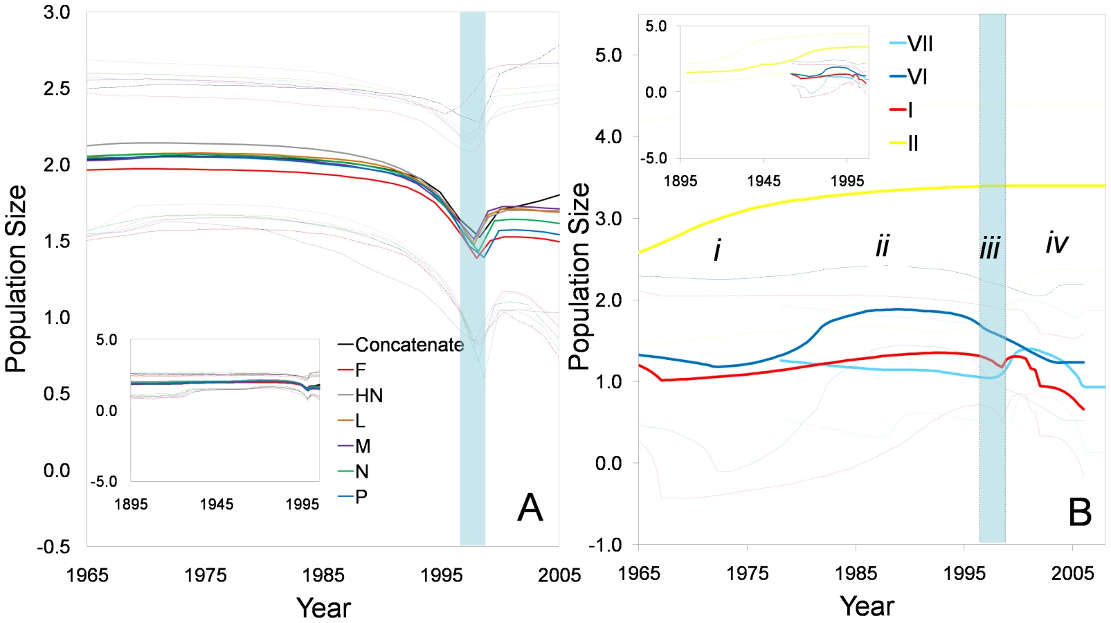

To determine if all genotypes exhibited the same population history observed for the composite genotype data set, we repeated the analysis based on 97 dated full length F genes, which provided a larger data set for this analysis. Isolates from genotype III, IV, and V were not included because limited numbers of dated sequences were available. The TMRCA for genotype II was estimated to be 1899±20 years, and the estimated evolutionary rates were between 0.3–1.1×10⁻⁴ substitutions/site/year, making this the slowest evolving aPMV-1 genotype (Table 2). Genotype I and VI emerged in the early 1960's and had higher evolutionary rates than genotype II. The most recently emerged strain was genotype VII, which dates to the late 1970's and had the highest evolutionary rate. BSP analyses based on the F gene demonstrated that each genotype had a unique population history. Prior to the emergence of genotype VII in the 1970s (phase i), genotype II showed an increase in population size (Figure 5B). After the emergence of genotype VII (phase ii) the population size of genotype VI began to increase, while that of genotypes I and VII were relatively constant. Phase iii depicts the time of the population bottleneck observed in Figure 5A, which was based on all genes in the composite genotype data set. Only genotypes I and VI show a trend for decreasing population size during this time. The last decade has been the most dynamical for the four genotypes of aPMV-1 (phase iv; Figure 5B). Genotypes I and VII showed a marginal increase in effective population size followed by a decline; genotype I has continued to decline, whereas genotype VII appears to have stabilized. Genotype VI showed continuous decline in population size in phase iv (Figure 5B). Although estimates of population sizes for genotypes I, VI, and VII, have some degree of overlaps at the 95% posterior limit, genotype II shows no sign of reduction in effective population size since its origin (Figure 5B).

Selection profiles
We compared the selection profiles on protein coding genes of genotypes I and II, which include strains that are circulating in the face of homotypic vaccination pressure (designated as the vaccine-related group), and genotypes III-VII (designated as the nonvaccine-related strains) (Table 3). Overall, the global rate of non-synonymous to synonymous substitutions (dN/dS) for all protein coding genes were less than 1, indicating purifying selection has been the major driving force in the evolution of class II aPMV-1 viruses. There were no codons identified to be under positive selection in M and L genes in either group (Table 3). However, there was a clear difference in the codon based selection profiles of N, P, F and HN genes between the vaccine - and nonvaccine-related groups. In the vaccine-related group, only the surface protein encoding genes, F and HN, had positively selected codons. Both groups had a single site identified in F; these were at codon 115 within the F0 cleavage site for the vaccine-related group and codon 28 in the signal peptide for the nonvaccine-related group. There were 3 positively selected sites identified in the HN gene in the vaccine-related group and one in the non vaccine-related group. In contrast, the P gene of nonvaccine-related genotypes III-VII had three sites predicted to be under positive selection but no sites were identified in the vaccine-related group. Thus, selection is focused on HN in vaccine-related groups and on P in nonvaccine-related genotypes and there are no shared sites under positive selection between the two groups.

Discussion
Our study explored the forces shaping the evolutionary history of class II aPMV-1 using available full genome sequences. We demonstrated that genotype affiliations based on individual genes and concatenated full length genomes of class II aPMV-1 were not consistent. This may account for discrepancies reported for genotype groupings that are based on partial or complete sequences of a single gene [13], [18], [43]. Topological incongruence among the gene trees reflects different evolutionary histories of each gene [44]; recombination is the most plausible explanation for this. The role of recombination in the evolution of aPMV-1, and negative sense RNA viruses in general, has been debated. For example, Sakaguchi et al. [30] and Toyoda et al, [31] reported consistent topological placement of different NDV strains in both F and HN gene trees. Seal et al. [45] also reported that there was no evidence of recombination among NDV M gene sequences. Although recombination is more common in positive-sense RNA viruses and can be explained by several genetic mechanisms [46], there is increasing evidence of homologous recombination in several non-segmented negative-sense RNA viruses [32], [47], [48], [49], [50]. Our approach differs from those used in previous studies of aPMV-1 evolution because we evaluated full genome sequences and tested individual recombinant regions with phylogeny-based incongruence tests. Thus, we show that all class II aPMV-1 genes have evidence of recombination breakpoints, that multiple recombination events are discernable in some isolates, and that both intragenic and intergenic recombination events are evident. We considered the possibility that the recombinants detected in our analysis were the result of laboratory artifacts, as has been previously suggested [32], [51]. Laboratory contamination is of concern because vaccine derived strains contributed the majority of the recombinant regions and these strains might have been present in laboratories sequencing field aPMV-1 isolates. The presence of unique nucleotide substitutions in the recombinant regions compared to the comparable region of the predicted parental genotypes suggests that these regions did not arise due to contamination with vaccine strains deposited in the sequence databases.
Our identification of recombinants derived from vaccine strains indicates that birds can be simultaneously infected with the live virus vaccine and other circulating aPMV-1 genotypes. Indeed vaccination is reported to protect poultry from disease but not always from infection with other strains [7], [8], [9]. Suboptimal vaccination strategies could also lead to birds becoming infected with both a vaccine strain and circulating genotype, which can alter viral virulence [16]. This is an important issue for poultry management. Lack of vaccine efficacy has not frequently been reported in the United States but other countries such as Nigeria [52], Korea [53], Taiwan [54], and China [34], [55] have experienced vaccine failures. Recombination between wild type virus and vaccine strains is not unique to aPMV-1; vaccine recombinants of bovine viral diarrhea virus (associated with fatal mucosal disease) [35], poliovirus (associated with paralytic poliomyelitis) [36], [37]; and infectious bursal disease virus [38] have all been reported. This raises concerns that modified live virus vaccines, although efficacious, may facilitate emergences of new strains with unpredictable phenotypes through recombination with circulating viruses.
The evolutionary rates presented here for class II aPMV-1 are compatible with the rates estimated for other RNA viruses (e.g. [56], [57], [58]), suggesting that class II aPMV-1 is also a rapidly evolving RNA virus. The relatively lower evolutionary rate for genotype II is consistent with the rate that was previously reported for avirulent NDV [33]. The larger effective population size, which counters the impact of genetic drift, is a possible explanation for lower evolutionary rates of genotype II. Based on these rate estimates, the TMRCA of this virus is estimated to be between 1868–1891, which is earlier than the first recorded outbreak of ND in Indonesia and England in the 1920's [2]. However, our data are in line with observations by Macpherson [59], who suggested that an outbreak of disease in domestic birds that occurred in Northwest Scotland from 1897–1898 was due to NDV.
The demographic history of class II aPMV-1 determined by Bayesian skyline plots indicated that there was an abrupt decline in population size during 1997–98. Although the factors responsible for such an abrupt decline in class II aPMV-1 are not known, the impact of a severe El-Nino event during that time frame [60] or the slaughter of millions of domestic fowl during the first outbreak of H5N1 avian influenza virus in 1997 [61], [62] could be possible explanations.
In contrast to the other genotypes, genotype II was not impacted by factors causing population decline in the late 1990s. We expected that vaccination, which started worldwide in the 1950s, should have limited the number of susceptible avian hosts, thus causing a bottleneck for this genotype. However, the impact of NDV vaccination is not seen in the BSP. It is possible that the data available in GenBank is insufficient to capture the population history of this genotype. However, a plausible explanation for the absence of a population bottleneck could be that genotype II NDV is maintained as an asymptomatic infection because it is continually introduced to susceptible populations as a modified live vaccine. Vaccination effectively prevents birds from developing disease when exposed to a virulent strain, but does not prevent shedding [7]. Thus, the number of available susceptible hosts may not dictate genotype II population demographics.
Although overall all genes of class II aPMV-1 are evolving under purifying selection consistent with other paramyxoviruses [33], [56], [63], distinctive profiles of positively selected codons were shown in both vaccine - and nonvaccine-related groups. Notably, P gene had the highest dN/dS ratio of any gene and had three sites predicted to be under selection in the nonvaccine-related group; there were no sites under selection in P gene in the vaccine related group. In contrast, HN had three sites predicted to be under selection in the vaccine related group. Of interest, codon 115 in the F cleavage domain is positively selected only in vaccine-related groups. Previous studies have reported that a single amino acid substitution at codon-115, which falls within the F0 cleavage site, resulted in a dramatic change from an avirulent infection to highly virulent NDV [64], [65], [66]. In contrast to the results reported in the present study, Miller et al [33] did not identify codon115 in F gene under positive selection. This is likely due to differences in the data sets because factors such as sequence length, sequence divergence, and the number of sequences can determine the ability to detect positively selected sites [67]. Thus, the vaccine-related genotypes I and II maintain a phenotypic mixture of strains with different infection and pathogenic potential and selection profiles.
Materials and Methods
Sequences data collection and phylogenetic analyses
A total of 54 complete genome sequences of class II aPMV-1 representing different avian hosts, geographic regions, year of isolation, and genotypes (based on previous published phylogenetic grouping) were retrieved from GenBank. The coding genome sequences were aligned using MEGA version 4 [68]. Six separate coding gene sequences datasets (for NP, P, M, F, HN and L genes) were generated (see Table S1) and a concatenated genome sequence from these six coding gene sequences was generated using Mesquite version 1.12 (http://mesquiteproject.org). Appropriate model of nucleotide substitution for each dataset was selected by the hierarchical likelihood ratio test implemented in Modeltest version 3.7 [69]. Maximum likelihood (ML) trees were reconstructed for all data sets using the heuristic search option, implementing stepwise addition with 100 random addition replicates and tree bisection-reconnection branch swapping in PAUP* version 4beta10 [70] and PHYML 3.412 [71] with 100 non-parametric bootstrapping replicates analyses. The inferred trees were visualized with FigTree version 1.12 (http://tree.bio.ed.ac.uk/software/figtree/) and the congruency of topology placement of class II aPMV-1 genotypes based on each gene and concatenated genome was tested using the Shimadoira, Hasegawa (SH) test [72] implemented in PAUP. The concatenated tree was constrained and tested versus other gene trees.
Putative recombination detection
The recombination predictions of the concatenated genome sequences were conducted with a suite of programs within the RDP3 package [39], [40]. The individual programs RDP [39], GENECONV [73], Bootscan [40], Maximum Chi [74], Chimaera [75], SiScan [76] and 3Seq [77], were implemented for the analysis. Since no single program provided optimal performance under all conditions, any event supported by five or more methods with p-values ≤10−5 was the criteria used for positive recombination breakpoints identification. The breakpoint position and the putative parental sequences were also determined.
Estimation of evolutionary rates and past population dynamics
Twenty six full length genome sequences for which year of isolation was available were used to infer evolutionary rate and dates using BEAST version 1.4.8 [41]. Demographic history of a population/species using multi-locus data, even from a small number of individuals, can precisely recover past bottlenecks in population size that cannot be characterized by analysis of a single locus [78]. Given this fact, estimates based on the whole genome sequence data are expected to be more reliable. To determine the population history of individual genotypes, 149 complete F gene sequences, which had dates of collection, were retrieved from GenBank. Phylogenetic analyses were done as described previously (Table S2). From the ML tree, a total of 97 isolates from genotype I, II, VI, and VII were selected to infer evolutionary rates and population dynamics. These included all available dated full length sequences from Genotypes I (24 sequences), II (28 sequences), and VI (23 sequences). The majority (95%) of genotype VII sequences were of Chinese origin. Thus, we included the seven non-Chinese sequences and picked an additional 15 sequences based on phylogenetic diversity to represent genotype VII in our analyses. The evolutionary rate (nucleotide substitutions per site per year) of each gene and concatenated genome was estimated using the Bayesian Markov chain Monte Carlo analyses (independent assumption of codivergence). Substitution models of both HKY + G4 and GTR + G6 with estimated base frequencies, gamma and invariable site portion were used, with uncertainty in the data reflected in the 95% high-probability density (HPD) intervals. Strict clock and uncorrelated exponential (UCED) relaxed clock models were attempted independently, and the best-fit clock model was determined to be UCED based on the Bayes Factor calculated from their posterior distributions (Table S3). The Coalescent Bayesian skyline plot (BSP) was used to infer the past population dynamics. The BSP was constructed using the growth rate and demographic parameters from the selected best-fit models. Bayesian Markov Chain Monte Carlo (BMCMC) analyses were run for 5–10×108 generations depending on each dataset. Convergence of trees was checked using Tracer v1.4.1 (http://beast.bio.ed.ac.uk/Tracer).
Selection analysis
Selection analyses were done based on datasets without putative recombinant sequences because recombination can result in falsely identifying positive selection [79]. The datasets were split into a vaccine-related group, which included strains from genotypes I and II, and nonvaccine-related group, which included strains from genotypes III, IV, V, VI, VII (see Table S2). Positively selected codons were detected using Fixed - Effect Likelihood (FEL) via the Datamonkey website (http://www.datamonkey.org/) and ML approach implemented in CODEML (PAML package version 3.15)[80]. For FEL analysis, p-values less than 0.05 were used to support positive selection. For PAML analysis, the likelihood ratio test was used to compare M1a, M7 and M8a models that assume no positive selection (ω<1) with those M2a and M8 models that assume positive selection (ω>1)[81].
Supporting Information
Zdroje
1. KaletaEF
BaldaufC
1988 Newcastle disease in free living and pet birds.
AlexanderDJ
Newcastle Disease Boston Kluwer Academic Publishers 197 246
2. SpradbrowPB
1987 Newcastle Disease - An Overview.
CoplandJW
Newcastle Disease in Poultry: A New Food Pellet Vaccine Melbourne Ramsay Ware Printing 12 18
3. AlexanderDJ
1988 Newcastle Disease Virus - An Avian Paramyxovirus.
AlexanderDJ
Newcastle Disease Boston Kluwer Academic Publishers 11 22
4. LeightonFA
HeckertRA
2007 Newcastle disease and related avian Paramyxoviruses.
ThomasNJ
HunterDB
AtkinsonCT
Infectious diseases of wild birds Oxford Blackwell Publishing 3 16
5. KimLM
KingDJ
SuarezDL
WongCW
AfonsoCL
2007 Characterization of class I Newcastle disease virus isolates from Hong Kong live bird markets and detection using real-time reverse transcription-PCR. J Clin Microbiol 45 1310 1314
6. HitchnerSB
1964 Control of Newcastle Disease in the United States by Vaccination.
HansonRP
Newcastle Disease Virus: An Evolving Pathogen/proceedings of an international symposium Madison The University of Wisconsin Press 85 98
7. KapczynskiDR
KingDJ
2005 Protection of chickens against overt clinical disease and determination of viral shedding following vaccination with commercially available Newcastle disease virus vaccines upon challenge with highly virulent virus from the California 2002 exotic Newcastle disease outbreak. Vaccine 23 3424 3433
8. MillerP
KingDJ
AfonsoC
SuarezD
2007 Antigenic differences among Newcastle disease virus strains of different genotypes used in vaccine formulation affect viral shedding after a virulent challenge. Vaccine 25 7238 7246
9. van BovenM
BoumaA
FabriTHF
KatsmaE
HartogL
2008 Herd immunity to Newcastle disease virus in poultry by vaccination. Avian Pathol 37 1 5
10. de LeeuwO
PeetersB
1999 Complete nucleotide sequence of Newcastle disease virus: evidence for the existence of a new genus within the subfamily Paramyxovirinae. J Gen Virol 80 131 136
11. MayoMA
2002 Virus taxonomy - Houston 2002. Arch Virol 147 1071 1076
12. StewardM
VipondIB
MillarNS
EmmersonPT
1993 RNA editing in Newcastle disease virus. J Gen Virol 74(Pt 12) 2539 2547
13. CzeglediA
UjvariD
SomogyiE
WehmannE
WernerO
2006 Third genome size category of avian paramyxovirus serotype 1 (Newcastle disease virus) and evolutionary implications. Virus Res 120 36 48
14. SealBS
WiseMG
PedersenJC
SenneDA
AlvarezR
2005 Genomic sequences of low-virulence avian paramyxovirus-1 (Newcastle disease virus) isolates obtained from live-bird markets in North America not related to commonly utilized commercial vaccine strains. Vet Microbiol 106 7 16
15. KattenbeltJA
StevensMP
GouldAR
2006 Sequence variation in the Newcastle disease virus genome. Virus Res 116 168 184
16. MillerPJ
DecaniniEL
AfonsoCL
2010 Newcastle disease: Evolution of genotypes and the related diagnostic challenges. Infect Genet Evol 10 26 35
17. WehmannE
CzeglediA
WernerO
KaletaEF
LomnicziB
2003 Occurrence of genotypes IV, V, VI and VIIa in Newcastle disease outbreaks in Germany between 1939 and 1995. Avian Pathol 32 157 163
18. KwonHJ
ChoSH
AhnYJ
SeoSH
ChoiKS
2003 Molecular epidemiology of Newcastle disease in Republic of Korea. Vet Microbiol 95 39 48
19. AldousEW
MynnJK
BanksJ
AlexanderDJ
2003 A molecular epidemiological study of avian paramyxovirus type 1 (Newcastle disease virus) isolates by phylogenetic analysis of a partial nucleotide sequence of the fusion protein gene. Avian Pathol 32 239 257
20. HuangZ
ElankumaranS
YunusAS
SamalSK
2004 A recombinant Newcastle disease virus (NDV) expressing VP2 protein of infectious bursal disease virus (IBDV) protects against NDV and IBDV. J Virol 78 10054 10063
21. LeeYJ
SungHW
ChoiJG
KimJH
SongCS
2004 Molecular epidemiology of Newcastle disease viruses isolated in South Korea using sequencing of the fusion protein cleavage site region and phylogenetic relationships. Avian Pathol 33 482 491
22. LiuXF
WanHQ
NiXX
WuYT
LiuWB
2003 Pathotypical and genotypical characterization of strains of Newcastle disease virus isolated from outbreaks in chicken and goose flocks in some regions of China during 1985–2001. Arch Virol 148 1387 1403
23. MaseM
ImaiK
SanadaY
SanadaN
YuasaN
2002 Phylogenetic analysis of Newcastle disease virus genotypes isolated in Japan. J Clin Microbiol 40 3826 3830
24. TsaiHJ
ChangKH
TsengCH
FrostKM
ManvellRJ
2004 Antigenic and genotypical characterization of Newcastle disease viruses isolated in Taiwan between 1969 and 1996. Vet Microbiol 104 19 30
25. MoyaA
ElenaSF
BrachoA
MirallesR
BarrioE
2000 The evolution of RNA viruses: A population genetics view. Proc Natl Acad Sci U S A 97 6967 6973
26. ElenaSF
SanjuanR
2005 Adaptive value of high mutation rates of RNA viruses: separating causes from consequences. J Virol 79 11555 11558
27. BruenTC
PossM
2007 Recombination in feline immunodeficiency virus genomes from naturally infected cougars. Virology 364 362 370
28. HerreweghAA
SmeenkI
HorzinekMC
RottierPJ
de GrootRJ
1998 Feline coronavirus type II strains 79–1683 and 79–1146 originate from a double recombination between feline coronavirus type I and canine coronavirus. J Virol 72 4508 4514
29. PalmenbergAC
SpiroD
KuzmickasR
WangS
DjikengA
2009 Sequencing and analyses of all known human rhinovirus genomes reveal structure and evolution. Science 324 55 59
30. SakaguchiT
ToyodaT
GotohB
InocencioNM
KumaK
1989 Newcastle disease virus evolution. I. Multiple lineages defined by sequence variability of the hemagglutinin-neuraminidase gene. Virology 169 260 272
31. ToyodaT
SakaguchiT
HirotaH
GotohB
KumaK
1989 Newcastle disease virus evolution. II. Lack of gene recombination in generating virulent and avirulent strains. Virology 169 273 282
32. HanGZ
HeCQ
DingNZ
MaLY
2008 Identification of a natural multi-recombinant of Newcastle disease virus. Virology 371 54 60
33. MillerPJ
KimLM
IpHS
AfonsoCL
2009 Evolutionary dynamics of Newcastle disease virus. Virology 391 64 72
34. QinZ
SunL
MaB
CuiZ
ZhuY
2008 F gene recombination between genotype II and VII Newcastle disease virus. Virus Res 131 299 303
35. BecherP
OrlichM
ThielHJ
2001 RNA recombination between persisting pestivirus and a vaccine strain: generation of cytopathogenic virus and induction of lethal disease. J Virol 75 6256 6264
36. GeorgescuMM
DelpeyrouxF
CrainicR
1995 Tripartite genome organization of a natural type 2 vaccine/nonvaccine recombinant poliovirus. J Gen Virol 76(Pt 9) 2343 2348
37. GuillotS
CaroV
CuervoN
KorotkovaE
CombiescuM
2000 Natural genetic exchanges between vaccine and wild poliovirus strains in humans. J Virol 74 8434 8443
38. HonCC
LamTT
YipCW
WongRT
ShiM
2008 Phylogenetic evidence for homologous recombination within the family Birnaviridae. J Gen Virol 89 3156 3164
39. MartinD
RybickiE
2000 RDP: detection of recombination amongst aligned sequences. Bioinformatics 16 562 563
40. MartinDP
WilliamsonC
PosadaD
2005 RDP2: recombination detection and analysis from sequence alignments. Bioinformatics 21 260 262
41. DrummondAJ
RambautA
2007 BEAST: Bayesian evolutionary analysis by sampling trees. BMC Evol Biol 7 214
42. DrummondAJ
RambautA
ShapiroB
PybusOG
2005 Bayesian coalescent inference of past population dynamics from molecular sequences. Mol Biol Evol 22 1185 1192
43. SealBS
1996 Analysis of Matrix Protein Gene Nucleotide Sequence Diversity Among Newcastle Disease Virus Isolates Demonstrates that Recent Disease Outbreaks Are Caused by Viruses of Psittacine Origin. Virus Genes 11 217 224
44. MaddisonWP
1997 Gene Trees in Species Trees. Syst Biol 46 523 536
45. SealBS
KingDJ
MeinersmannRJ
2000 Molecular evolution of the Newcastle disease virus matrix protein gene and phylogenetic relationships among the paramyxoviridae. Virus Res 66 1 11
46. LaiMM
1992 RNA recombination in animal and plant viruses. Microbiol Mol Biol Rev 56 61 79
47. ChareER
GouldEA
HolmesEC
2003 Phylogenetic analysis reveals a low rate of homologous recombination in negative-sense RNA viruses. J Gen Virol 84 2691 2703
48. GeueL
ScharesS
SchnickC
KliemtJ
BeckertA
2008 Genetic characterisation of attenuated SAD rabies virus strains used for oral vaccination of wildlife Vaccine 26 3227 3235
49. McCarthyAJ
ShawMA
GoodmanSJ
2007 Pathogen evolution and disease emergence in carnivores. Proc Biol Sci 274 3165 3174
50. WittmannTJ
BiekR
HassaninA
RouquetP
ReedP
2007 Isolates of Zaire ebolavirus from wild apes reveal genetic lineage and recombinants. Proc Natl Acad Sci U S A 104 17123 17127
51. AfonsoCL
2008 Not so fast on recombination analysis of Newcastle disease virus. J Virol 82 9303
52. OkoyeJO
ShoyinkaSV
1983 Newcastle disease in a vaccinated flock which had experienced subclinical infectious bursal disease. Trop Anim Health Prod 15 221 225
53. ChoSH
KimSJ
KwonHJ
2007 Genomic sequence of an antigenic variant Newcastle disease virus isolated in Korea. Virus Genes 35 293 302
54. ChenJP
WangCH
2002 Phylogenetic analysis of Newcastle disease virus in Taiwan. J Microbiol Immunol Infect 35 223 228
55. TanLT
XuHY
WangYL
QinZM
SunL
2008 Molecular characterization of three new virulent Newcastle disease virus variants isolated in China. J Clin Microbiol 46 750 753
56. PadhiA
PossM
2009 Population dynamics and rates of molecular evolution of a recently emerged paramyxovirus, avian metapneumovirus subtype C. J Virol 83 2015 2019
57. PomeroyLW
BjornstadON
HolmesEC
2008 The evolutionary and epidemiological dynamics of the paramyxoviridae. J Mol Evol 66 98 106
58. SmithGJ
VijaykrishnaD
BahlJ
LycettSJ
WorobeyM
2009 Origins and evolutionary genomics of the 2009 swine-origin H1N1 influenza A epidemic. Nature 459 1122 1125
59. MacphersonLW
1956 Some observations on the epizootiology of New Castle Disease. Can J Comp Med Vet Sci 20 155 168
60. KovatsRS
BoumaMJ
HainesA
1999 World Health Organization - Sustainable Development and Healthy Environments: El Niño and Health. Geneva
61. BridgesCB
LimW
Hu-PrimmerJ
SimsL
FukudaK
2002 Risk of influenza A (H5N1) infection among poultry workers, Hong Kong, 1997-1998. J Infect Dis 185 1005 1010
62. ChanPK
2002 Outbreak of avian influenza A(H5N1) virus infection in Hong Kong in 1997. Clin Infect Dis 34 Suppl 2 S58 64
63. HolmesEC
2009 The Evolution and Emergence of RNA Viruses. Great Britain Oxford University Press 254
64. GouldAR
KattenbeltJA
SelleckP
HanssonE
Della-PortaA
2001 Virulent Newcastle disease in Australia: molecular epidemiological analysis of viruses isolated prior to and during the outbreaks of 1998-2000. Virus Res 77 51 60
65. PeetersBP
deLeeuwOS
KochG
GielkensAL
1999 Rescue of Newcastle disease virus from cloned cDNA: evidence that cleavability of the fusion protein is a major determinant for virulence. J Virol 73 5001 5009
66. WestburyH
2001 Newcastle disease virus: an evolving pathogen? Avian Pathol 30 5 11
67. AnisimovaM
BielawskiJP
YangZ
2001 Accuracy and power of the likelihood ratio test to detect adaptive molecular evolution. Mol Biol Evol 18 1585 1592
68. TamuraK
DudleyJ
NeiM
KumarS
2007 MEGA4: Molecular Evolutionary Genetics Analysis (MEGA) software version 4.0. Mol Biol Evol 24 1596 1599
69. PosadaD
CrandallKA
1998 MODELTEST: testing the model of DNA substitution. Bioinformatics 14 817 818
70. SwoffordDL
2002 PAUP*: phylogenetic analysis using parsimony (*and other methods), version 4. Sunderland, Massachusetts Sinauer Associates
71. GuindonS
GascuelO
2003 A simple, fast, and accurate algorithm to estimate large phylogenies by maximum likelihood. Syst Biol 52 696 704
72. ShimodairaH
HasegawaM
1999 Letter to the Editor Multiple Comparisons of Log-Likelihoods with Applications to Phylogenetic Inference. Mol Biol Evol 16 1114 1116
73. PadidamM
SawyerS
FauquetCM
1999 Possible emergence of new geminiviruses by frequent recombination. Virology 265 218 225
74. SmithJM
1992 Analyzing the mosaic structure of genes. J Mol Evol 34 126 129
75. PosadaD
CrandallKA
2001 Evaluation of methods for detecting recombination from DNA sequences: computer simulations. Proc Natl Acad Sci U S A 98 13757 13762
76. GibbsMJ
ArmstrongJS
GibbsAJ
2000 Sister-scanning: a Monte Carlo procedure for assessing signals in recombinant sequences. Bioinformatics 16 573 582
77. BoniMF
PosadaD
FeldmanMW
2007 An exact nonparametric method for inferring mosaic structure in sequence triplets. Genetics 176 1035 1047
78. HeledJ
DrummondAJ
2008 Bayesian inference of population size history from multiple loci. BMC Evol Biol 8 289
79. AnisimovaM
NielsenR
YangZ
2003 Effect of recombination on the accuracy of the likelihood method for detecting positive selection at amino acid sites. Genetics 164 1229 1236
80. YangZ
1997 PAML: a program package for phylogenetic analysis by maximum likelihood. Bioinformatics 13 555 556
81. YangZ
NielsenR
GoldmanN
PedersenAM
2000 Codon-substitution models for heterogeneous selection pressure at amino acid sites. Genetics 155 431 449
Štítky
Hygiena a epidemiologie Infekční lékařství LaboratořČlánek vyšel v časopise
PLOS Pathogens
2010 Číslo 4
- Parazitičtí červi v terapii Crohnovy choroby a dalších zánětlivých autoimunitních onemocnění
- Vakcíny proti klíšťové encefalitidě
- Kdy je nejlepší očkovat
- Možné vedlejší účinky očkování
- Imunogenita vakcín
Nejčtenější v tomto čísle
- The Effect of Vaccination on the Evolution and Population Dynamics of Avian Paramyxovirus-1
- Reconstitution of SARS-Coronavirus mRNA Cap Methylation
- Increased Monocyte Turnover from Bone Marrow Correlates with Severity of SIV Encephalitis and CD163 Levels in Plasma
- Deficiencies in Jasmonate-Mediated Plant Defense Reveal Quantitative Variation in Pathogenesis
Zvyšte si kvalifikaci online z pohodlí domova
Mazová zátka a její řešení
nový kurzVšechny kurzy