-
Články
- Vzdělávání
- Časopisy
Top články
Nové číslo
- Témata
- Kongresy
- Videa
- Podcasty
Nové podcasty
Reklama- Kariéra
Doporučené pozice
Reklama- Praxe
Hybridní postupy v léčbě pseudoaneurysmat oblouku aorty – kazuistika
Hybrid Procedures in the Treatment of Pseudoaneurysms of the Aortic Arch – Case Report
Classical surgical therapy of dilatation disorders of the aortic arch require extracorporeal circulation, selective brain perfusion and/or deep hypothermia and it is still associated with very high mortality and morbidity. Endovascular therapy has until recently indicated only been in cases when the disease did not affect the area of the origins of the main branches within the aortic arch. We are presenting a case report of a 68 year female patient with a vascular anomaly (arteria lusoria) and 2 pseudoaneurysms of the aortic arch between the origins of arteria carotis communis on the right and arteria carotis communis on the left, respectively between a. carotis communis on the left and arteria subclavia on the left, when we took advantage of a hybrid procedure in the therapy. The patient was treated by creating a new branching of the aortal arch using a prosthesis from the ascendant aorta and subsequently by an introduction of 2 stent-grafts to the aortic arch using femoral arteries.
Key words:
aortic arch – aneurysm – hybrid therapy – stent-graft
Autoři: V. Kuntscher 1; V. Třeška 1; M. Novák 2; F. Šlauf 2; B. Bohuslav 1; J. Křižan 1; R. Šulc 1; J. Moláček 1
Působiště autorů: Surgical Clinic, University Hospital Plzeň-Lochotín, Head of Dept. : Prof. MUDr. V. Třeška, DrSc. 1; Radiodiagnostic Clinic, University Hospital Plzeň, Head of Dept. : Assoc. Prof. MUDr. B. Kreuzberg, Ph. D. Medical Faculty in Plzeň, Charles University in Prague 2
Vyšlo v časopise: Rozhl. Chir., 2008, roč. 87, č. 7, s. 384-387.
Kategorie: Monotematický speciál - Původní práce
Souhrn
Klasická chirurgická léčba dilatací oblouku aorty vyžaduje provedení v mimotělním oběhu, selektivní cerebrální perfuzi a/nebo hlubo-kou hypotermii, a přesto je stále zatížena vysokou mortalitou a morbiditou. Endovaskulární léčba byla až donedávna indikována pouze v případech, že onemocnění nepostihovalo oblasti odstupů hlavních větví v oblouku aorty. Autoři předkládají kazuistiku 68leté pacientky s cévní anomálií (arteria lusoria) a 2 pseudoaneuryzmaty oblouku aorty mezi odstupy arteria carotis communis vpravo a arteria carotis communis vlevo a mezi a. carotis communis vlevo a arteria subclavia vlevo, kdy byl při léčbě použit hybridní postup. Bylo vytvořeno nové větvení aortického oblouku s použitím protézy z ascendentní aorty a poté byl do aortického oblouku zaveden stent s použitím štěpů femorálních arterií.
Klíčová slova:
oblouk aorty – aneuryzma – hybridní terapie – stentINTRODUCTION
Conventional surgical therapy of dilatation disorders within the aortic arch using extracorporeal circulation (cardiopulmonary bypass /CPB/) and deep hypothermia with circulatory arrest (deep hypotermic circulatory arrest /DHCA/) is still associated with very high morbidity and mortality. Hospitalization mortality and morbidity is reported in between 10–38% [1, 2, 3]. The main causes of high morbidity and mortality are mainly significant systemic inflammatory reaction and severe myocardial lesions, mainly in patients from risk subgroups after CPB [4] and more than 5% incidence of permanent neurolo - gical consequences after DHCA [5].
Charles Dotter performed the first stenting of an artery in an experiment on a dog in the early 1960s [6] and A. Balko performed a remodelling of an arterial aneurysm using a stent-graft for the first time in the 1980s [7]. Since then endovascular therapy developed into one of methods for a safe therapy of the diseases in various areas of the arterial system; dilatation disorders affecting the aortic arch and originating arteries were and they still are considered a contraindication for the usage of endovascular prostheses due to the risks following from their closure [8, 9, 10].
CLINICAL PAPER
Our patient is a 68 year old woman who was admitted on 14/6/2005 to the University Hospital in Plzeň after 14 days of increasing retrosternal pain and pain in the back and progressive dyspnoea. The patient has a history of arterial hypertension treated from 2004; she has been using hormonal substitution therapy for a long time for a hypofunction of the thyroid gland; the father of the patient had an aneurysm of the subrenal aorta.
The cause of the complaints was determined to be 2 pseudoaneurysms of the aortic arch between the origins of the main arteries (Figure 1); the patient was also diagnosed as having an anomalous origin of arteria subclavia on the right as the last artery of the arch – a. lusoria. After introduction of controlled hypotension the subjective complaints receded; there were even no objective signs of progression; we indicated the patient for an elective procedure. On 18/7/2005 we proceeded with surgery. To ensure vascular access in the case that extra-corporeal circulation was necessary and for the second phase of the operation for the introduction of the stent-graft, we started the procedure with preparation of the arteries in the right groin. After that we performed medial sternotomy and after the opening of the pericardium, we exposed the ascending aorta, on which we placed a bandage for safe anchoring of the proximal part of the stent-graft using a 12.5 cm long Dacron band so that the diameter of the aorta in the site of the stent-graft anchorage was 39 to 40 mm. After that we placed a wall vascular clamp on the ascending aorta and created a proximal anastomosis with bifurcation Dacron wrapped prosthesis 14/7 to a longitudinal aortotomy (Figure 2).
Obr. 1. Pseudoaneurysms of the aortic arch Obr. 1. Pseudoaneurysma oblouku aorty 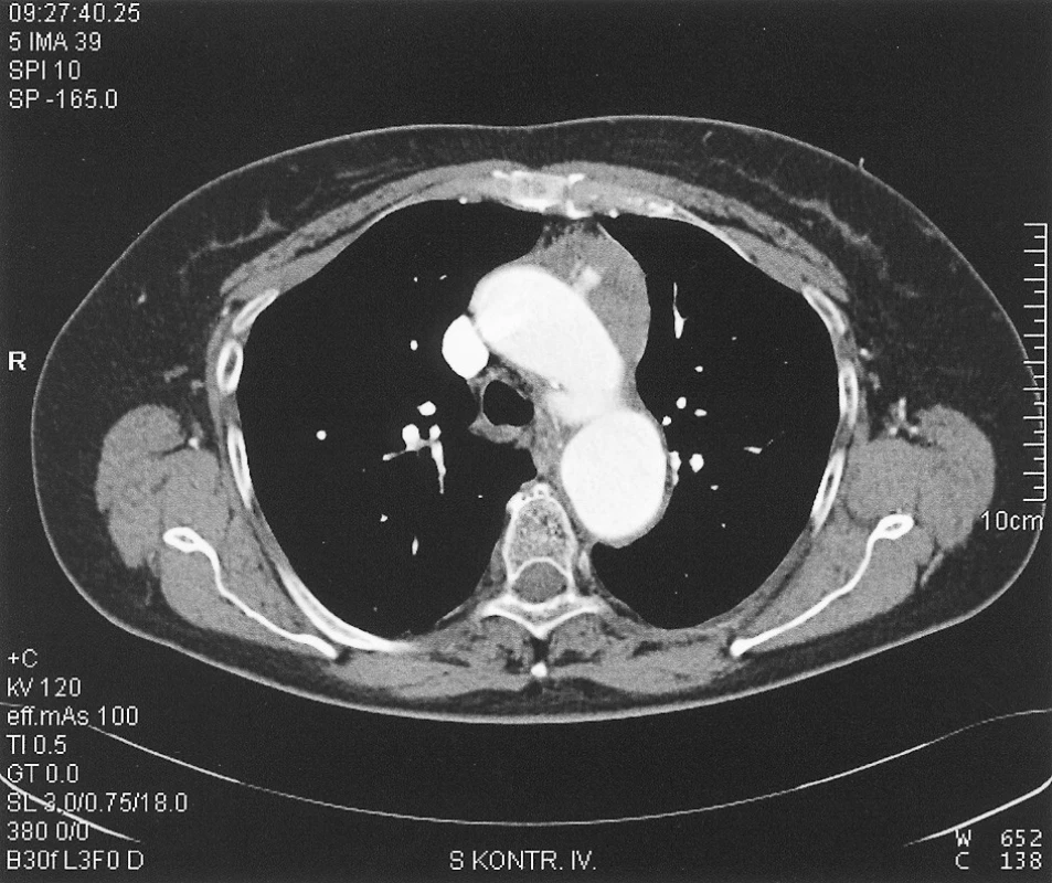
Obr. 2. Wall clamp and longitudinal aortotomy Obr. 2. Aortická svorka a longitudinální aortotomie 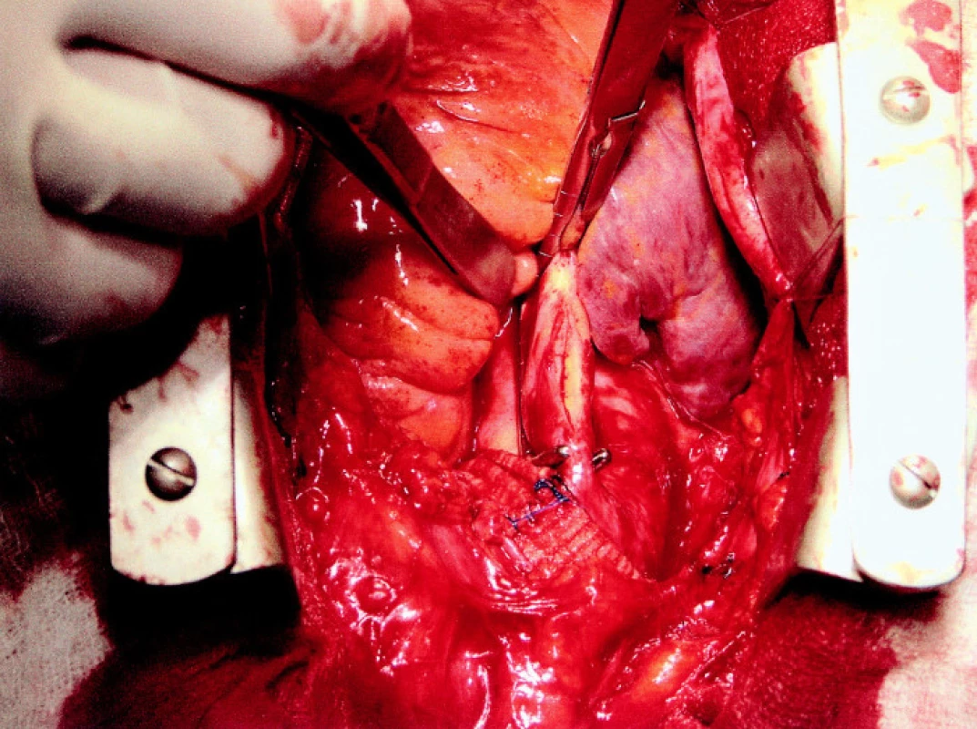
After creating this anastomosis and removal of a wall clamp, we administered systemic heparin – 7,500 IU i.v.
The branches of the reconstruction were located ventrally from the anonymous vein and we gradually created anastomoses end-to-side with arteria carotis communis; initially on the right side and after restoration of flow and exchange of the clamps to the left, the anastomosis was also created on the left side. Using the supraclavicular approach, after cutting m. scalenus ant. and preservation of the phrenic nerve, we prepared the arteria subclavia on the right side (resp. arteria lusoria). In order to prevent the risk of oesophageal compression during the patient’s vascular anomaly, we interrupted the arteria lusoria centrally from the arteria vertebralis, the central stump was closed and then the peripheral stump was connected end-to-end with a 7 mm wrapped Dacron prosthesis (the remnant of a branch from the bifurcation prosthesis was used). The prosthesis was placed dorsally from vena jugularis interna and the right branch of the aorto-bicarotic reconstruction was connected to it side-to-end. We proceeded the same way on the left side; we did not interrupt the arteria subclavia, but we used an end-to-side connection. We verified the function of all parts of such an aorto-bicarotico-bisubclavian reconstruction (Figure 3).
Obr. 3. Final structure: aorto-bicarotic-bisubclavian reconstruction Obr. 3. Konečný stav: aorto-bikaroticko-bisubklaviální rekonstrukce 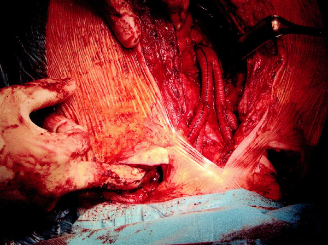
Using the pre-prepared arteria femoralis communis on the right, we introduced 2 stentgrafts to the aortic arch, the longer of which was proximally anchored in the site of the ascending aorta bandage. Both pseudoaneurysms were excluded with the introduction of the stent-graft; there were no signs of any leak or other complications observed during a control injection (Figure 4).
Obr. 4. Peroperation angiography after introduction of stent-grafts Obr. 4. Peroperační angiografie po zavedení stentů 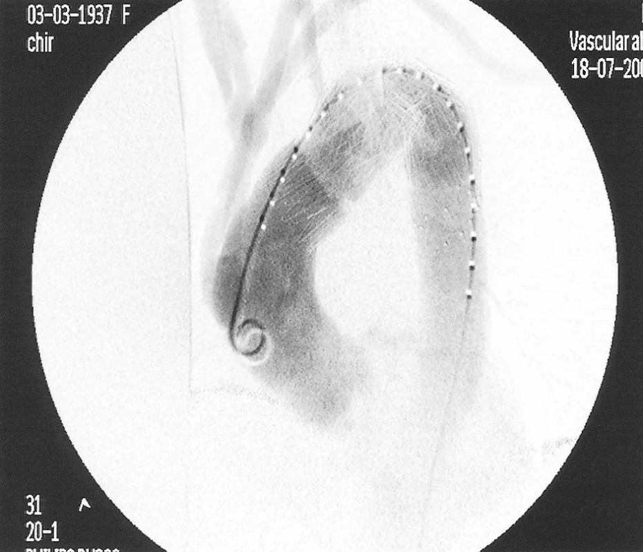
We completed the procedure with a suture of the wounds in anatomic layers after drainage of the mediastinum.
The patient required 2 weeks of supportive pulmonary ventilation after the procedure (for respiratory insufficiency, it was necessary to re-intubate through the nose after the first trial of extubation); the condition required l electrocardioversion for atrial fibrillation with fast response of ventricles in the first post-operation week; sinus rhythm was restored permanently after cardioversion. The recovery was fast and smooth after complete weaning from the ventilator and the patient was discharged on 11/8/2005 to home care.
On 4/10/2005 she underwent the first post-operation follow up and also a follow up CTAG examination was performed (Figure 5). The patient attends inspections reguralry. She has no neurological deficit, no complaints; the follow up graphical examination reveals normal function of the reconstruction.
Obr. 5. 3D CTAG image of the condition at the post-operation follow up after 3 months Obr. 5. 3D CTAG obraz stavu při kontrole 3 měsíce po operaci 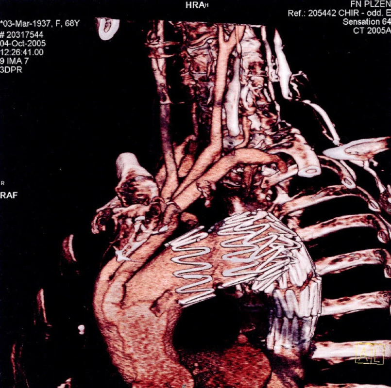
DISCUSSION
Classical surgical therapy of dilatation disorders within the aortic arch is associated with a very high risk. Peri-operation mortality and morbidity is reported in 10–38%; the risk of severe permanent neurological lesion is at least 5%. A significant portion of consequences corresponds with the need to use CPB and DHCA. A combined approach to these diseases with replantation of the aortic branches to another part of the aorta and exclusion of the dilated part of the aorta with introduction of a stent-graft enables avoidance of CPB and DHCA completely and, according to our opinion, this decreases the operation risk significantly. Despite the fact that there is little experience with this procedure worldwide, the operation risk can be estimated at 1–2% mortality and from 2 to 5 % early postoperation morbidity based on the evaluation of the results of intra-thoracic vascular surgical procedures performed for occlusive disease of the arteries within the aortic arch and after an evaluation of the results of endovascular prostheses in other localisations within aorta. Definite conclusions will not be available until there are a sufficient number of such procedures performed.
The paper was published in the framework of the investigation of the Medical Faculty in Plzeň MZM 0021620819.
Práce je publikována v rámci výzkumného záměru Lékařské fakulty v Plzni MZM 0021620819.
MUDr. V. Kuntscher, Ph.D.
Chirurgická klinika Fakultní nemocnice
Alej Svobody 80
304 60 Plzeň
e-mail: kuntscher@fnplzen.cz
Zdroje
1. Kazui, T., Washiyama, N., Muhammad, B. A., Terada, H., Yamashita, K., Takinami, M., Tamiya, Y. Total arch replacement using aortic arch branched grafts with the aid of antegrade selective cerebral perfusion. Ann. Thorac. Surg., 70 (2000) (1), pp. 3–8.
2. Gottardi, R., Lammer, J., Grimm, M., Czerny, M. Entire rerouting of the supraaortic branches for endovascular stent-graft placement of an aortic arch aneurysm. Eur. J. Cardiothorac. Surg., 29 (2006), pp. 258–260.
3. Fiřt, P., Hejnal, J., Vaněk, I. Výduť aortalního oblouku. In: Cévní chirurgie. Avicenum, 1991, pp. 303–306.
4. Czerny, M., Baumer, H., Kilo, J., Lassnigg, A., Hamwi, A., Vukovich, T., Wolner, E., Grimm, M. Inflammatory response and myocardial injury following coronary artery bypass grafting with or without cardiopulmonary bypass. Eur. J. Cardiothorac. Surg., 17 (2000) (6), pp. 737–742.
5. Czerny, M., Fleck, T., Zimpfer, D., Dworschak, M., Hofmann, W., Hutschala, D., Dunkler, D., Ehrlich, M., Wolner, E., Grabenwoger, M. Risk factors of mortality and permanent neurologic injury in patients undergoing ascending aortic and arch repair. J. Thorac. Cardiovasc. Surg., 26 (2003) (5).
6. Dotter, C. T. Transluminally-placed coilspring and endarterial tube grafts: long term patency in canine popliteal artery. Invest. Radiol., (1969) (5), pp. 329–332.
7. Balko, A., Piasecky, G. J., Shah, D. M., Carney, W. I., Hopkins, R. W., Jackson, B. T. Transfemoral placement of intraluminal polyuretane prosthesis for abdominal aortic aneurysm. J. Surg. Res., (1986) (4), pp. 305–309.
8. Pavcnik, D., Ferko, A., Krajina, A. Endovaskulární výkony na hrudní aortě. In: A. Ferko, A. Krajina: Arteriální aneuryzmata. ATD, 1999, pp. 53–63.
9. Brunkwall, J., Gawenda, M., Sudkamp, M., Zahringer, M. Current indication for endovascular treatment of thoracic aneurysms. J. Cardiovasc. Surg. (Torino), 44 (2003), (3), pp. 465–470.
10. Sunder-Plassmann, L., Orend, K. H. Stentgrafting of the thoracic aorta – complications. J. Cardiovasc. Surg. (Torino) 46, (2005) (2), pp. 121–130.
Štítky
Chirurgie všeobecná Ortopedie Urgentní medicína
Článek Expertní činnost v chirurgiiČlánek Reexpanzný pľúcny edém, ako komplikácia drenáže hrudníka pri spontánnom pneumotoraxe – kazuistikaČlánek Stenty – paliativní a kurativní ošetření jícnu. Sedmileté zkušenosti chirurgického pracovištěČlánek Rekonstrukce po gastrektomiiČlánek Komentář k článku
Článek vyšel v časopiseRozhledy v chirurgii
Nejčtenější tento týden
2008 Číslo 7- Metamizol jako analgetikum první volby: kdy, pro koho, jak a proč?
- Nejlepší kůže je zdravá kůže: 3 úrovně ochrany v moderní péči o stomii
- Stillova choroba: vzácné a závažné systémové onemocnění
- Metamizol v léčbě různých bolestivých stavů – kazuistiky
-
Všechny články tohoto čísla
- Expertní činnost v chirurgii
- Zkušenosti s radiofrekvenční termoablací mozkových nádorů
- Ischemické poškození míchy následkem tupého poranění hrudníku – kazuistika
- Reexpanzný pľúcny edém, ako komplikácia drenáže hrudníka pri spontánnom pneumotoraxe – kazuistika
- Pacientka s fibrosarkomem srdce. Kazuistika
- Stenty – paliativní a kurativní ošetření jícnu. Sedmileté zkušenosti chirurgického pracoviště
- Pseudoaneurysma arteria hepatica manifestující se hemobilií jako komplikace laparoskopické cholecystektomie
- Ojedinělé případy liposarkomů retroperitonea obrovských rozměrů
- Rekonstrukce po gastrektomii
- Masivní hemotorax po kanylaci v. subclavia – kazuistika
- Ruptura šlachy m. pectoralis maior a anabolické steroidy v anamnéze – kazuistika
- Hybridní postupy v léčbě pseudoaneurysmat oblouku aorty – kazuistika
- Komentář k článku
- Zemřel doc. MUDr. Ladislav Krušina, CSc.
- Rozhledy v chirurgii
- Archiv čísel
- Aktuální číslo
- Informace o časopisu
Nejčtenější v tomto čísle- Ruptura šlachy m. pectoralis maior a anabolické steroidy v anamnéze – kazuistika
- Ojedinělé případy liposarkomů retroperitonea obrovských rozměrů
- Reexpanzný pľúcny edém, ako komplikácia drenáže hrudníka pri spontánnom pneumotoraxe – kazuistika
- Stenty – paliativní a kurativní ošetření jícnu. Sedmileté zkušenosti chirurgického pracoviště
Kurzy
Zvyšte si kvalifikaci online z pohodlí domova
Autoři: prof. MUDr. Vladimír Palička, CSc., Dr.h.c., doc. MUDr. Václav Vyskočil, Ph.D., MUDr. Petr Kasalický, CSc., MUDr. Jan Rosa, Ing. Pavel Havlík, Ing. Jan Adam, Hana Hejnová, DiS., Jana Křenková
Autoři: MUDr. Irena Krčmová, CSc.
Autoři: MDDr. Eleonóra Ivančová, PhD., MHA
Autoři: prof. MUDr. Eva Kubala Havrdová, DrSc.
Všechny kurzyPřihlášení#ADS_BOTTOM_SCRIPTS#Zapomenuté hesloZadejte e-mailovou adresu, se kterou jste vytvářel(a) účet, budou Vám na ni zaslány informace k nastavení nového hesla.
- Vzdělávání


