-
Články
- Vzdělávání
- Časopisy
Top články
Nové číslo
- Témata
- Kongresy
- Videa
- Podcasty
Nové podcasty
Reklama- Kariéra
Doporučené pozice
Reklama- Praxe
Viral Protein Inhibits RISC Activity by Argonaute Binding through Conserved WG/GW Motifs
RNA silencing is an evolutionarily conserved sequence-specific gene-inactivation system that also functions as an antiviral mechanism in higher plants and insects. To overcome antiviral RNA silencing, viruses express silencing-suppressor proteins. These viral proteins can target one or more key points in the silencing machinery. Here we show that in Sweet potato mild mottle virus (SPMMV, type member of the Ipomovirus genus, family Potyviridae), the role of silencing suppressor is played by the P1 protein (the largest serine protease among all known potyvirids) despite the presence in its genome of an HC-Pro protein, which, in potyviruses, acts as the suppressor. Using in vivo studies we have demonstrated that SPMMV P1 inhibits si/miRNA-programmed RISC activity. Inhibition of RISC activity occurs by binding P1 to mature high molecular weight RISC, as we have shown by immunoprecipitation. Our results revealed that P1 targets Argonaute1 (AGO1), the catalytic unit of RISC, and that suppressor/binding activities are localized at the N-terminal half of P1. In this region three WG/GW motifs were found resembling the AGO-binding linear peptide motif conserved in metazoans and plants. Site-directed mutagenesis proved that these three motifs are absolutely required for both binding and suppression of AGO1 function. In contrast to other viral silencing suppressors analyzed so far P1 inhibits both existing and de novo formed AGO1 containing RISC complexes. Thus P1 represents a novel RNA silencing suppressor mechanism. The discovery of the molecular bases of P1 mediated silencing suppression may help to get better insight into the function and assembly of the poorly explored multiprotein containing RISC.
Published in the journal: . PLoS Pathog 6(7): e32767. doi:10.1371/journal.ppat.1000996
Category: Research Article
doi: https://doi.org/10.1371/journal.ppat.1000996Summary
RNA silencing is an evolutionarily conserved sequence-specific gene-inactivation system that also functions as an antiviral mechanism in higher plants and insects. To overcome antiviral RNA silencing, viruses express silencing-suppressor proteins. These viral proteins can target one or more key points in the silencing machinery. Here we show that in Sweet potato mild mottle virus (SPMMV, type member of the Ipomovirus genus, family Potyviridae), the role of silencing suppressor is played by the P1 protein (the largest serine protease among all known potyvirids) despite the presence in its genome of an HC-Pro protein, which, in potyviruses, acts as the suppressor. Using in vivo studies we have demonstrated that SPMMV P1 inhibits si/miRNA-programmed RISC activity. Inhibition of RISC activity occurs by binding P1 to mature high molecular weight RISC, as we have shown by immunoprecipitation. Our results revealed that P1 targets Argonaute1 (AGO1), the catalytic unit of RISC, and that suppressor/binding activities are localized at the N-terminal half of P1. In this region three WG/GW motifs were found resembling the AGO-binding linear peptide motif conserved in metazoans and plants. Site-directed mutagenesis proved that these three motifs are absolutely required for both binding and suppression of AGO1 function. In contrast to other viral silencing suppressors analyzed so far P1 inhibits both existing and de novo formed AGO1 containing RISC complexes. Thus P1 represents a novel RNA silencing suppressor mechanism. The discovery of the molecular bases of P1 mediated silencing suppression may help to get better insight into the function and assembly of the poorly explored multiprotein containing RISC.
Introduction
Most eukaryotes, including plants, make use of a well-conserved RNA silencing mechanism to regulate many essential biological processes, ranging from development and control of physiological activities, to responses to abiotic and biotic stress, in particular antiviral defense [1], [2].
Antiviral defense in plants begins with the activity of RNase III type Dicer-Like (DCL) enzymes, which target viral RNAs [3], [4]. Concerted action of the DCL4, DCL2, DCL3 and occasionally DCL1 enzymes results in the appearance of 21–24 nt small interfering RNAs (siRNAs), the central components of the RNA silencing pathway [4], [5]. These viral siRNAs subsequently loaded to endogenous AGO proteins, which are catalytic component of RNA-induced silencing complex (RISC) [6], [7]. AGO1 and AGO7 are suggested to be involved in antiviral silencing [8], [9], [10] although previous study failed to detect viral siRNAs in tagged AtAGO1 [11]. It has been also shown that AGO7 favors less structured RNA targets, while AGO1 is capable of targeting viral RNAs with more compact structures [9]. AGO proteins are responsible for targeting RISC to viral genomes (either RNA or DNA), and exert their action either through cleavage or inhibition of translation [12]. The RNA-dependent RNA polymerases (RDRs) of the host also play important roles in antiviral RNA silencing, being involved in production of secondary viral siRNA [13], [14], [15], [16], [17], [18].
Viruses have evolved suppressors to counteract the RNA-silencing defense of the host [1], [2], [19]. The more than 35 viral silencing-suppressor families so far identified use different strategies to inhibit RNA silencing [2], [20]. Sequestering siRNAs by siRNA-binding suppressors is a very common way to inhibit RISC assembly [21], [22], but other mechanisms have been described, such as inhibiting the biogenesis of 21 nt siRNA species [4], [20], [23]. Other suppressors inhibit RNA silencing through protein-protein interaction. The 2b protein of CMV strain Fny is suggested to inhibit RISC activity via physical interaction with the PAZ domain of the plant AGO1 protein [10]. Polerovirus P0 suppressor protein has been suggested to target PAZ domain of AGO1 and directing its degradation [24], [25].
The Potyviridae is the largest family of plant RNA viruses; in most members, the single-stranded RNA genome is about 10 kb in size and encodes a single polyprotein that is processed into at least 9 mature proteins [26] (Figure 1). In the genus Potyvirus, the multifunctional HC-Pro (helper component-proteinase) was the first viral product to be recognized as a silencing suppressor [27], [28], [29]. The genome of Cucumber vein yellowing virus (CVYV), genus Ipomovirus, family Potyviridae, lacks HC-Pro but contains two P1-type proteases [30], properties shared by at least one other ipomovirus [31]. In CVYV, the second P1 cistron (P1b) was found to suppress RNA silencing [30] with a mode of action resembling that of the HC-Pro of potyviruses [32]. Interestingly, the type member of the genus Ipomovirus, Sweet potato mild mottle virus (SPMMV), possesses an HC-Pro region and a single large P1 serine protease [33].
Fig. 1. Genome structure of SPMMV ipomovirus and sequence peculiarities of the N-terminal part of its polyprotein which includes the P1 protein. 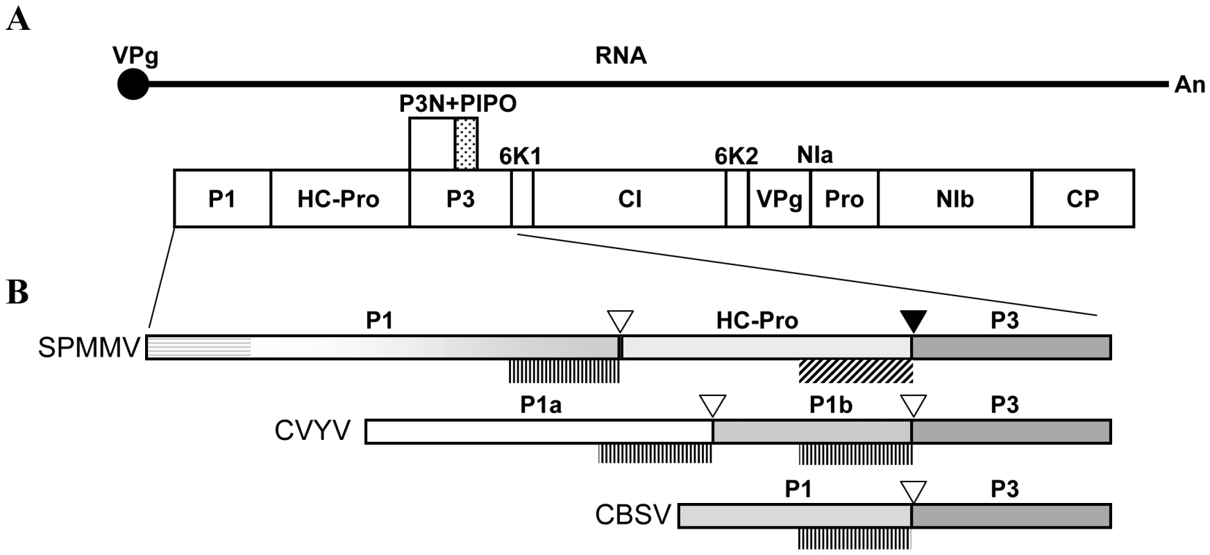
(A) Graphical representation of the genome organization of viruses of the family Potyviridae. The ssRNA genome of a generic potyvirid is depicted as a solid line with a 5′ covalently linked VPg and a 3′ polyA tail. The viral polyprotein is shown as a large box divided in the different mature protein products, and the small dotted box above represents the alternative P3-embeded ORF known as PIPO, expressed after a frameshift. (B) Details of genomic variants found in the N-terminal part of ipomoviruses. Examples of individual viruses (acronyms at the left) of each type of genomic organization are shown, and the relative sizes of proteins are represented proportionally. The proteinase domains and cleavage sites are indicated respectively by plotted vertical lines (below) and empty triangles (above) in the case of P1-like serine proteinases, or by oblique lines (below) and solid triangles (above) for the HC-Pro-like cysteine proteinase in SPMMV. Pattern consistency is used to represent homologies, and the gradational scale in the C-terminal part of SPMMV P1 indicates that it presents partial homology in this region to the P1b of CVYV (and also to the P1b of SqVYV, not shown), and to the P1 of CBSV, while the horizontal lines represents a characteristic N-terminal extension with partial homology to the N-terminal part of the P1 protein of the potyvirus SPFMV. The peculiarities of the SPMMV genome that incorporates the largest P1 region among all known members of the family together with a typical HC-Pro region (Figure 1), prompted us to study how this virus might deal with the RNA-silencing machinery in its hosts.
In the present study, we show that the large P1 protein of SPMMV possesses silencing-suppressor activity, while its HC-Pro protein does not, on its own. Using various reporter systems, we show that in vivo P1 inhibits target RNA cleavage mediated by RISC complexes loaded with either endogenous miRNA or with virus-derived siRNA. Moreover, suppression activity mapped to the N-terminal half of P1, a region containing three WG/GW motifs that mimics AGO-binding linear peptide motif conserved both in metazoans and plants [34], [35]. We have also determined that the WG/GW motifs at the very N-terminal end in P1 are required for AGO1 binding and for silencing-suppression, suggesting that P1 may use the conserved WG/GW motif binding surface of Ago proteins to inhibit RISC activity.
Results
A partial sequence of 3633 nucleotides was obtained from a plant infected with SPMMV African isolate 130 after RT-PCR amplification with primers designed to flank the first two cistrons of the viral polyprotein. The N-terminal part of this sequence shares structure with the only SPMMV whole genome sequence available in databanks [33]. It presents a large P1 cistron encoding 743 amino acids (15 residues more than the published sequence, starting at position 362), followed by a 453 amino acid HC-Pro cistron, which is more similar in size to other potyviral HC-Pros. The expected cleavage sites and the corresponding residues for the active sites of P1 and HC-Pro proteases [36] could be recognized in the sequence. However, SPMMV HC-Pro lacks the conserved FRNK box characteristic of potyviral HC-Pros and required for small RNA binding and symptom development [37]. These characteristics make SPMMV unique among the Potyviridae, including other ipomoviruses (Figure 1). Sequences are compared in Figure S1.
P1 is the silencing suppressor of SPMMV
To investigate whether P1 and/or HC-Pro serve (s) as RNA silencing suppressor for SPMMV, we used the standard Agrobacterium coinfiltration assay [22]. The complete cistrons for P1 and HC-Pro were cloned into binary vectors and the resulting expression constructs were transferred into A. tumefaciens. Cultures of A. tumefaciens able to express GFP from a 35S-promoter GFP binary plasmid were mixed with cultures transformed with our SPMMV constructs before infiltration into Nicotiana benthamiana leaves. In this assay both fluorescence and RNA analysis identified SPMMV P1, but not SPMMV HC-Pro, as the suppressor of RNA silencing (Figure 2A). Weak fluorescence and low GFP mRNA levels were observed in patches infiltrated with the pBin61 empty vector (negative control), and strong suppressor activity and increased GFP mRNA level were detected in patches infiltrated with a construct expressing the P1b of CVYV (positive control) [32].
Fig. 2. SPMMV P1 suppresses RNA silencing by mechanisms that diverge from other viral suppressors. 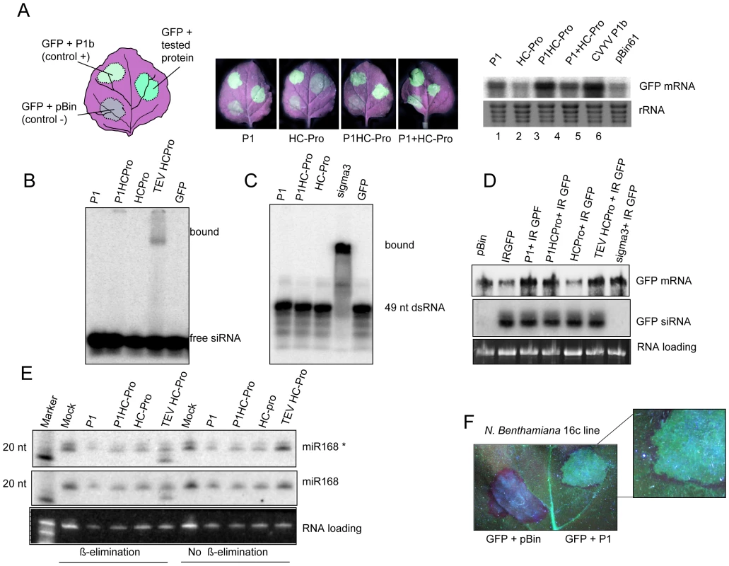
(A) P1 and HC-Pro proteins of SPMMV were agroinfiltrated into N.benthamiana leaves (according to the schematic drawing shown at the left) along with a reporter 35S-GFP construct to visualize (central panels) and to analyze by northern blot (right panel) their effects on RNA silencing. The P1b suppressor of CVYV was used as positive control, while an empty pBin61 served as negative control. (B) 32P-labeled ds 21nt siRNA was incubated with extracts of leaves infiltrated with constructs expressing SPMMV P1, SPMMV HC-Pro, both P1 and HC-Pro of SPMMV in cis (P1HC-Pro), TEV HC-Pro and GFP. Bound complexes were electrophoretically separated on a native gel. (C) 32P-labeled ds 49 nt RNA was incubated with extracts of leaves infiltrated with P1, P1HC-Pro, and HC-Pro of SPMMV, Reovirus sigma3 and GFP. Bound complexes were electrophoretically separated on a native gel. (D) RNA preparations from leaves of 16c transgenic lane of N. benthamiana plants expressing GFP infiltrated with SPMMV P1, SPMMV HC-Pro and TEV HC-Pro were analyzed by northern blot with GFP-specific probes for the presence of mRNA and siRNA (upper and central panels); loading charge is shown at the lower panel. (E) Analysis of 3′ methylation of miR168 in the presence of the indicated viral suppressors. RNA samples with or without oxidation and ß-elimination were separated in 12% polyacryamide gel and then blotted. Small RNA blot was hybridized with LNA oligonucleotide probes detecting miR168 star (miR168*) and miR168 mature strands. Faster miRNA bands indicate the non-methylated miRNAs. (F) Detail of a leaf from a N. benthamiana 16c plant under UV light at 8 days after co-agroinfiltration with the reporter 35S-GFP and pBin empty vector (left) or SPMMV P1 (right). The development of the red halo around the agroinfiltrated patch indicated that SPMMV P1 did not abolish movement of the signal. SPMMV P1 differs in mode of action from RNA-silencing suppressors of related viruses
Experiments designed to compare in parallel the suppression activity of SPMMV P1 with that of a suppressor from a potyvirus, the HC-Pro protein of Tobacco etch virus (TEV) were performed next. First, we checked in vitro if SPMMV proteins could bind either typical 21 nt ds siRNAs, or longer dsRNAs. Extracts of N. benthamiana leaves infiltrated with Agrobacterium strains expressing different suppressors were tested for siRNA binding with labeled 21 nt ds siRNA, and the complexes were resolved on a native gel. As expected, TEV HC-Pro bound ds siRNA, while SPMMV P1 and HC-Pro did not show any siRNA binding activity (Figure 2B). The same extracts were then incubated with a labeled 49 nt dsRNA, and the putative complexes were analyzed on a native gel. In this case, formation of the expected RNA-protein complex only occurred between the 49 nt dsRNA and the Sigma3 protein of a Reovirus [38] used as positive control, but no complexes were detected in any of the other samples from constructs of P1 and HC-Pro of SPMMV (Figure 2C).
Next, we tested if P1 inhibits small RNA processing in leaves of transgenic N. benthamiana line 16C, expressing a GFP transgene, that were coinfiltrated with an Agrobacterium strain harboring a GFP inverted repeat (GFP-IR) construct. To this end, patches infiltrated with constructs expressing SPMMV P1, SPMMV HC-Pro (individual proteins), or SPMMV P1HC-Pro (a construct containing both proteins in cis), or with TEV HC-Pro, were analyzed after 3 days for the presence of GFP mRNA and siRNAs by Northern blotting. No reductions in siRNA processing from the GFP-IR were observed in all expressed proteins (Figure 2D), in contrast to the complete abolition observed in the positive control, which was the dsRNA-binding Sigma3 protein of Reovirus [38]. The 16c plants agroinfiltrated were also observed under UV light at 8 days after agroinfiltration to monitor the spreading of silencing signal. Importantly we found that the presence of SPMMV P1 did not abolish movement of the signal, and therefore silencing of the transgene around the agroinfiltrated area was observed (Figure 2F).
We also checked the capacity of the different viral proteins to inhibit in vivo 3′ modifications of small RNAs by the HEN1 methyltransferase [39], [40]. We expressed P1 along with different silencing suppressor proteins, and the 3′ end methylation status of the mature and star strands of miR168 were then evaluated by oxidation and beta elimination followed by Northern blotting of total RNA samples. Consistently with our previous results [41], TEV HC-Pro inhibited the 3′ methylation of both strands of the endogenous miR168. However , SPMMV P1 and HC-Pro had no effect on HEN1 mediated 3′ modification. (Figure 2 E).
SPMMV P1 inhibits miRNA and siRNA programmed RISC activity
Our findings showed that in contrast to several other silencing suppressors SPMMV P1 does not interfere with the initial steps of the silencing pathway. Thus we hypothesized that it might compromise assembled RISC activity. Active RISC complexes are known to contain ss siRNA and are licensed to cleave the target RNA in a sequence specific manner [42]. Recently, we developed assays based on the transient expression of sensor constructs to test the effect of RNA-silencing suppressors on miRNA and siRNA loaded active RISC. Using these assays we have previously demonstrated that silencing suppressors with ds siRNA binding capacity such as the HC-Pro of potyviruses does not have any effect on miRNA and siRNA loaded RISCs in planta [22], [43].
To determine whether SPMMV P1 might inhibit miRNA loaded RISC complexes, we agroinfiltrated GFP171.1 and GFP171.2 sensor constructs [44] with or without the viral suppressors. In these sensors a full complementary miR171 binding site was placed downstream of the STOP codon of GFP ORF allowing miR171-mediated silencing of the GFP171.1 mRNA, while GFP171.2 carried a mutant miR171 target site, which is refractory to miR171-driven RNA silencing [44]. In this experiment the control construct used was TEV HC-Pro. At two days postinfiltration, GFP fluorescence was evaluated under UV light, and then the infiltrated patches were used for RNA and protein isolation. Consistent with previous results, miR171-driven RNA silencing downregulated GFP171.1, but not GFP171.2 at both the RNA and protein level. Strikingly, comparable GFP fluorescence and GFP mRNA and protein were detected in samples infiltrated with GFP171.1+P1 and GFP171.2+P1, indicating that SPMMV P1 efficiently inhibited miR171 loaded active RISC complexes (Figure 3 A, B). As expected for the control, TEV HC-Pro did not inhibit miR171 mediated degradation of GFP171.1 mRNA [22].
Fig. 3. SPMMV P1 inhibits miRNA and viral siRNA loaded RISCs. 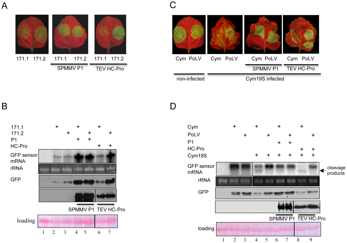
(A) Constructs expressing tagged SPMMV P1 and TEV HC-Pro proteins were agroinfiltrated with GFP-171.1 and GFP-171.2 sensors. GFP expression was visualized under UV light. (B) RNA gel blot and immunoblot analysis of miRNA sensor RNAs and GFP protein isolated from infiltrated patches. Immunoblot analysis of expression of the different silencing suppressor proteins isolated from infiltrated leaves are also shown. (C) GFP fluorescence of Cym 19S-infected recovering leaves and non-infected leaves infiltrated with GFP-Cym and GFP-PolV sensors and SPMMV P1 and TEV HC-Pro proteins. (D) RNA gel blot and immunoblot analysis of miRNA sensor RNAs and GFP protein isolated from infiltrated patches. Immunoblot analysis of expression of the different silencing suppressor proteins isolated from infiltrated leaves are also shown. Next, we investigated if SPMMV P1 inhibits viral siRNA-loaded active RISC complexes. A previously described system which exploits N. benthamiana plants infected with Cymbidium ringspot virus (CymRSV) 19 Stop mutant (Cym19S) was used [45]. Cym19S, not expressing the ds siRNA binding silencing suppressor p19, permits a strong RNA silencing response against the virus to be initiated and maintained by enabling viral siRNAs to be loaded into RISC complexes, leading to the recovery of the initially infected plant [45], [46]. At 14–18 dpi of plants carrying Cym19S, the first systemic leaves showed recovery as a consequence of the remarkable amount of active RISCs loaded with siRNAs derived from the virus [22]. Messenger RNAs expressed from the sensor construct GFP-Cym, in which GFP ORF is fused with a ∼200 bp portion of the CymRSV, could be targeted and cleaved by RISC complexes containing Cym19S-derived siRNAs, while GPF-PoLV, in which GFP fused with a ∼200 bp region of Pothos latent virus, a virus unrelated to CymRSV, cannot be cleaved, and was used as a negative control [43]. Recovering leaves of Cym19S-infected plants were infiltrated with GFP-Cym and GFP-PoLV alone or with the indicated silencing suppressors. At 2 days post-agroinfiltration (dpa), efficiency of RNA silencing was monitored by visual examination followed by Northern and Western blotting of RNA and protein samples isolated from infiltrated patches (Figure 3C,D).
When the agroinfiltration was performed only with sensors, Northern analysis using a GFP probe detected a shorter hybridizing band, diagnostic for RISC cleavage of the mRNA expressed from the GFP-Cym construct mediated by viral siRNA, while the hybridizing band remained intact in the case of the GFP-PoLV sensor. As expected, the control TEV HC-Pro was not competent to inhibit ss viral siRNA-loaded active RISC complexes, so the GFP-Cym sensor RNA was cleaved [22]. Remarkably, the Northern and Western analyses showed that GFP mRNA and protein levels were similar in GFP-Cym+P1 and in GFP-PoLV+P1 infiltrated samples, and no cleavage product of GFP was detected in the GFP-Cym+P1 infiltrated sample, suggesting that SPMMV P1 efficiently inhibited the slicing activity of the viral siRNA loaded RISC complexes (Figure 3 C,D).
P1 targets the RISC complex and interacts with AGO1
The RISC complex is of high molecular weight (>669 kD) in animals [47], [48], contains the catalytic AGO protein, and has intrinsic small-RNA-dependent target cleavage activity. In plants such as N. benthamiana, transiently expressed or endogenous AGO1 protein co-fractionates in extracts with small RNAs, and can be found in at least two distinct complexes of above 669 kD and 158 kD [49]. In addition, it was reported that high molecular weight complexes containing viral siRNAs exhibited nuclease activity in vitro and preferentially targeted homologous viral sequences [50]. Having established that P1 inhibits active RISC, we hypothesized that inhibition of RISC requires physical interaction of P1 with AGO1 and small-RNA-containing complexes. To investigate this, we first tested whether P1 co-fractionates with AGO1 and small RNAs on a gel filtration column. N-terminally HA-tagged SPMMV P1 (HA-P1), 6×myc-tagged AGO1 of Arabidopsis thaliana (myc-AGO1) [10] and GFP-IR were co-expressed in N. benthamiana leaves. At 3 dpi, extracts prepared from infiltrated leaves were fractionated on a Superdex 200HR column. Small RNAs were extracted from each fraction and analyzed by Northern blotting, and the AGO1 and P1 protein contents of fractions were monitored by Western blotting using antibodies raised against the HA and myc tags. Consistent with previous results, GFP siRNAs and miR159 were fractionated in two distinct complexes peaking at >669 and 158 kD, although they appeared in all fractions which also contained AGO1 (Figure 4A). We experienced technical difficulty in separating the protein peaks, and this might reflect the disintegration of the large complexes during chromatography or more likely due to limited availability of the other RISC components. Despite these problems, the infiltrated myc-AGO1 co-fractionated clearly with small RNAs suggesting that GFP siRNAs had been loaded into the myc-AGO1-containing complexes. Interestingly, SPMMV P1 co-fractionated mainly with the 669 kD, but not with the smaller 158 kD myc-AGO1 and small RNA-containing complexes (Figure 4 A).
Fig. 4. SPMMV P1 cofractionates with AtAGO1 and small RNAs. 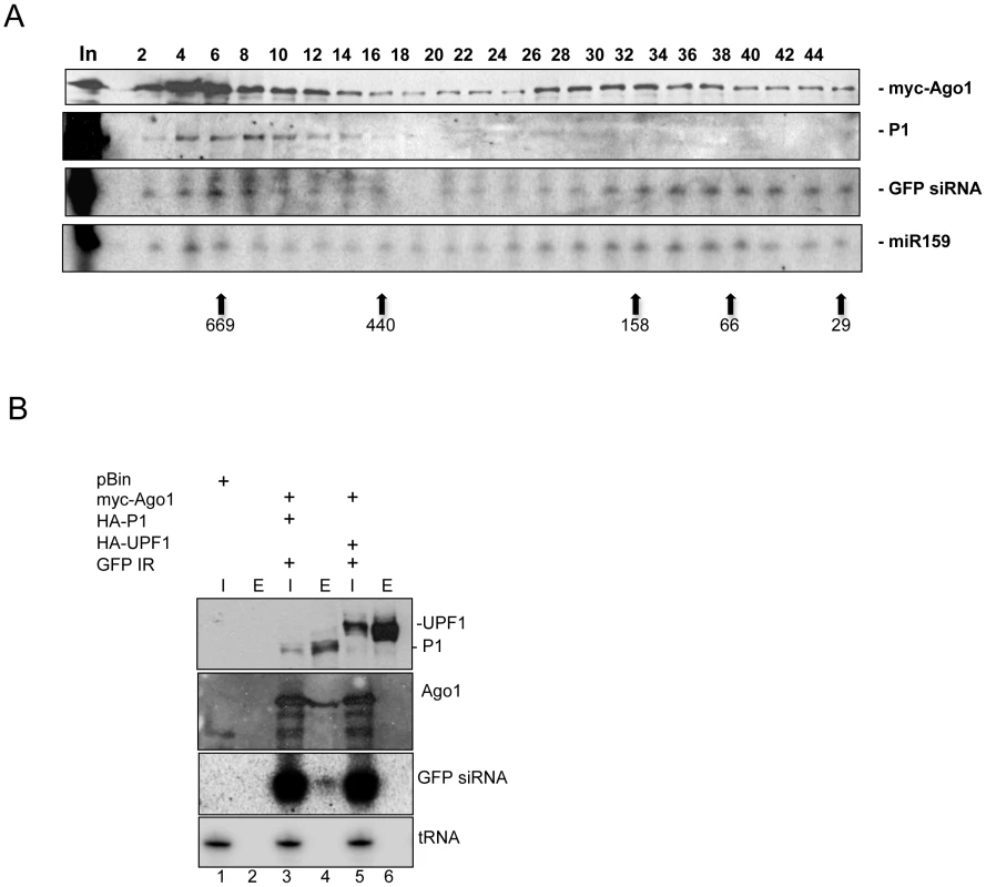
(A) Western analysis of even fractions of extracts of leaves infiltrated with HA-P1, 6×myc-AtAGO1 and GFP-IR. Anti-myc antibody was used to detect AtAGO1 and anti-HA antibody to detect P1. Northern analysis of RNA isolated from odd fractions. A positive sense GFP RNA probe was used to detect GFP siRNAs processed from GFP-IR. Arrows indicate the elution position of protein molecular weight markers for all panels. The two expected peaks of AtAGO1 correspond to fractions with relatively larger amounts of protein. (B) HA-tagged proteins were immunoprecipitated from extracts of leaves infiltrated with HA-P1+6×myc-AtAGO1+GFP-IR and HA-UPF1+6×myc-AtAGO1+GFP-IR. Myc and HA tagged proteins and GFP siRNAs were detected as in panel (A). tRNA was detected by tRNA specific oligonucleotide probe. Inputs (I) and eluates (E) are indicated above each lane. Next, we investigated if co-fractionation of P1 with myc-AGO1 and small RNAs was due to physical interaction. To test this, we agroinfiltrated HA-P1 with myc-AGO1 and GFP-IR. As a negative control, myc-AGO1 and GFP-IR were agroinfiltrated with HA-UPF1, which is known not to be involved in RISC formation. At 3 dpi, extracts were prepared from infiltrated leaves, and HA-tagged proteins were immunoprecipitated (IP) with an anti-HA antibody. Inputs and eluates of IPs were tested for proteins by Western blotting and for GFP siRNA in Northern blots. The results showed that HA-P1 and HA-UPF1 were expressed at comparable levels and could be efficiently immunoprecipitated from extracts. Importantly, we found that myc-AGO1 coimmunoprecipitated with HA-P1, but not with HA-UPF1, confirming that the interaction between HA-P1 and myc-AGO1 is specific (Figure 4B). Moreover, we found that GFP siRNAs, but not tRNA coimmunoprecipitated exclusively with HA-P1 and myc-AGO1, strongly suggesting that P1 interacts with small RNA-loaded AGO1.
Taken together, we showed that siRNAs derived from GFP-IR became incorporated into myc-AGO1, and that the P1 silencing suppressor specifically interacted with GFP siRNA-loaded myc-AGO1. These results, along with earlier data proving that P1 inhibits si - and miRNA programmed RISC, suggest that the large complex (669 kD) containing AGO1 and small RNAs corresponds to the plant RISC complex.
The N-terminal 383 amino acids of P1 contains the silencing suppressor domain
P1 contains an extended N-terminal region and a protease domain at its C-terminal end, similar to the P1b of other ipomoviruses [30]; Text S1). To determine the minimal region required for silencing suppression, we constructed P1 mutant truncated from the C-terminal end but retaining the first (N-terminal) 383 aa, designated as HA-P11-383 (Figure 5A). To evaluate its silencing-suppressor activity, the mutant was co-expressed with GFP-171.1 in N. benthamiana plants. Visual examination under UV light and analysis of GFP-171.1 sensor RNA and GFP expression showed that HA-P11-383 was an effective silencing suppressor although lacking the entire C-terminal protease domain. Then, we checked the interaction between the deletion mutant of P1 and AtAGO1. To test for interaction, we immunoprecipitated myc-AGO1 from extracts of infiltrated leaves expressing myc-AGO1 and GFP-IR with HA-P1 and HA-P11-383. We used the pBIN61 empty vector as negative control. We found that HA-P11-383 interacted with myc-AGO1 as strongly as wt P1. We concluded that P1 may be composed of two functional domains, the silencing suppressor domain is located at the N-terminal part, and the C-terminal part of P1 contains the protease domain. However, the protease activity of P1 was not analyzed.
Fig. 5. The N-terminal 383 amino acids bears the silencing suppressor domain of SPMMV P1. 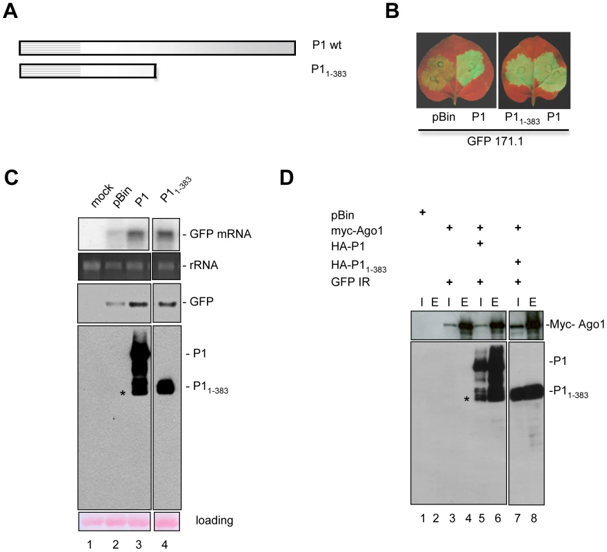
(A) Graphical representation of P1 deletion mutants. (B) P1 deletion mutants and P1 wt were coinfiltrated with mir171.1. pBIN was used as a negative control. Silencing suppressor activity of the mutants was compared to P1wt under UV light. (C) RNA and protein analysis of infiltrated patches. (D) Interaction of P1 wt and deletion mutants with AtAGO1. 6×myc-AtAGO1 was immunoprecipitated with anti-myc beads. Inputs (I) and eluates (E) were tested for 6×myc-AtAGO1 and for HA-tagged P1 wt and deletion mutants. Stars indicate the non-specific degradation products of full length P1. The P1 WG/GW motifs are required for suppressor activity and AGO1 binding
Our results showed that the N-terminal end of the P1 is required for Ago binding. Further inspection of the N-terminal end of P1 revealed repeating tryptophan-glycine/glycine-tryptophan residues (WG/GW) (Figure 6A and Figure S2), which are identical to the core amino acids of the WG/GW motifs recently found in Argonaute binding proteins, such as Tas3 and RNA Pol V (El-Shami et al, 2007; Till et al, 2007). Furthermore, analysis of amino acid composition revealed that the regions neighboring the WG/GW residues in P1 are rich in alanine, serine, glutamic acid, asparagine and aspartic acid, providing a context similar to that described for Tas3 and RNA Pol V proteins [35], [51].
Fig. 6. Three WG/GW motifs in the N-terminal part of SPMMV P1 are essential for silencing suppressor activity and AGO1 binding. 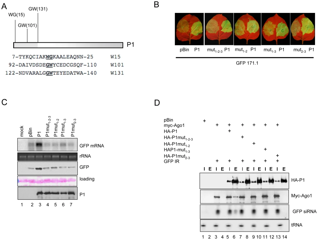
(A) Graphical representation of the WG/GW signatures in P1. Numbers represent the position of tryptophan residues in P1. (B) P1wt and the double and triple mutants were coinfiltrated with miR171.1 sensor. GFP expression representing silencing suppressor activity of the mutants was compared to that of P1wt. (C) RNA gel blot and immunoblot analysis of infiltrated patches. (D) Interaction of P1wt and point mutants with AtAGO1. HA-tagged P1wt and double and triple mutants were immunoprecipitated with anti-HA beads. Inputs (I) and eluates (E) were tested for 6×myc-AtAGO1, HA-tagged P1wt and double and triple mutants by western blotting. GFP siRNAs were detected by northern blotting. This observation prompted us to investigate the significance of the tryptophan residues at the N-terminal end of P1 in silencing suppressor activity. For this, we generated single, double and triple mutants of P1 by replacing tryptophan (W) by alanine (A) residue(s) at positions 15, 101 and 131, individually and in all double (3) and triple (1) combinations, by site-directed mutagenesis. Silencing-suppressor activity of the HA-tagged P1 single mutants was compared to the HA-P1wt, co-infiltrated with the GFP-171.1 sensor construct. Expression analysis of the GFP marker gene reflecting the strength of suppression of RNA silencing showed that suppressor activity of any of the single mutants was not reduced significantly, suggesting that presence of the remaining two tryptophan residues were sufficient to maintain the silencing suppressor activity of P1 (data not shown). In contrast, the suppressor activities of double and triple mutants were greatly reduced. Consistently, wt P1 and the mutants were expressed at comparable level (Figure 6 B,C).
To test whether the WG/GW motifs are required for RNA silencing-suppression because they contribute to AGO1 binding, we tested the interactions between AtAGO1 and P1 double and triple mutants. Myc-AGO1 and GFP-IR were co-infiltrated with double and triple mutants of HA-P1 in line GFP16c/RDR6i N. benthamiana plants. As positive control, we used HA-P1wt and as negative control, myc-AGO1 and GFP-IR were infiltrated without P1 wt. At 3 dpi, extracts of infiltrated leaves were used to immunoprecipitate HA-tagged P1 wt and mutant proteins. Western analysis showed that HA-tagged proteins were expressed at comparable levels and were successfully immunoprecipitated. However, probing Western and Northern blots to detect myc-AGO1 protein and GFP siRNA derived from GFP-IR revealed that myc-AGO1 protein and GFP siRNAs were specifically co-immunoprecipitated with P1 wt, but not with any of the double or triple mutants of P1 (Figure 6D).
These results showed that changing at least two out of three tryptophan residues to alanine in the WG/GW motifs of P1 abolished its silencing suppressor activity. Moreover, our analysis showed that the ability of P1 to bind AGO1 depends on the presence of these motifs, suggesting a correlation between AGO1 binding and its activity as silencing suppressor.
P1 binds endogenous miRNA-loaded AGO1 through direct interaction in vivo
AGO1 of A. thaliana is involved both in the miRNA and the antiviral RNA silencing pathways [10], [11], [52]. Our results showed that P1 interacts with AGO1 to inhibit active miRNA and siRNAs loaded RISC.
To get better insight into the mechanism of inhibition of RISC mediated by P1, we performed immunoprecipitations with small RNA-loaded RISCs against P1 (wild type) and P1mut1-2-3 (triple mutant)-infiltrated leaf extracts. RNA samples from inputs and immunoprecipitates were probed for presence of two endogenous miRNAs (Figure 7A). The results showed that mature miR159 and miR319 specifically co-immunoprecipitated with wt P1, but not with P1mut1-2-3. In addition, we found only mature miRNAs in the eluates of P1 immunoprecipitates, and the star strands for miR159 and mir319 could not be detected in inputs, or in eluates. Thus our results indicated that P1 interacts with endogenous RISC complexes loaded with single-stranded miRNAs (Figure 7A). To test whether P1 interacts directly or indirectly with AGO1 we performed in vitro pull-down assays using recombinant MBP-AGO1366-1048 containing the PAZ-MID-PIWI domains and the 6×His-P11-383 N-terminal fragment of wt P1. The results showed that MBP-AGO1366-1048 binds wt 6×His-P11-383 efficiently, while the triple mutant P11-383 was bound less strongly by AGO1 protein (Figure 7B). This result strongly suggests a direct interaction between P1 and AGO1 proteins in vivo as well.
Fig. 7. Interaction between P1 and AGO1. 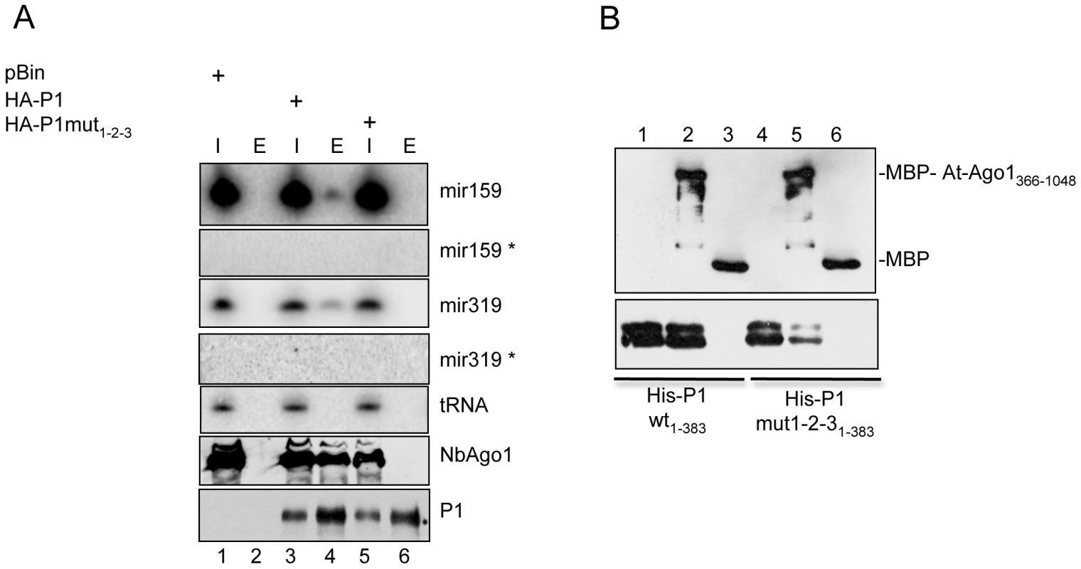
(A) P1 interacts with endogenous miRNA loaded Nb-AGO1. Extracts of leaves infiltrated with pBIN, wt HA-P11-383 and HA-P1mut1-2-31-383 were immunoprecipitated with HA-beads. Inputs (I) and eluates (E) were tested for wt P11-383, P1mut1-2-3 and for miR319 and mir159. (B) In vitro pull-down assays of MBP-AtAGO1366-1048 with wt and triple mutant of 6×His-P11-383. We used 2 mg of MBP-AtAGO1366-1048 or MBP to be mixed with 4 mg of 6×His-P1383 and 6×His-P1mut1-2-31-383 in a buffer containing 30mM TRIS pH 7.5, 150mM NaCl, 5mM MgCl2 and 10% glycerol for 1 hour at 4°C. Then MBP-tagged proteins were isolated on 30 ml maltose resin, washed three times with the same buffer and eluted with 2×SDS loading buffer. Proteins were detected by western blotting using anti-MBP and anti-HIS antibodies. Discussion
Available data suggest that virtually all plant viruses encode at least one suppressor and more than 35 individual viral silencing suppressor families have been identified to date. However, the mechanisms of action have been explored only for a few [2], [20]. Among the best-characterized RNA silencing suppressors, the p19 protein of tombusviruses and the HC-Pro protein of potyviruses share the ability to bind and sequester siRNAs, which are the most conserved element of the silencing machinery. SiRNA binding has been postulated as a common and effective strategy to counteract plant defenses against viruses [22]. However, further studies have shown that many viruses use other strategies and can adapt unrelated proteins to target and interfere with different steps in the silencing pathway. For instance, inhibition of siRNA generation by TCV p38 has been described [4], as has the targeting of AGO proteins, the conserved catalytic components of RISC, by viral proteins through protein-protein interactions [10], [25], [53].
In this work we explored the mechanism of silencing suppression mediated by the SPMMV P1 silencing suppressor protein, which interferes with miRNA and siRNA driven RISC activity by binding to the AGO1 subunit of RISC complexes through its WG/GW motifs conserved also in Argonaute binding cellular proteins.
Characterization of P1 as RNA-silencing suppressor
SPMMV is the only ipomovirus that has a typical potyvirid genome structure with P1 and HC-Pro regions in the C-terminal part of the polyprotein [33]. Other ipomoviruses with available complete sequences do not possess HC-Pro regions [31], [54], [55]. Despite the presence of an HC-Pro in SPMMV, we have found that the role of RNA-silencing suppressor is played by P1.
To better understand the molecular basis of P1-mediated silencing suppression, we analyzed the effect of P1 on different steps of the RNA silencing pathway. We found that P1 does not seem to interfere with the biogenesis of either transgene-derived siRNAs or endogenous miRNAs, since we observed that the accumulation of GFP-IR-derived siRNAs was mostly unaltered in the presence or absence of P1 (Figure 2D). Similarly, accumulation of endogenous miRNAs and their 3′ methylation status were not influenced by the expression of P1, in contrast to the well known TEV HC-Pro suppressor, which binds ds siRNA and miRNA intermediates and partially inhibits their 3′ methylation [22], [41] (Figure 2E). We also showed that P1 failed to bind short and long ds RNAs, in contrast to potyviral HC-Pro and the reovirus sigma3, which efficiently bind ds siRNAs and long dsRNAs, respectively (Figure 2B,C). Thus, this mechanism of silencing suppression seems to be unique among virus-encoded silencing suppressors identified so far.
P1 inhibits active RISC by interacting with AGO1 loaded with siRNA and miRNA
We tested the effect of P1 on miRNA - and viral siRNA-activated RISC complexes using GFP sensor constructs (Figure 3) and it turned out that P1 efficiently inhibited both types of activated RISC in vivo. Moreover, we showed that transiently expressed AGO1 protein was found in a large (>667kD) and an approximately 158 kD protein complexes in size. Interestingly, P1 was found co-fractionating only with the large AGO1 containing complex with GFP siRNAs. This results distinguishes P1 from previously studied silencing suppressors, because it does not inhibit RISC assembly, like small RNA binding suppressors, nor inhibits RISC assembly by promoting degradation of AGO proteins, as it was found in the case of P0 protein of poleoviruses [25], [49], [53]. The 2b protein of CMV FNY strain was also shown to interact with AGO1 in vivo and in vitro and to inhibit RISC activity in vitro [10]. However, it is not known whether 2b prevents RISC assembly or inhibits siRNA loaded RISC by AGO1 binding. In addition, a recent report revealed that 2b proteins of Tomato aspermy virus (TAV) and the FNY strain of CMV bind 21nt ds small RNAs [56], [57], so it is not clear, whether 2b protein of cucumoviruses inhibits RNA silencing through siRNA binding, interacting with AGO1 or both. In contrast, P1 binds RISC by interacting the AGO1 subunit of RISC loaded with si - or miRNAs, as shown by our immunoprecipitation studies (Figure 4 and 7). Importantly, P1 interacted with AGO1 containing mature miRNAs but not their star strand; this adds support to our hypothesis that P1 interacts with the AGO1 component of active RISC complexes, and is in line with the efficient inhibition of GFP-sensor silencing by P1. Our cumulative evidence strongly suggests that the P1 interaction with AGO1 is a direct physical interaction. Finally, using mutant P1 proteins with their silencing suppressor activity compromised/abolished, we obtained evidence that silencing suppression and AGO1 binding are linked.
P1 uses the conserved Ago-hook to bind Argonaute
The WG/GW motifs located at N-terminal part of P1 strongly resemble the evolutionarily conserved GW linear peptide motifs shared by different silencing-related proteins used as “Ago hooks” to interact with Argonaute proteins [35], [58]. Such WG/GW motifs have been described in proteins from different organisms, such as in the largest subunit NRPD1b of the RNA polymerase V in plants [51], the P body-localized human protein GW182 [59], [60], [61], and the Tas3 homologue of the GW182 RITS complex component in yeast [62], [63]. All these proteins can interact with Argonaute proteins [35]. Recently, an RdDM effector KTF1 containing abundant WG/GW motifs and SPT5-like domains has also been identified as an AGO4 binding element [64], [65]. Similarly to cellular WG/GW proteins, our analysis of P1 mutants indicates that the tryptophan residues are essential for interaction with AGO1 and are strictly required for silencing suppressor function (Figure 6).
Possible models of P1 action
Recent results showed that the AGO-binding domains and the effector domain of GW182 paralogs map in different parts of the proteins [66]. Thus, the modular architecture of the WG/GW proteins that allowed the evolution of Ago-binding elements with positive effects on different RNA silencing pathways, like RNA Pol V, Tas3, GW182 and KTF1 [35], [51], [64], [65], [66], could have been mimicked by a viral protein, although in the case of P1 the effect is negative/suppressive. An attractive possibility to explain the negative effect exerted by P1 on the silencing machinery could be its capacity to outcompete essential AGO1 interacting components of RISC, although in plants these hypothetical AGO1 interactors have not been identified yet. Further experiments will be required to test this possibility and to identify which endogenous elements might be displaced by P1.
We can also postulate alternative explanations for the action of P1. Since small RNA-dependent target cleavage by RISC requires base-pairing between the small RNA and the target RNA, the presence of P1 as an AGO1 interactor might result in covering the small RNA-binding groove of AGO1, thus interfering with base-pairing between the small RNA and the target RNA. This latter possibility is really plausible, because precluding base-pairing between the target RNA and the small RNA would inhibit translation as well, and indeed the importance of translation inhibition in plants has recently been highlighted [12]. In agreement with this, our results with viral siRNA-loaded RISC complexes (Figure 3C and D) show that target cleavage activity did not always correlate with GFP expression (compare lanes 4 and 8 in Figure 3 D); this may indirectly indicate that translational inhibition is hampered by P1 in our system as well.
The efficient binding of AGO1, and inhibition of its function by P1, shown by our experiments suggest that this suppressor might have evolved to bind AGO1 protein with high affinity to inhibit its function. Independently of its final mode of action during suppression, P1 is another example of the extraordinary adaptation of viruses, which are able to target highly conserved key elements of the antiviral silencing response, to be able to complete their infectious cycle. In our study we analyzed only AGO1 as P1 interactor, and we don't know whether P1 is able to interact with other plant AGO proteins. Interestingly, P1 failed to show any effect (Peter Moffett, personal communication) when assayed in an R gene-induced anti-viral response test that is dependent on AGO4-like but not AGO1-like activity [67], suggesting that P1 is not able to target all AGOs.
Implications of P1-mediated silencing suppression in SPMMV pathogenicity
Our results can also help to explain the pathology of SPMMV, either alone or in synergism with other viruses. Generally speaking, defeat of the host RNA silencing response by a virus equipped with a silencing suppressor requires a high concentration of the suppressor in infected cells, above both the dissociation constant of the suppressor with its target and the intracellular concentration of the target molecule [68]. In SPMMV-infected cells, both existing and de novo assembled RISCs, including miRNA - and viral siRNA-loaded RISCs, should be considered as potential targets for P1. At early stages of infection, the concentration of existing active RISC might be much greater than that of the de novo viral siRNA-loaded active RISC, and this would lead to the sequestration of P1 mainly by existing active RISC complexes. Consequently, we may hypothesize that the newly formed viral siRNA-loaded RISC could escape from suppression, resulting in low SPMMV titre, mild transient symptoms, and recovery of the plant. However, in cases where plants have additionally been infected with other viruses such as SPCSV, the initial antiviral response might be suppressed by the two-component silencing suppressor system of SPCSV [69], [70] resulting in a much higher titre of SPMMV, which in turn might allow a high concentration of P1 in infected cells. P1 might then efficiently suppress RISCs loaded with both endogenous small RNAs and antiviral RNAs, which would then lead to the synergistic sweet potato disease. High accumulation of SPMMV has indeed been observed in mixed infection with SPCSV, resulting in a severe disease [71]. It is likely that symptom aggravation comes from the fact that both pathogens encode suppressors with complementary effects.
Materials and Methods
Virus and plant materials
The African isolate SPMMV-130 was kindly provided by Jari Valkonen (University of Helsinki, Finland) in a sweet potato plant, and maintained in N. tabacum Xanthi plants. The complete P1 and HC-Pro regions of the virus were RT-PCR amplified from total nucleic acid extracts using primers 5′CCTCTAGAATGGGGAAATCCAAACTCACTTAC3′ and 5′GTCCCGGGTCAATAGAATTGTATCTGTTTAAGTTTACTAG3′ for P1, and 5′CCTCTAGAATGGCAAGTTCTGTTGTACCCAATTTC3′ and 5′GTCCCGGGTCAACCAACCTTATAGGTTAACATCTCAC3′ for HC-Pro, and cloned, using the restriction sites highlighted in bold, into competent plasmids for sequencing. The two viral genes were cloned into pBIN-derived constructs for transient expression in N. benthamiana leaves. Variants incorporating the tagging element HA were also prepared. Plants were kept in a greenhouse at 22°C under a photoperiod of 12 h/12 h light/dark. Infiltration assays were performed on expanded N. benthamiana leaves of plants about 21 days old.
A. tumefaciens infiltration
N. benthamiana leaves were infiltrated essentially as previously described [46]. Agrobacterium strains harboring 35S-GFP, GPF-IR, GFP171.1, GFP171.2, GFP-Cym, GFP-PoLV and miR171c precursor were infiltrated with OD600 = 0.1. SPMMV 35S-HC-Pro, HA-UPF1 [72], 6×myc-AtAGO1 [10] and RNA-silencing suppressors such as 35S-P1, 35S-P1HC-Pro, HA-P1 were infiltrated with OD600 = 0.2–0.3. Infiltrated patches were used for total RNA and protein isolation, then analyzed by Northern and western blotting.
RNA isolation and hybridization analysis
Total RNA was isolated using TRIZOL reagent. RNA was analyzed on 37% formaldehyde containing agarose gels as described [46]. Small RNAs were analyzed on 12% arcylamide 8M urea gels. RNA isolation from column fractions also was described earlier [73]. Briefly, equal volume of 2×PK buffer was added to each fractions and Proteinase K at final concentration of 80ng/µl. Samples were incubated at 55°C for 15 min. Then, RNA was extracted by phenol-chlorophorm and precipitated with 2,5 volumes of ethanol. After recovering, RNA was resuspended in 50% formamide containing buffer and loaded on 12% arcylamide and 8M urea gels. The gels were blotted and hybridized with riboprobes to detect small RNAs or random primed DNA probes for conventional Northern blots.
Analysis of the methylation status of small RNAs
Total RNA samples were oxidized, ß-eliminated and detected as described in [41]. Briefly, a total of 10 µg total RNA was dissolved in 17.5 ml borax buffer, pH 8.6, 50 mM boric acid and 2.5 ml 0.2 M sodium periodate was added. The reaction mixture was incubated for 10 min at room temperature in the dark and, after addition of 2µl of glycerol, incubation was repeated. The mixture was lyophilized, dissolved in 50 ml borax buffer, pH 9.5 (33.75 mM borax, 50 mM boric acid, pH adjusted by NaOH) and incubated for 90 min at 45°C. RNA species were then separated on 12% denaturing PAGE blotted and hybridized using 32P labeled LNA oligonucleitide probes [74] as described above.
Immunoprecipitation and immunoblotting
Extracts for immunoprecipitation were prepared in IP buffer containing 30mM TRIS (pH 7.5), 150mM NaCl, 5 mM MgCl2, 5mM DTT and 10% glycerol, then incubated for 1 hour at 4°C with beads containing anti-HA (Roche) or anti-myc antibody (Sigma). The beads were then washed with IP buffer. Half of the eluates were used for RNA isolation as described [73]. Commercially available antibodies were used for detecting GFP (Roche), HA-tag (Roche), myc-tag (Sigma), His-tag (Amersham Biosciences) MBP-tag (Sigma). For AGO1 detection we used previously described anti-peptide antibody against N. benthamiana AGO1 [49].
Gel filtration chromatography
Plant extracts for gel filtration were prepared in IP buffer and fractionation was carried out similarly as described earlier [73]. Briefly, 200 µl of extracts were loaded on the Superdex 200HR column and washed with IP buffer with 0.5 ml/min. 25 fractions were collected and after vortexing them for equilibration, each fraction were divided into two for RNA and protein isolation. RNA was isolated, as described above. Proteins were precipitated with 4 volumes of acetone and collected by centrifugation, then solubilized in 2×Laemmli buffer. Proteins were detected by western blotting.
Site-directed mutagenesis
Site-directed mutagenesis was performed using the Quickchange site-directed mutagenesis kit (Stratagene) according the manufacturer's instructions to generate single, double and triple mutants with oligonucleotides listed in Text S1. Deletion mutants were prepared by PCR using oligonucleotides P1-5′ 5′GGGGATCCCTAGAATGGGGAAATCCAAACTC3′, and P1-383 5′GCGGATCCTCAATCATCAACTTGTGCGTTTAGGGA3′. All mutants were verified by sequencing.
GenBank accession numbers for new viral nucleotide sequence: GQ353374 and for complete SPMMV sequence: NC_003797.
Supporting Information
Zdroje
1. BaulcombeD
2004 RNA silencing in plants. Nature 431 356 363
2. DingSW
VoinnetO
2007 Antiviral Immunity Directed by Small RNAs. Cell 130 413 426
3. BoucheN
LauresserguesD
GasciolliV
VaucheretH
2006 An antagonistic function for Arabidopsis DCL2 in development and a new function for DCL4 in generating viral siRNAs. Embo J 25 3347 3356
4. DelerisA
Gallego-BartolomeJ
BaoJ
KasschauKD
CarringtonJC
2006 Hierarchical action and inhibition of plant Dicer-like proteins in antiviral defense. Science 313 68 71
5. MoissiardG
VoinnetO
2006 RNA silencing of host transcripts by cauliflower mosaic virus requires coordinated action of the four Arabidopsis Dicer-like proteins. Proc Natl Acad Sci U S A 103 19593 19598
6. VaucheretH
2008 Plant ARGONAUTES. Trends Plant Sci 13 350 358
7. HutvagnerG
SimardMJ
2008 Argonaute proteins: key players in RNA silencing. Nat Rev Mol Cell Biol 9 22 32
8. MorelJB
GodonC
MourrainP
BeclinC
BoutetS
2002 Fertile hypomorphic ARGONAUTE (ago1) mutants impaired in post-transcriptional gene silencing and virus resistance. Plant Cell 14 629 639
9. QuF
YeX
MorrisTJ
2008 Arabidopsis DRB4, AGO1, AGO7, and RDR6 participate in a DCL4-initiated antiviral RNA silencing pathway negatively regulated by DCL1. Proc Natl Acad Sci U S A 105 14732 14737
10. ZhangX
YuanYR
PeiY
LinSS
TuschlT
2006 Cucumber mosaic virus-encoded 2b suppressor inhibits Arabidopsis Argonaute1 cleavage activity to counter plant defense. Genes Dev 20 3255 3268
11. BaumbergerN
BaulcombeDC
2005 Arabidopsis ARGONAUTE1 is an RNA Slicer that selectively recruits microRNAs and short interfering RNAs. Proc Natl Acad Sci U S A 102 11928 11933
12. BrodersenP
Sakvarelidze-AchardL
Bruun-RasmussenM
DunoyerP
YamamotoYY
2008 Widespread translational inhibition by plant miRNAs and siRNAs. Science 320 1185 1190
13. QiX
BaoFS
XieZ
2009 Small RNA deep sequencing reveals role for Arabidopsis thaliana RNA-dependent RNA polymerases in viral siRNA biogenesis. PLoS ONE 4 e4971
14. QuF
YeX
HouG
SatoS
ClementeTE
2005 RDR6 has a broad-spectrum but temperature-dependent antiviral defense role in Nicotiana benthamiana. J Virol 79 15209 15217
15. SchwachF
VaistijFE
JonesL
BaulcombeDC
2005 An RNA-dependent RNA polymerase prevents meristem invasion by potato virus X and is required for the activity but not the production of a systemic silencing signal. Plant Physiol 138 1842 1852
16. VoinnetO
2008 Use, tolerance and avoidance of amplified RNA silencing by plants. Trends Plant Sci 13 317 328
17. DonaireL
WangY
Gonzalez-IbeasD
MayerKF
ArandaMA
2009 Deep-sequencing of plant viral small RNAs reveals effective and widespread targeting of viral genomes. Virology
18. WangXB
WuQ
ItoT
CilloF
LiWX
RNAi-mediated viral immunity requires amplification of virus-derived siRNAs in Arabidopsis thaliana. Proc Natl Acad Sci U S A 107 484 489
19. BurgyanJ
2008 Role of Silencing Suppressor Proteins. Methods Mol Biol 451 69 79
20. CsorbaT
PantaleoV
BurgyanJ
2009 RNA silencing: an antiviral mechanism. Adv Virus Res 75 35 71
21. ChapmanEJ
ProkhnevskyAI
GopinathK
DoljaVV
CarringtonJC
2004 Viral RNA silencing suppressors inhibit the microRNA pathway at an intermediate step. Genes Dev 18 1179 1186
22. LakatosL
CsorbaT
PantaleoV
ChapmanEJ
CarringtonJC
2006 Small RNA binding is a common strategy to suppress RNA silencing by several viral suppressors. Embo J 25 2768 2780
23. HaasG
AzevedoJ
MoissiardG
GeldreichA
HimberC
2008 Nuclear import of CaMV P6 is required for infection and suppression of the RNA silencing factor DRB4. EMBO J 27 2102 2112
24. PazhouhandehM
DieterleM
MarroccoK
LechnerE
BerryB
2006 F-box-like domain in the polerovirus protein P0 is required for silencing suppressor function. Proc Natl Acad Sci U S A 103 1994 1999
25. BaumbergerN
TsaiCH
LieM
HaveckerE
BaulcombeDC
2007 The Polerovirus silencing suppressor P0 targets ARGONAUTE proteins for degradation. Curr Biol 17 1609 1614
26. Urcuqui-InchimaS
HaenniAL
BernardiF
2001 Potyvirus proteins: a wealth of functions. Virus Res 74 157 175
27. AnandalakshmiR
PrussGJ
GeX
MaratheR
MalloryAC
1998 A viral suppressor of gene silencing in plants. Proc Natl Acad Sci U S A 95 13079 13084
28. BrignetiG
VoinnetO
LiWX
JiLH
DingSW
1998 Viral pathogenicity determinants are suppressors of transgene silencing in Nicotiana benthamiana. Embo J 17 6739 6746
29. KasschauKD
CarringtonJC
1998 A counterdefensive strategy of plant viruses: suppression of posttranscriptional gene silencing. Cell 95 461 470
30. ValliA
Martin-HernandezAM
Lopez-MoyaJJ
GarciaJA
2006 RNA silencing suppression by a second copy of the P1 serine protease of Cucumber vein yellowing ipomovirus, a member of the family Potyviridae that lacks the cysteine protease HCPro. J Virol 80 10055 10063
31. LiW
HilfME
WebbSE
BakerCA
AdkinsS
2008 Presence of P1b and absence of HC-Pro in Squash vein yellowing virus suggests a general feature of the genus Ipomovirus in the family Potyviridae. Virus Res 135 213 219
32. ValliA
DujovnyG
GarciaJA
2008 Protease activity, self interaction, and small interfering RNA binding of the silencing suppressor p1b from cucumber vein yellowing ipomovirus. J Virol 82 974 986
33. ColinetD
KummertJ
LepoivreP
1998 The nucleotide sequence and genome organization of the whitefly transmitted sweetpotato mild mottle virus: a close relationship with members of the family Potyviridae. Virus Res 53 187 196
34. TillS
LejeuneE
ThermannR
BortfeldM
HothornM
2007 A conserved motif in Argonaute-interacting proteins mediates functional interactions through the Argonaute PIWI domain. Nat Struct Mol Biol 14 897 903
35. TillS
LadurnerAG
2007 RNA Pol IV plays catch with Argonaute 4. Cell 131 643 645
36. AdamsMJ
AntoniwJF
FauquetCM
2005 Molecular criteria for genus and species discrimination within the family Potyviridae. Arch Virol 150 459 479
37. ShibolethYM
HaronskyE
LeibmanD
AraziT
WasseneggerM
2007 The conserved FRNK box in HC-Pro, a plant viral suppressor of gene silencing, is required for small RNA binding and mediates symptom development. J Virol 81 13135 13148
38. LichnerZ
SilhavyD
BurgyanJ
2003 Double-stranded RNA-binding proteins could suppress RNA interference-mediated antiviral defences. J Gen Virol 84 975 980
39. HuangY
JiL
HuangQ
VassylyevDG
ChenX
2009 Structural insights into mechanisms of the small RNA methyltransferase HEN1. Nature 461 823 827
40. RamachandranV
ChenX
2008 Small RNA metabolism in Arabidopsis. Trends Plant Sci 13 368 374
41. LozsaR
CsorbaT
LakatosL
BurgyanJ
2008 Inhibition of 3′ modification of small RNAs in virus-infected plants require spatial and temporal co-expression of small RNAs and viral silencing-suppressor proteins. Nucleic Acids Res 36 4099 4107
42. MatrangaC
TomariY
ShinC
BartelDP
ZamorePD
2005 Passenger-strand cleavage facilitates assembly of siRNA into Ago2-containing RNAi enzyme complexes. Cell 123 607 620
43. PantaleoV
SzittyaG
BurgyanJ
2007 Molecular Bases of Viral RNA Targeting by Viral Small Interfering RNA-Programmed RISC. J Virol 81 3797 3806
44. ParizottoEA
DunoyerP
RahmN
HimberC
VoinnetO
2004 In vivo investigation of the transcription, processing, endonucleolytic activity, and functional relevance of the spatial distribution of a plant miRNA. Genes Dev 18 2237 2242
45. SzittyaG
MolnarA
SilhavyD
HornyikC
BurgyanJ
2002 Short defective interfering RNAs of tombusviruses are not targeted but trigger post-transcriptional gene silencing against their helper virus. Plant Cell 14 359 372
46. SilhavyD
MolnarA
LucioliA
SzittyaG
HornyikC
2002 A viral protein suppresses RNA silencing and binds silencing-generated, 21 - to 25-nucleotide double-stranded RNAs. Embo J 21 3070 3080
47. HockJ
WeinmannL
EnderC
RudelS
KremmerE
2007 Proteomic and functional analysis of Argonaute-containing mRNA-protein complexes in human cells. EMBO Rep 8 1052 1060
48. PhamJW
PellinoJL
LeeYS
CarthewRW
SontheimerEJ
2004 A Dicer-2-dependent 80s complex cleaves targeted mRNAs during RNAi in Drosophila. Cell 117 83 94
49. CsorbaT
LozsaR
HutvagnerG
BurgyanJ
Polerovirus protein P0 prevents the assembly of small RNA-containing RISC complexes and leads to degradation of ARGONAUTE1. Plant J
50. OmarovRT
CiomperlikJJ
ScholthofHB
2007 RNAi-associated ssRNA-specific ribonucleases in Tombusvirus P19 mutant-infected plants and evidence for a discrete siRNA-containing effector complex. Proc Natl Acad Sci U S A 104 1714 1719
51. El-ShamiM
PontierD
LahmyS
BraunL
PicartC
2007 Reiterated WG/GW motifs form functionally and evolutionarily conserved ARGONAUTE-binding platforms in RNAi-related components. Genes Dev 21 2539 2544
52. QiY
DenliAM
HannonGJ
2005 Biochemical specialization within Arabidopsis RNA silencing pathways. Mol Cell 19 421 428
53. BortolamiolD
PazhouhandehM
MarroccoK
GenschikP
Ziegler-GraffV
2007 The Polerovirus F box protein P0 targets ARGONAUTE1 to suppress RNA silencing. Curr Biol 17 1615 1621
54. JanssenD
MartinG
VelascoL
GomezP
SegundoE
2005 Absence of a coding region for the helper component-proteinase in the genome of cucumber vein yellowing virus, a whitefly-transmitted member of the Potyviridae. Arch Virol 150 1439 1447
55. MbanzibwaDR
TianY
MukasaSB
ValkonenJP
2009 Cassava brown streak virus (Potyviridae) encodes a putative Maf/HAM1 pyrophosphatase implicated in reduction of mutations and a P1 proteinase that suppresses RNA silencing but contains no HC-Pro. J Virol 83 6934 6940
56. ChenHY
YangJ
LinC
YuanYA
2008 Structural basis for RNA-silencing suppression by Tomato aspermy virus protein 2b. EMBO Rep 9 754 760
57. Diaz-PendonJA
LiF
LiWX
DingSW
2007 Suppression of antiviral silencing by cucumber mosaic virus 2b protein in Arabidopsis is associated with drastically reduced accumulation of three classes of viral small interfering RNAs. Plant Cell 19 2053 2063
58. KarlowskiWM
ZielezinskiA
CarrereJ
PontierD
LagrangeT
Genome-wide computational identification of WG/GW Argonaute-binding proteins in Arabidopsis. Nucleic Acids Res
59. Behm-AnsmantI
RehwinkelJ
DoerksT
StarkA
BorkP
2006 mRNA degradation by miRNAs and GW182 requires both CCR4:NOT deadenylase and DCP1:DCP2 decapping complexes. Genes Dev 20 1885 1898
60. EulalioA
HuntzingerE
IzaurraldeE
2008 GW182 interaction with Argonaute is essential for miRNA-mediated translational repression and mRNA decay. Nat Struct Mol Biol 15 346 353
61. LiuJ
RivasFV
WohlschlegelJ
YatesJR3rd
ParkerR
2005 A role for the P-body component GW182 in microRNA function. Nat Cell Biol 7 1261 1266
62. BuhlerM
VerdelA
MoazedD
2006 Tethering RITS to a nascent transcript initiates RNAi - and heterochromatin-dependent gene silencing. Cell 125 873 886
63. VerdelA
JiaS
GerberS
SugiyamaT
GygiS
2004 RNAi-mediated targeting of heterochromatin by the RITS complex. Science 303 672 676
64. Bies-EtheveN
PontierD
LahmyS
PicartC
VegaD
2009 RNA-directed DNA methylation requires an AGO4-interacting member of the SPT5 elongation factor family. EMBO Rep 10 649 654
65. HeXJ
HsuYF
ZhuS
WierzbickiAT
PontesO
2009 An effector of RNA-directed DNA methylation in arabidopsis is an ARGONAUTE 4 - and RNA-binding protein. Cell 137 498 508
66. LazzarettiD
TournierI
IzaurraldeE
2009 The C-terminal domains of human TNRC6A, TNRC6B, and TNRC6C silence bound transcripts independently of Argonaute proteins. RNA 15 1059 1066
67. BhattacharjeeS
ZamoraA
AzharMT
SaccoMA
LambertLH
2009 Virus resistance induced by NB-LRR proteins involves Argonaute4-dependent translational control. Plant J 58 940 951
68. ZamorePD
2004 Plant RNAi: How a viral silencing suppressor inactivates siRNA. Curr Biol 14 R198 200
69. CuellarWJ
KreuzeJF
RajamakiML
CruzadoKR
UntiverosM
2009 Elimination of antiviral defense by viral RNase III. Proc Natl Acad Sci U S A 106 10354 10358
70. KreuzeJF
SavenkovEI
CuellarW
LiX
ValkonenJP
2005 Viral class 1 RNase III involved in suppression of RNA silencing. J Virol 79 7227 7238
71. MukasaSB
RubaihayoPR
ValkonenJPT
2006 Interactions between a crinivirus, an ipomovirus and a potyvirus in coinfected sweetpotato plants. Plant Pathology 55 458 467
72. KerenyiZ
MeraiZ
HiripiL
BenkovicsA
GyulaP
2008 Inter-kingdom conservation of mechanism of nonsense-mediated mRNA decay. EMBO J 27 1585 1595
73. LakatosL
SzittyaG
SilhavyD
BurgyanJ
2004 Molecular mechanism of RNA silencing suppression mediated by p19 protein of tombusviruses. Embo J 23 876 884
74. ValocziA
HornyikC
VargaN
BurgyanJ
KauppinenS
2004 Sensitive and specific detection of microRNAs by northern blot analysis using LNA-modified oligonucleotide probes. Nucleic Acids Res 32 e175
75. ValliA
Lopez-MoyaJJ
GarciaJA
2007 Recombination and gene duplication in the evolutionary diversification of P1 proteins in the family Potyviridae. J Gen Virol 88 1016 1028
76. SakaiJ
MoriM
MorishitaT
TanakaM
HanadaK
1997 Complete nucleotide sequence and genome organization of sweet potato feathery mottle virus (S strain) genomic RNA: the large coding region of the P1 gene. Arch Virol 142 1553 1562
Štítky
Hygiena a epidemiologie Infekční lékařství Laboratoř
Článek The Role of Coupled Positive Feedback in the Expression of the SPI1 Type Three Secretion System inČlánek A Systems Immunology Approach to Plasmacytoid Dendritic Cell Function in Cytopathic Virus Infections
Článek vyšel v časopisePLOS Pathogens
Nejčtenější tento týden
2010 Číslo 7- Stillova choroba: vzácné a závažné systémové onemocnění
- Jak souvisí postcovidový syndrom s poškozením mozku?
- Perorální antivirotika jako vysoce efektivní nástroj prevence hospitalizací kvůli COVID-19 − otázky a odpovědi pro praxi
- Diagnostika virových hepatitid v kostce – zorientujte se (nejen) v sérologii
- Infekční komplikace virových respiračních infekcí – sekundární bakteriální a aspergilové pneumonie
-
Všechny články tohoto čísla
- The Mouse Resistance Protein Irgm1 (LRG-47): A Regulator or an Effector of Pathogen Defense?
- Leprosy and the Adaptation of Human Toll-Like Receptor 1
- Intergenomic Arms Races: Detection of a Nuclear Rescue Gene of Male-Killing in a Ladybird
- The Role of Chemokines during Viral Infection of the CNS
- Bottlenecks and the Maintenance of Minor Genotypes during the Life Cycle of
- DNA Damage Triggers Genetic Exchange in
- The Role of Coupled Positive Feedback in the Expression of the SPI1 Type Three Secretion System in
- Uropathogenic Modulates Immune Responses and Its Curli Fimbriae Interact with the Antimicrobial Peptide LL-37
- Biogenesis of the Inner Membrane Complex Is Dependent on Vesicular Transport by the Alveolate Specific GTPase Rab11B
- A Spatio-Temporal Analysis of Matrix Protein and Nucleocapsid Trafficking during Vesicular Stomatitis Virus Uncoating
- Hepatitis B Virus Polymerase Blocks Pattern Recognition Receptor Signaling via Interaction with DDX3: Implications for Immune Evasion
- Quasispecies Theory and the Behavior of RNA Viruses
- Bid Regulates the Pathogenesis of Neurotropic Reovirus
- Distinct Roles for Dectin-1 and TLR4 in the Pathogenesis of Keratitis
- Unexpected Inheritance: Multiple Integrations of Ancient Bornavirus and Ebolavirus/Marburgvirus Sequences in Vertebrate Genomes
- Balanced Nuclear and Cytoplasmic Activities of EDS1 Are Required for a Complete Plant Innate Immune Response
- Adaptation of Hepatitis C Virus to Mouse CD81 Permits Infection of Mouse Cells in the Absence of Human Entry Factors
- An Outer Membrane Receptor of Involved in Zinc Acquisition with Vaccine Potential
- Inositol Hexakisphosphate-Induced Autoprocessing of Large Bacterial Protein Toxins
- Plus- and Minus-End Directed Microtubule Motors Bind Simultaneously to Herpes Simplex Virus Capsids Using Different Inner Tegument Structures
- Distinct Pathogenesis and Host Responses during Infection of by and
- Can Bacteria Evolve Resistance to Quorum Sensing Disruption?
- RNA Virus Replication Complexes
- PPARγ and LXR Signaling Inhibit Dendritic Cell-Mediated HIV-1 Capture and -Infection
- The Virulence Protein SopD2 Regulates Membrane Dynamics of -Containing Vacuoles
- Genome-Wide Mutagenesis Reveals That ORF7 Is a Novel VZV Skin-Tropic Factor
- Adaptive Evolution of Includes Retroviral Insertion and Positive Selection at Two Clusters of Residues Flanking the Substrate Groove
- A Systems Immunology Approach to Plasmacytoid Dendritic Cell Function in Cytopathic Virus Infections
- Transduction of Human T Cells with a Novel T-Cell Receptor Confers Anti-HCV Reactivity
- Identification of GBV-D, a Novel GB-like Flavivirus from Old World Frugivorous Bats () in Bangladesh
- Virus-Infection or 5′ppp-RNA Activates Antiviral Signal through Redistribution of IPS-1 Mediated by MFN1
- HIV gp41 Engages gC1qR on CD4+ T Cells to Induce the Expression of an NK Ligand through the PIP3/H2O2 Pathway
- Oxidation of Helix-3 Methionines Precedes the Formation of PK Resistant PrP
- Protection from the 2009 H1N1 Pandemic Influenza by an Antibody from Combinatorial Survivor-Based Libraries
- Murine Gamma-Herpesvirus 68 Hijacks MAVS and IKKβ to Initiate Lytic Replication
- Viral Protein Inhibits RISC Activity by Argonaute Binding through Conserved WG/GW Motifs
- TOPO3α Influences Antigenic Variation by Monitoring Expression-Site-Associated Switching in
- Functional Genetic Diversity among Complex Clinical Isolates: Delineation of Conserved Core and Lineage-Specific Transcriptomes during Intracellular Survival
- The Meningococcal Vaccine Candidate Neisserial Surface Protein A (NspA) Binds to Factor H and Enhances Meningococcal Resistance to Complement
- Network Modeling Reveals Prevalent Negative Regulatory Relationships between Signaling Sectors in Arabidopsis Immune Signaling
- Endothelial Galectin-1 Binds to Specific Glycans on Nipah Virus Fusion Protein and Inhibits Maturation, Mobility, and Function to Block Syncytia Formation
- Extreme CD8 T Cell Requirements for Anti-Malarial Liver-Stage Immunity following Immunization with Radiation Attenuated Sporozoites
- Integration Preferences of Wildtype AAV-2 for Consensus Rep-Binding Sites at Numerous Loci in the Human Genome
- Epigenetic Analysis of KSHV Latent and Lytic Genomes
- Vaccinia Virus–Encoded Ribonucleotide Reductase Subunits Are Differentially Required for Replication and Pathogenesis
- PLOS Pathogens
- Archiv čísel
- Aktuální číslo
- Informace o časopisu
Nejčtenější v tomto čísle- RNA Virus Replication Complexes
- Virus-Infection or 5′ppp-RNA Activates Antiviral Signal through Redistribution of IPS-1 Mediated by MFN1
- Functional Genetic Diversity among Complex Clinical Isolates: Delineation of Conserved Core and Lineage-Specific Transcriptomes during Intracellular Survival
- Extreme CD8 T Cell Requirements for Anti-Malarial Liver-Stage Immunity following Immunization with Radiation Attenuated Sporozoites
Kurzy
Zvyšte si kvalifikaci online z pohodlí domova
Autoři: prof. MUDr. Vladimír Palička, CSc., Dr.h.c., doc. MUDr. Václav Vyskočil, Ph.D., MUDr. Petr Kasalický, CSc., MUDr. Jan Rosa, Ing. Pavel Havlík, Ing. Jan Adam, Hana Hejnová, DiS., Jana Křenková
Autoři: MUDr. Irena Krčmová, CSc.
Autoři: MDDr. Eleonóra Ivančová, PhD., MHA
Autoři: prof. MUDr. Eva Kubala Havrdová, DrSc.
Všechny kurzyPřihlášení#ADS_BOTTOM_SCRIPTS#Zapomenuté hesloZadejte e-mailovou adresu, se kterou jste vytvářel(a) účet, budou Vám na ni zaslány informace k nastavení nového hesla.
- Vzdělávání


