-
Články
- Vzdělávání
- Časopisy
Top články
- Témata
- Kongresy
- Videa
- Podcasty
Nové podcasty
Reklama- Kariéra
Doporučené pozice
Reklama- Praxe
Epidemiological Characteristics of 2009 (H1N1) Pandemic Influenza Based on Paired Sera from a Longitudinal Community Cohort Study
Background:
While patterns of incidence of clinical influenza have been well described, much uncertainty remains over patterns of incidence of infection. The 2009 pandemic provided both the motivation and opportunity to investigate patterns of mild and asymptomatic infection using serological techniques. However, to date, only broad epidemiological patterns have been defined, based on largely cross-sectional study designs with convenience sampling frameworks.Methods and Findings:
We conducted a paired serological survey of a cohort of households in Hong Kong, recruited using random digit dialing, and gathered data on severe confirmed cases from the public hospital system (>90% inpatient days). Paired sera were obtained from 770 individuals, aged 3 to 103, along with detailed individual-level and household-level risk factors for infection. Also, we extrapolated beyond the period of our study using time series of severe cases and we simulated alternate study designs using epidemiological parameters obtained from our data. Rates of infection during the period of our study decreased substantially with age: for 3–19 years, the attack rate was 39% (31%–49%); 20–39 years, 8.9% (5.3%–14.7%); 40–59 years, 5.3% (3.5%–8.0%); and 60 years or older, 0.77% (0.18%–4.2%). We estimated parameters for a parsimonious model of infection in which a linear age term and the presence of a child in the household were used to predict the log odds of infection. Patterns of symptom reporting suggested that children experienced symptoms more often than adults. The overall rate of confirmed pandemic (H1N1) 2009 influenza (H1N1pdm) deaths was 7.6 (6.2–9.5) per 100,000 infections. However, there was substantial and progressive increase in deaths per 100,000 infections with increasing age from 0.66 (0.65–0.86) for 3–19 years up to 220 (50–4,000) for 60 years and older. Extrapolating beyond the period of our study using rates of severe disease, we estimated that 56% (43%–69%) of 3–19 year olds and 16% (13%–18%) of people overall were infected by the pandemic strain up to the end of January 2010. Using simulation, we found that, during 2009, larger cohorts with shorter follow-up times could have rapidly provided similar data to those presented here.Conclusions:
Should H1N1pdm evolve to be more infectious in older adults, average rates of severe disease per infection could be higher in future waves: measuring such changes in severity requires studies similar to that described here. The benefit of effective vaccination against H1N1pdm infection is likely to be substantial for older individuals. Revised pandemic influenza preparedness plans should include prospective serological cohort studies. Many individuals, of all ages, remained susceptible to H1N1pdm after the main 2009 wave in Hong Kong.
: Please see later in the article for the Editors' Summary
Published in the journal: . PLoS Med 8(6): e32767. doi:10.1371/journal.pmed.1000442
Category: Research Article
doi: https://doi.org/10.1371/journal.pmed.1000442Summary
Background:
While patterns of incidence of clinical influenza have been well described, much uncertainty remains over patterns of incidence of infection. The 2009 pandemic provided both the motivation and opportunity to investigate patterns of mild and asymptomatic infection using serological techniques. However, to date, only broad epidemiological patterns have been defined, based on largely cross-sectional study designs with convenience sampling frameworks.Methods and Findings:
We conducted a paired serological survey of a cohort of households in Hong Kong, recruited using random digit dialing, and gathered data on severe confirmed cases from the public hospital system (>90% inpatient days). Paired sera were obtained from 770 individuals, aged 3 to 103, along with detailed individual-level and household-level risk factors for infection. Also, we extrapolated beyond the period of our study using time series of severe cases and we simulated alternate study designs using epidemiological parameters obtained from our data. Rates of infection during the period of our study decreased substantially with age: for 3–19 years, the attack rate was 39% (31%–49%); 20–39 years, 8.9% (5.3%–14.7%); 40–59 years, 5.3% (3.5%–8.0%); and 60 years or older, 0.77% (0.18%–4.2%). We estimated parameters for a parsimonious model of infection in which a linear age term and the presence of a child in the household were used to predict the log odds of infection. Patterns of symptom reporting suggested that children experienced symptoms more often than adults. The overall rate of confirmed pandemic (H1N1) 2009 influenza (H1N1pdm) deaths was 7.6 (6.2–9.5) per 100,000 infections. However, there was substantial and progressive increase in deaths per 100,000 infections with increasing age from 0.66 (0.65–0.86) for 3–19 years up to 220 (50–4,000) for 60 years and older. Extrapolating beyond the period of our study using rates of severe disease, we estimated that 56% (43%–69%) of 3–19 year olds and 16% (13%–18%) of people overall were infected by the pandemic strain up to the end of January 2010. Using simulation, we found that, during 2009, larger cohorts with shorter follow-up times could have rapidly provided similar data to those presented here.Conclusions:
Should H1N1pdm evolve to be more infectious in older adults, average rates of severe disease per infection could be higher in future waves: measuring such changes in severity requires studies similar to that described here. The benefit of effective vaccination against H1N1pdm infection is likely to be substantial for older individuals. Revised pandemic influenza preparedness plans should include prospective serological cohort studies. Many individuals, of all ages, remained susceptible to H1N1pdm after the main 2009 wave in Hong Kong.
: Please see later in the article for the Editors' SummaryIntroduction
Influenza A infection causes substantial morbidity and mortality each year [1]. Periodically, novel human strains emerge, spread rapidly, and cause increased incidence of infection, as was the case with the novel 2009 strain of H1N1 pandemic influenza (H1N1pdm) [2]. However, because many influenza infections are either asymptomatic or cause only mild symptoms, it is difficult to measure infection, rather than clinical disease, across a population [3]. With only clinical data, establishing robust rates of severe disease per infection is difficult. Also, it is not possible to establish traditional risk factors for infection. Hence, it is challenging to generate evidence-based advice for individuals and policy makers about the value of interventions designed to reduce the chance of infection, such as vaccination, social distancing, and other nonpharmaceutical interventions.
Previous community-based serological surveys of populations outside Hong Kong have established a broad consistent pattern for the 2009 influenza pandemic, namely, high rates of infection in school-aged children relative to younger adults and lower rates in older adults: Australia [4]–[7]; Belgium [8]; China [9]; Costa Rica [8]; England and Wales [10]; Germany [8]; India [11]; Japan [8]; New Zealand [12]; Scotland [13]; Singapore [14]; Thailand [15]; and the United States of America [8],[16]. Also, our own previous work has established a similar age-specific pattern of infection during the first wave in Hong Kong and per-infection mortality rates that escalated sharply with age, [17]. However, previous studies rely almost exclusively on noncohort designs and convenient recruitment, thus leaving a number of important issues unaddressed. For example, accurately estimating low attack rates in older individuals is challenging without paired sera, thus preventing robust estimates of severe disease in older individuals. Also, the absence of individual-level data other than age and sex prevents the investigation of straightforward hypotheses about possible risk factors for infection.
Here, we describe a longitudinal community cohort study of the main wave of the 2009 (H1N1) influenza pandemic in Hong Kong, with a design somewhat similar to the seroepidemiological components of the Tecumseh [18] and Seattle [19] studies. A substantive difference between our design and these two previous studies was that we attempted to recruit a representative sample of all households from a large well-mixed population, rather than restricting ourselves to a convenient sample of households with school-aged children.
Methods
Ethics Statement
All study protocols were approved by The Institutional Review Board of the University of Hong Kong/Hospital Authority Hong Kong West Cluster.
Baseline Telephone Recruitment
Households were approached to take part in the study on the basis of their fixed-line telephone number. Numbers were obtained from two sources, either directly from random calling of residential landline numbers for Hong Kong (the direct group), or from a subgroup of participants that had already completed a parallel study of risk behaviors [20] and had indicated that they would be willing to be called again (the parallel group; Table S1). Members of the parallel study had themselves been recruited using random fixed-line phone numbers by the same team of call centre operatives.
We attempted to “bracket” the main wave of the pandemic by obtaining blood samples as soon as possible and then collecting follow-up samples when the peak of transmission had passed (Figure 1). The first baseline sample was taken on 4 July 2009 and the last on 19 September 2009. We started to follow-up individuals when clinical surveillance suggested that a substantial peak in transmission had passed. The first follow-up sample was taken on 11 November 2009 and the last taken on 6 February 2010. We invited participants to follow-up appointments in the order that they had attended for the baseline visit. However, if participants were unavailable for the initial appointment, we offered them as many further opportunities as required for them to attend (within the period of the study). The detailed pattern of recruitment timing, by individual, is presented in Figure S1.
Fig. 1. Timing of study recruitment relative to the time series of hospitalized cases in Hong Kong, by week of onset. 
Colors are coded for age groups in both charts: red, 3–19 y; green, 20–39 y; blue, 40–59 y; and magenta 60 y and older. (A) Shows the timing of recruitment of members of the study. (B) Shows the time series of hospitalized cases in Hong Kong, by week of onset. When the phone was answered, we attempted to speak to an individual from the household who was at least 18 y old and who normally slept in the household for at least 5 nights per week. We explained the objectives of the study and asked the respondent if they and members their household were interested in participating. If the respondent agreed, we asked them to estimate the number of members of the household who would participate and we made an appointment for the household to visit the study clinic. The respondent was informed that at least one member of the household would be required to give a blood sample in order for the household to be eligible to enter the study.
Clinic Visits
On arrival at the study clinic, each individual was given an information sheet and the opportunity to question a member of the study team. Informed consent was obtained individually for either full participation in the study, or for participation without giving a blood sample. For children aged 8–17, we obtained written consent from both the child and their parent or guardian. For children aged 2–7, written consent was obtained from the parent or guardian. A questionnaire was administered and ∼8 ml whole blood was obtained. Participating households were given a tympanic thermometer. Households were allowed to keep the thermometer at the end of the study. Participants who gave a blood sample were compensated with 100 HKD (≈US$13). Incentives were given as either supermarket vouchers (adults) or book tokens (children).
Reporting of Symptoms
Participants were asked to report when any member of the household was experiencing two or more of: fever (>37.5°C, temperature measured only when a fever was suspected), cough, sputum, sore throat, runny nose, or myalgia. Participants were offered three methods of reporting. First, we asked them to phone the study team directly to report symptoms as soon as possible. Second, we asked them to fill out a paper diary with the day and type of the symptoms. Third, during a follow-up interview, we asked them if they had experienced any symptoms between baseline and follow-up and, if so, what symptoms they had experienced. For each mode of reporting, we constructed three types of symptomatic episode: acute respiratory infection (ARI, all reported episodes of symptoms), influenza-like illness (ILI, fever plus cough or sore throat), and fever alone.
Laboratory Techniques
Blood samples were refrigerated at 4°C in the clinic and transferred (<1 h) using a cool box to the study laboratory later that evening. The next morning, samples were centrifuged at 1,500 rpm for 10 min and the sera extracted. The sera were frozen to −30°C for storage. For testing, sera were thawed and then heat inactivated at 56°C for 30 min. Replicate serum dilutions were mixed with 100 tissue culture infectious dose 50 (TCID50) of A/California/4/2009 (H1N1pdm) for 2 h and then transferred onto preformed monolayers of Madin-Darby Canine Kidney (MDCK) cells grown in 96-well microtitre plates. The plates were incubated at 37°C in 5% CO2 for 3 d. Neutralization of virus cytopathogenic effect (CPE) was observed under an inverted microscope to determine the highest serum dilution that neutralized ≥50% of the wells. A virus back titration, positive controls, and negative controls were included in each assay. The sensitivity of the test method was benchmarked using a standard positive control serum 09/194 provided by the National Institute for Biological Standards and Control, Centre for Health Protection, London. Neutralization tests, rather than hemagglutination tests, were chosen for assaying antibody responses to pandemic influenza H1N1 virus because neutralization tests are more sensitive for patients with virologically confirmed pandemic H1N1 infection [21].
Individuals were classified as seroconverters if there was a 4-fold or greater rise in their neutralization titre between baseline and follow-up. Initially, all samples were screened at dilutions of 1∶20 and 1∶40 with baseline and follow-up serum samples tested in parallel in the same set of assays. If exposure status or seroconversion status from the screening dilutions was unambiguous after these screening assays, no further titrations were performed. For all other pairs of sera, antibody titration was performed in 2-fold dilutions from 1∶10 to 1∶1,280.
Data on Severe Cases
From the start of May 2009, patients admitted to public hospitals in Hong Kong with acute respiratory illness were routinely tested for H1N1pdm using reverse transcription (RT)-PCR. Every case for which a test was conducted was entered into an information management system administered by the Hospital Authority (eFlu). This system was integrated with the Hong-Kong–wide network for electronic notes and assigned a unique identifier based on Hong Kong identification numbers. In Hong Kong, 90% of inpatient bed days are in the public system [22]. We cross-referenced every positive test result with admissions and discharge data for the entire public system to identify individuals who had tested positive and subsequently been admitted to intensive care units (ICU) or who had died while in hospital. We removed duplicate hospital episode records, keeping the record closest to, but after, the positive test.
Inferring Rates of Severe Disease
Estimating the number of infections per severe case would be straightforward if recruitment and follow-up had occurred during short periods of time and antibody titres rose immediately after infection. However, because of the rolling nature of recruitment and follow-up and the delay in the rise of antibody titres after infection, we developed a simple likelihood-based framework to estimate the number of infections per severe case for each age group, where a severe case could be an individual admitted to hospital, one admitted to an ICU, or a fatal case. Inference for a specific combination of age group and level of severity was independent of other combinations. Effectively, the proportion of an age group infected was equal to the number of severe cases, divided by the probability that an infection resulted in a severe case, expressed as a proportion of the total number of people in that age group (with adjustment for rising titres). Confidence intervals based on this approach reflect uncertainty arising from the size of the study and do not reflect other sources of uncertainty such as the variability in the speed with which antibody titres rise and the overall percentage of individuals whose antibodies rise after infection (we assumed 100%). Therefore, our results may slightly underestimate the number of infections. Details are given in Text S1.
Simulation Study
In order to investigate alternate study designs and to validate our estimates of the rate of severe disease per infection, we developed a simulation of exactly the stochastic process assumed by the likelihood calculation above. For any given study protocol, we summed the number of actual severe cases between baseline and follow-up (for each individual, adjusting for rising titres) and then chose randomly between individuals being infected or not infected on the basis of the probability of severe infection. Thus, we could use the same likelihood framework to analyse results from the simulation studies as was used for the actual data.
Results
Study Population
Paired sera were obtained from 770 individuals living in 469 study households. Our response rates were (for households): 1.8% of all residential landlines selected (n = 26,205) and 3.7% of all households in which an eligible adult completed the initial call (n = 12,834) (Figure S2; Table S1). We compared the overall population of Hong Kong with the study group from which paired sera were obtained (Table S2). It is reassuring that our study population was representative of children aged 3–19 and adults of 60 y and older. The distribution of adults in the study between the ages of 20 and 59 was skewed towards older individuals, compared with the Hong Kong population in general. Women were more likely to take part in the study than were men, as were those with a bachelor's degree education or higher.
Age and the Risk of Infection
The infection attack rate declined with increasing age. For those aged: 3–19, the attack rate was 39% (95% confidence interval 31%–49%); 20–39 y, 8.9% (5.3%–14.7%); 40–59 y, 5.3% (3.5%–8.0%); and 60 y or older, 0.77% (0.18%–4.2%). The attack rate in the oldest group had wide confidence intervals because only a single infection was observed in 131 participants. Differences in rates of seroconversion could not be explained by baseline titres, which were similar across age groups (Figure S3; Table S3). For example, five of 112 individuals ages 3–19 y had baseline titres of 1∶40 compared with eight of 131 individuals aged 60 y or older.
In order to fully capture the influence of age on the risk of infection, we used the Akaike Information Criterion (AIC) to compare three alternative regression models: 20-y age classes, AIC = 414.1; linear age, AIC = 413.0; and a restricted cubic spline model, AIC = 407.7. We considered spline fits with between 3 and 8 knots: the 5-knot curve was best able to explain the data. The fitted spline function corresponded well with age-based rolling average of infection incidence and shows: a sharp drop in risk of infection ages older than school age, followed by a plateau for middle ages, before another sharp drop for older adults (Figure 2A).
Fig. 2. Age and risk of infection. 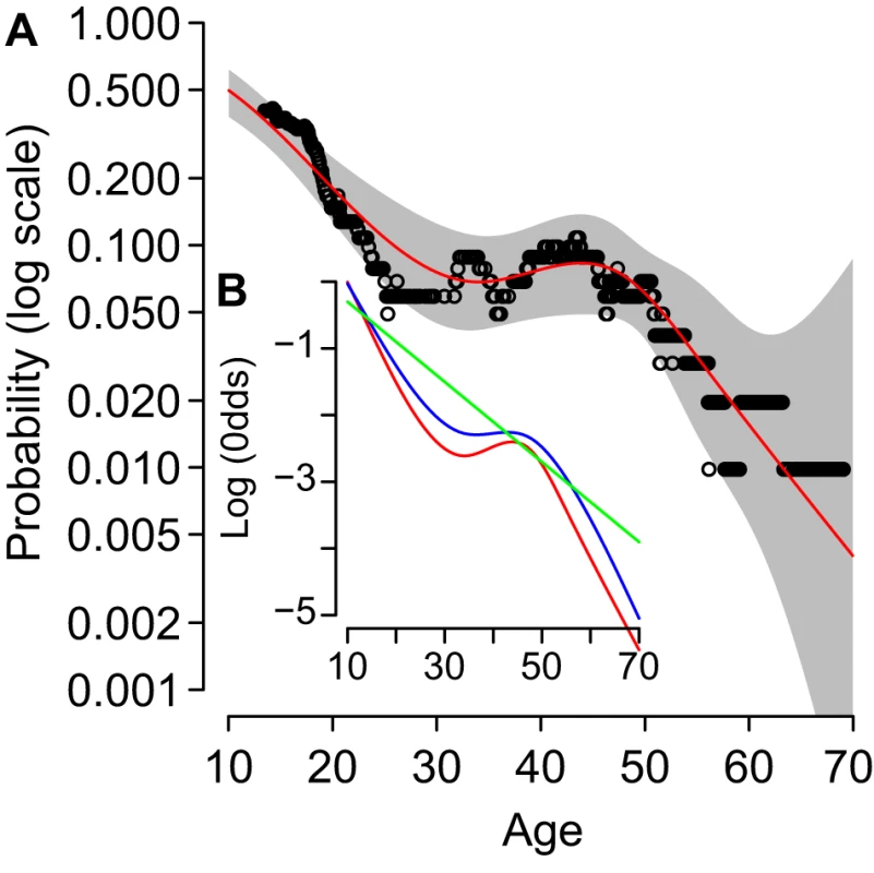
(A) Shows the average probability of infection for median age (x-axis) in rolling windows of 100 study participants (black circles), the best-fit probability of infection (red line, univariate restricted cubic spline), and 95% confidence intervals (grey area). (B) Shows the log-odds, relative to age 1, for: the same best-fit univariate spline fit as in (A) (red); the spline age model adjusted for the presence of a child in the household (blue); and a linear model adjusted for the presence of a child (green, best-fitting model A in Table 1). The presence of a child in the household explained the plateau in the age-risk of infection in these data. We adjusted the spline model using a binary variable for the presence or absence of a child in the household and compared the shape of the odds ratio curve (Figure 2B, blue line, AIC = 406.1) with the unadjusted model: the odds ratio curve was more linear, with less pronounced turning points. Therefore, we also fitted the linear age model, adjusted for the presence or absence of a child. We found that the adjusted linear model was a more parsimonious explanation for the data (Figure 2B, green line; Table 1, model A, AIC = 405.5, ΔAIC = 0). The age-adjusted odds ratio for infection for those in a household without children was 0.39 (0.21–0.73), relative to those living in households with children.
Tab. 1. Risk factors for infection with 2009 H1N1 pandemic influenza for 667 participants of the study for whom paired sera were tested and for whom complete information was available. 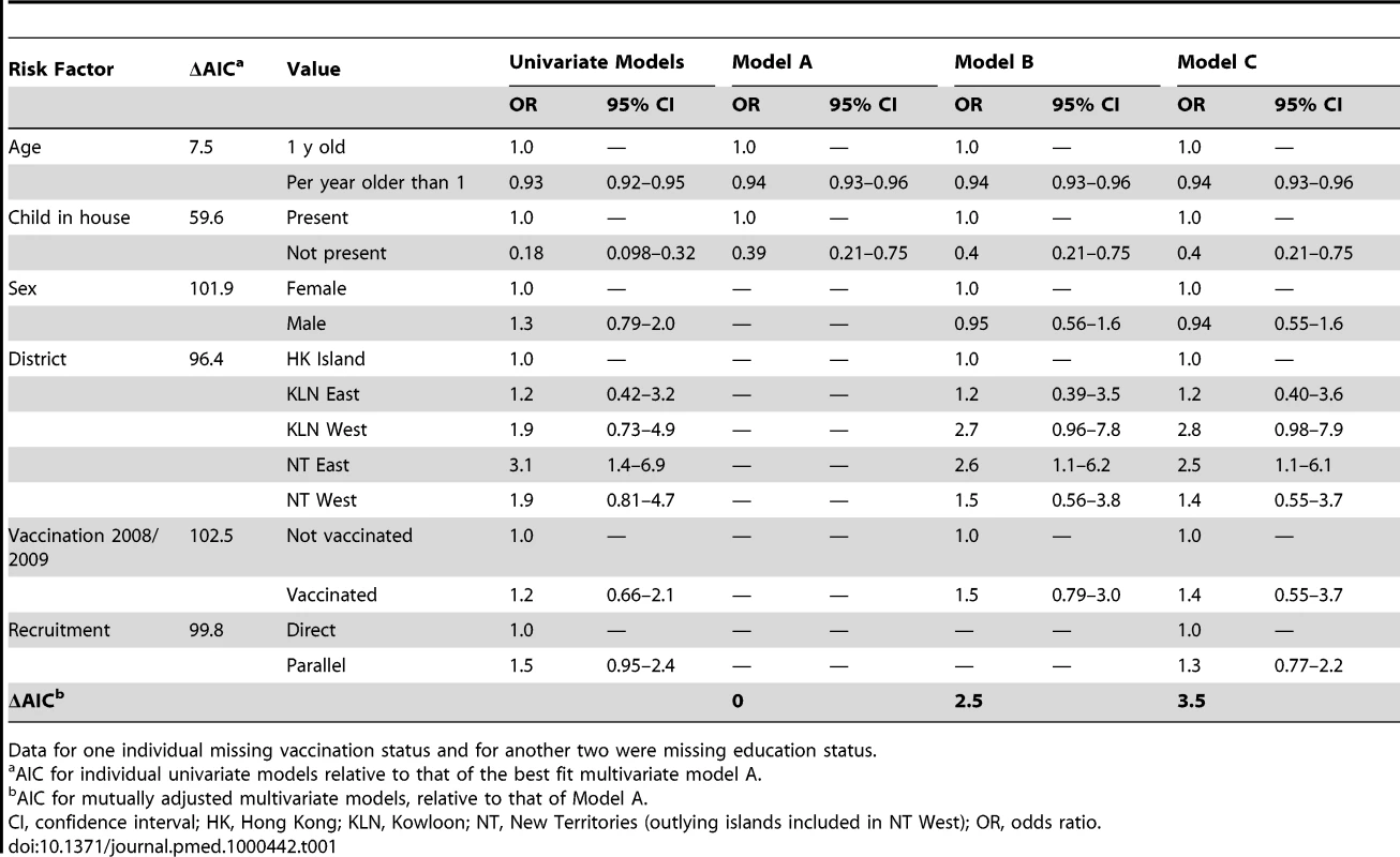
Data for one individual missing vaccination status and for another two were missing education status. Other Risk Factors
We had substantive interest in six other potential risk factors, in addition to age and the presence or absence of a child in the household. In order to efficiently prioritize model selection, we calculated the AIC for all possible regression models (63 nonempty subsets of six risk factors) combined with linear age and the presence or absence of a child in the household. None of the 63 models had a lower AIC than model A. Only three additional risk factors appeared in models not substantially different from model A on the AIC scale (ΔAIC <3): district of residence, sex, and status for 2008/2009 influenza vaccination (Table 1). Even though univariate analyses suggested association in some cases, the following three risk factors did not appear in models close to the best model: household size (best ΔAIC = 7.4), profession (best ΔAIC = 6.4), and level of education (best ΔAIC = 5.4).
Residents of New Territories East had an increased risk of infection during the study period, even after adjusting for other risk factors of interest (Table 1, model B, ΔAIC = 2.5), with an odds ratio of 2.6 (1.1–6.2) relative to residents of Hong Kong Island. Although sex was included in a number of well-fitting models, the odds ratio for males relative to females was close to unity in the adjusted model, suggesting that correlation between sex and other risk factors in the study group was somewhat different from that in the wider population. Although not statistically significant, estimates of the adjusted odds ratio for vaccine status suggest that those who reported being vaccinated in 2008/2009 were at an increased risk of infection.
In order to control for possible bias from the two alternate sources of recruitment (the direct group or the parallel group), we included the source of recruitment as a possible confounder in a final mutually adjusted regression model, model C. This model scored slightly worse on the AIC scale than did model B (ΔAIC = 3.5), with only very minor changes in estimated odds ratios and confidence intervals. Although recruitment from the parallel group was associated with an increased risk of infection, the estimated odds ratio was not significantly different from unity.
We also considered a baseline titre of 1∶40 or greater as a risk factor for infection (despite this variable being a component of the outcome variable, Table S3). Even though a low baseline titre was protective, the small number of raised titres observed ensured that this variable had little explanatory power (ΔAIC for model A with baseline titre added as a binary variable = 1.1).
Individual-level data from the serological survey are provided as Dataset S1 with the fields defined in Table S4.
Symptoms
Rates of reported symptoms were low, but varied substantially by definition and by mode of reporting (Figure 3). In general, when reporting by phone or by symptom diary, between 10% and 20% of seroconverters reported having experienced symptoms. The only exception was the reporting of ARI by diary, which was considerably higher. Rates of reporting by follow-up interview were considerably higher. Febrile symptoms for seroconverting children were reported by phone more often than for adults who seroconverted (Table 2). Of the 41 children seroconverters in the study, ten reported by phone that they had experienced a fever and, of those, seven reported ILI. In contrast only one of 40 adult seroconverters in the study had telephoned us to report a febrile illness (and also ILI).
Fig. 3. Absolute levels of symptom reporting. 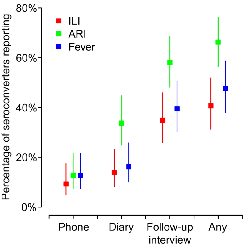
Three different definitions of symptoms were used: ILI, acute respiratory infection, or fever (see main text for details). Symptoms were reported by one of: study participants phoning into the study phone line, by symptom diary, or at follow-up interview. We also report an all-inclusive rate: the percentage of seroconverters that reported symptoms by any of the three modes. 95% confidence bounds are based on the binomial distribution. Tab. 2. Symptoms reported by study participants by infection status, symptom definition, and method of reporting. 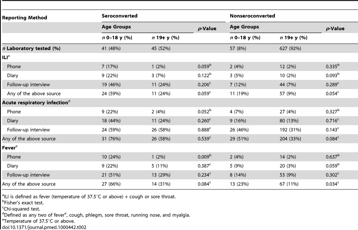
ILI is defined as fever (temperature of 37.5°C or above) + cough or sore throat. Rates of Severe Disease
The overall rate of confirmed H1N1pdm-associated deaths was 7.6 (6.2–9.5) per 100,000 infections. Rates of severe disease increased with age (Figure 4A). Although rates of hospitalization per infection were not substantially different for the younger three age groups (∼1%), individuals aged 60 and older were at slightly increased risk. However, rates of mortality increased substantially with age. The risk of death per infection for 3–19 y olds was 1.3 (1.0–1.7) per 100,000 while the risk for individuals aged 60 or older was 220 (50–4,000) per 100,000. Wide confidence intervals for 60 y and older were driven by the single observed infection in that group. However, the general trend of rapidly increasing mortality with age can be seen clearly across the other adult age groups.
Fig. 4. (A) Overall and age-specific estimated rates of severe disease per infection; squares for hospitalization, triangles for admission to ICUs, and circles for death. 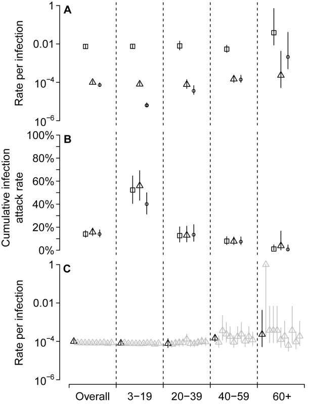
(B) Estimated cumulative attack rates for infection up to the end of January 2010. Three separate estimates of cumulative infection attack rate are given for each age group, on the basis of the three levels of severity, with symbols as per (A). (C) Comparison of estimates of rates of ICU admission per infection from the current study (black triangles, as per (A)) with estimates of the same statistic from ten simulations of an alternate, nonbracketing, study design (see text). Cumulative Attack Rate up to End January 2010
Although we aimed to obtain bracketing sera (Figure 1), there was substantial transmission outside of the period of our study. Therefore, we used the total number of confirmed severe cases to estimate the age-specific cumulative infection attack rates for a longer period of time than was captured by our study (Figure 4B). Between 27 April 2009 and 7 February 2010, in Hong Kong, there were 7,981 hospitalizations, 109 admissions to ICU, and 70 deaths among laboratory confirmed cases of H1N1pdm. Using the rates of severe disease per infection, these totals suggest cumulative infection attack rates of: 14% (11%–17%), based on hospitalization; 16% (13%–19%), based on ICU admission; and 14% (12%–18%), based on deaths.
Simulation of a Nonbracketing Design
We simulated an alternate trial design in which 500 samples were obtained from each of the four age groups during the week containing 1 June 2009 and the 500 follow-up samples were obtained from each age group during the week containing 30 September 2009 (Figure 4C). In this trial design, fewer infections and many fewer severe cases were observed. However, despite the reduced power of the alternate design, overall rates of severity are well estimated and the pattern of increasing severity with age is readily apparent. Even though no infections were observed in some of the simulated studies in the 60 and older age class, it was still possible to establish a relatively high lower bound for rates of severe disease. Although we present results for ICU as an example, the results for admission to hospital and death are not substantively different.
Discussion
The main wave of the 2009 (H1N1) pandemic infected many more children than it did adults. These differences are not explained by baseline antibody titres to H1N1pdm, but could be explained partly by social mixing patterns of the population in these different age strata. However, given that social mixing patterns within the 20–60-y age range do not exhibit substantial variation [23], and that we have controlled for the presence of a child in the household, it is plausible that increasing age leads to decreased susceptibility independently of mixing and titres to H1N1pdm, possibly as a result of repeated seasonal influenza infections, but by a mechanism not detectable by assays for neutralizing antibody. Whether this reflects antibody that protects by mechanisms other than neutralization, such as antibody-dependent cell cytotoxicity or cell-mediated immunity, remains worthy of investigation.
While older adults had low infection rates, those individuals infected developed severe disease much more frequently. Further, our results suggest that individuals over 60 y experience very high absolute rates of severe outcomes, with approximately one reported and positively tested death for every 200 infections. If continuing waves of H1N1pdm infection are driven by antigenic drift, and if that drift decreases the efficiency of the cross-protection currently possessed by older adults, it is likely that future waves could have higher overall mortality than initial waves. Surveillance of clusters of severe disease in older adults should be prioritized because this may be the first clear signal of a significant antigenic evolutionary event. The efficacy of alternate vaccine formulations in preventing infection in older individuals should be assessed as a matter of priority [24].
The low rate of ILI reported by phone and symptom diary for seroconverters in this study is consistent with results from an independent parallel study of household contacts of children in Hong Kong [21]. However, much higher rates of symptoms were reported in military personal in Singapore [25]. These differences may be driven by the age distributions of the different cohorts. The Singapore cohort was much younger: our comparison of symptom reporting by age suggests that reduced level of symptom reporting observed in adults was likely due to a reduced rate of experiencing symptoms, rather than just to a reduced propensity to report. The proportion of uninfected children that reported febrile illness was similar to that of uninfected adults. However, our results do suggest that adults are slightly less likely to report symptoms during a follow-up interview than are children. In general, rates of reporting were much higher but less specific when participants were asked a direct question during the follow-up interview compared with more passive symptom diaries or participant call-in. Future studies attempting to address rates of illness associated with influenza infection should attempt more intensive prospective follow-up of participants to minimize potential recall bias.
Our study has a number of limitations. Firstly, we did not measure incidence in children aged 2 and lower, who are much more likely to be admitted to hospital for acute respiratory infection than other age groups, but less likely to be infected with pandemic influenza than older children [26]. Given our focus on the use of paired sera, this shortcoming was unavoidable. The incorporation of data from cross-sectional samples from young children is the topic of ongoing investigation. Also, our cases are defined by an observed 4-fold rise in titre, which may not have occurred for all infections; and we will not have captured all severe cases as some H1N1pdm cases will have entered the private hospital system (although this is likely to have occurred much more rarely than the average rate of ∼10% for all types of admission). We suggest that the combined effect of imperfect test sensitivity and imperfect severe case matching will have generated only minor biases in our estimates of infection rates and severe disease rates and that the two effects will have acted in opposite directions.
In order to extrapolate from the period of our study to the full period of the pandemic in Hong Kong, we made the assumption that the testing process for individuals who became hospitalized was consistent. This is almost certainly not the case for all hospitalized individuals. In particular, anecdotal evidence suggests that less severe hospitalized cases were less likely to be tested for H1N1pdm after the end of September. Analysis of the rate of admission to ICU per positive hospital admission supports the anecdotal evidence (unpublished data). Therefore, it is reassuring that estimates of the overall and age-specific attack rates based on the three different outcome measures (hospitalization, admission to ICU, and death) are largely consistent.
We cannot exclude the possibility of substantial sampling bias in our serological survey. We were only able to successfully obtain paired sera from an average of 1.6 individuals in ∼2% of households initially identified by random telephone number selection. Although similar in many respects, after using common demographic characteristics to compare the study population with the wider Hong Kong population (age, sex, district, and education), we cannot exclude the possibility that individuals more likely to take part in our study had a different probability of infection than the population at large. However, we suggest that the potential impact of sampling bias in our results (and the value of evidence presented here in general) should be assessed on a result-by-result basis against the background of other reported community surveys of the 2009 influenza pandemic.
For the 2009 Hong Kong pandemic season, the current results add substantially to our earlier work [17] in a number of ways. Using an entirely different sampling scheme, the current study confirms the general pattern of sharply decreasing age-specific rates of infection for a similar period of the epidemic, for the age ranges contained in both studies: thus strengthening the case for rapid cross-sectional serological studies on the basis of convenient samples [17] at the same time as suggesting that serious sampling biases were not present in either study. Also, to a certain extent, multiple recruitment groups in the current study provided a proxy for propensity to take part: those recruited via the parallel study had already agreed to complete one telephone questionnaire and to be contacted again for other studies. Having agreed for a third time to take part, by enrolling in the main serological survey, it seems reasonable to assume that parallel study recruits are from a subset of the population more likely to take part in this type of study. Although our univariate estimate of the odds ratio for the parallel recruitment group was greater than one, compared with the direct group, the inclusion of recruitment source as a covariate did not improve the parsimony of our multivariate regression model. Also, the strength of the odds ratio for the parallel group in the univariate model was reduced substantially when adjusting for our epidemiological variables of interest. Therefore, although it is certainly possible that propensity to take part in the paired serological study was correlated with the risk of infection, comparison with our previously published cross-sectional study and comparison of our two recruitment groups suggests that sample bias was of considerably lesser influence on infection than variables we were able to measure directly, such as age and the presence of a child in the household.
The current study, by recruiting from a wide age range using the same sampling framework and a paired sera outcome, allows us to add to the available literature in a number of other ways. We present important data on infection rates and severity for those aged 60 y and older that were not reported in our previous study [17]: even the single observed 4-fold rise in titre out of 131 paired samples is valuable. In an age group typically at high risk from influenza morbidity and mortality, these data allow an informative upper bound for the absolute risk of infection and, hence, an informative lower bound for the absolute risk of severe outcomes. Without paired samples, obtaining accurate bounds for estimates of low rates of incidence from cross-sectional samples is problematic. When trying to estimate low rates of incidence with cross-sectional data, statistical noise becomes significant in the numerator: to overcome this noise, large sample sizes are required. Good evidence for high absolute risk in a particular age group may be of substantial public health value for the prioritization of interventions.
With good data on other potential risk factors for infection, we were able to show how the presence of a child in the household could explain an apparent age plateau in risk of infection, while variables such as education and profession did not appear to be risk factors once adjusted for age. This type of traditional risk-factor analysis is not possible with unlinked samples for which only the following variables are usually available: age, sex, and clinic location. Similarly, our analysis of home district (using only a single clinic location) suggests that micro-scale spatial heterogeneities persisted for longer than might have been expected in a large well-connected population. For the period of our study, residents of one district (New Territories East) appeared to be at substantially greater risk of infection than were residents of other districts. It is possible that the overall level of transmission was higher in that one district than in other districts, or that the epidemic occurred sooner there than it did elsewhere. Further, it seems possible that, in Hong Kong, spatial decorrelation took a long time to occur or never did occur. Individual-based models of respiratory infections, parameterized with the commuting patterns of adults [27], and also those parameterized with explicit school locations [28],[29], suggest much more rapid spread at small scales in large populations. Had the pandemic strain been more severe, good knowledge of small-scale spatiotemporal patterns could have been of value in optimizing the provision of key health care facilities and the timing of rolling school closures.
Our results can be compared with serology-based studies of influenza incidence in other populations during the 2009 (H1N1) pandemic [4]–[16]. In England and Wales, a study of cross-sectional clinical samples found substantial increases in the proportion of younger children with titres 1∶32 or greater between a 2008 baseline (n = 1,403) and sample taken in September 2009 (n = 1,954), thus giving valuable early evidence that the infection attack rate was high in some age groups and, hence, that the rate of severe cases per infection in the most affected age groups was likely to be low [10]. As already mentioned above, the consistency of our results for Hong Kong between the current study and our previous cross-sectional study [17] validates the use of convenient clinical samples during the early stages of a pandemic as a useful tool for the estimation of incidence in high-incidence groups. However, a high degree of cross-reactivity in hemagglutination inhibition assays in sera from adults in many studies introduced considerable statistical noise and prevented reliable estimates of attack rates in older age groups. In Singapore, sera were collected from four groups: an existing sample of healthy adults (n = 838), military personnel (n = 1,213), staff from an acute care hospital (n = 558), and staff and residents of a long-term care facility (n = 300) [14]. Although an overall infection attack rate of 13% was observed in this study, it is difficult to generalize these results because no subgroup contained school-aged children and many of the infection events occurred in the military substudy.
It is more difficult to compare our community-wide results with historical studies such as the Tecumseh [18] and Seattle [19] study. Both were designed to efficiently obtain viral samples from households with children and, therefore, did not attempt to recruit from childless households. Also, information on their precise sampling framework for households with children is difficult to obtain. However, future analyses of household-level data from the current study and follow-up waves should permit a like-for-like comparison between the subgroups in the Hong Kong study and canonical historical studies of respiratory infection.
Our simulation results show that a larger paired-sera cohort study with a shorter follow-up period could have generated—more rapidly—similar data to those presented here. We suggest that this revised design would be a valuable addition to revised pandemic preparedness plans for a small subset of large well-connected global cities. Sentinel hospitals could be established in early-affected populations to help ensure that the testing process remains consistent for ICU cases throughout the epidemic curve. Given that (a) many believe the 2009 response to have been overzealous and (b) the severity of the next pandemic strain is not known, there appears to be a substantial risk that the public health impact of the next pandemic will be underestimated. Therefore, revised preparedness plans should prioritize reactive studies that can rapidly and reliably distinguish between 2009 (H1N1)-like strains (∼1∶10,000 infection fatality rate) and more severe pandemics. If the next pandemic strain were similar in all other respects, but had an infection fatality rate of ∼1∶1,000; we could reasonably expect peak demand on key health care services such as ICU to be ten times greater than that observed during 2009/2010 [30].
Supporting Information
Zdroje
1. ThompsonWW
ShayDK
WeintraubE
BrammerL
BridgesCB
2004 Influenza-associated hospitalizations in the United States. JAMA 292 1333 1340
2. ShindeV
BridgesC
UyekiT
ShuB
BalishA
2009 Triple-reassortant swine influenza A (H1) in humans in the United States, 2005-2009. N Engl J Med 360 2616 2625
3. Van KerkhoveMD
AsikainenT
BeckerNG
BjorgeS
Desenclos J-C, et al. 2010 Studies needed to address public health challenges of the 2009 H1N1 influenza pandemic: insights from modeling. PLoS Med 7 e1000275 doi:10.1371/journal.pmed.1000275
4. DowseGK
SmithDW
KellyH
BarrI
LaurieKL
2011 Incidence of pandemic (H1N1) 2009 influenza infection in children and pregnant women during the 2009 influenza season in Western Australia - a seroprevalence study. Med J Aust 194 68 72
5. GilbertGL
CretikosMA
HuestonL
DoukasG
O'TooleB
2010 Influenza A (H1N1) 2009 antibodies in residents of New South Wales, Australia, after the first pandemic wave in the 2009 southern hemisphere winter. PLoS ONE 5 e12562 doi:10.1371/journal.pone.0012562
6. GrillsN
PiersLS
BarrI
VaughanLM
LesterR
2010 A lower than expected adult Victorian community attack rate for pandemic (H1N1) 2009. Aust N Z J Public Health 34 228 231
7. McvernonJ
LaurieK
NolanT
OwenR
IrvingD
2010 Seroprevalence of 2009 pandemic influenza A(H1N1) virus in Australian blood donors, October - December 2009. Euro Surveill 15
8. TsaiTF
PedottiP
HilbertA
LindertK
HohenbokenM
2010 Regional and age-specific patterns of pandemic H1N1 influenza virus seroprevalence inferred from vaccine clinical trials, August-October 2009. Euro Surveill 15
9. DengY
PangXH
YangP
ShiWX
TianLL
2011 Serological survey of 2009 H1N1 influenza in residents of Beijing, China. Epidemiol Infect 139 52 58
10. MillerE
HoschlerK
HardelidP
StanfordE
AndrewsN
2010 Incidence of 2009 pandemic influenza A H1N1 infection in England: a cross-sectional serological study. Lancet 375 1100 1108
11. TandaleBV
PawarSD
GuravYK
ChadhaMS
KoratkarSS
2010 Seroepidemiology of pandemic influenza A (H1N1) 2009 virus infections in Pune, India. BMC Infect Dis 10 255
12. BandaranayakeD
HuangQS
BissieloA
WoodT
MackerethG
2010 Risk factors and immunity in a nationally representative population following the 2009 influenza A(H1N1) pandemic. PLoS ONE 5 e13211 doi:10.1371/journal.pone.0013211
13. AdamsonWE
MaddiS
RobertsonC
McDonaghS
MolyneauxPJ
2010 2009 pandemic influenza A(H1N1) virus in Scotland: geographically variable immunity in Spring 2010, following the winter outbreak. Euro Surveill 15
14. ChenMIC
LeeVJM
Lim W-Y, BarrIG
LinRTP
2010 2009 influenza A(H1N1) seroconversion rates and risk factors among distinct adult cohorts in Singapore. JAMA 303 1383 1391
15. PrachayangprechaS
MakkochJ
PayungpornS
ChieochansinT
VuthitanachotC
2010 Serological analysis of human pandemic influenza (H1N1) in Thailand. J Health Popul Nutr 28 537 544
16. ZimmerSM
CrevarCJ
CarterDM
StarkJH
GilesBM
2010 Seroprevalence following the second wave of Pandemic 2009 H1N1 influenza in Pittsburgh, PA, USA. PLoS ONE 5 e11601 doi:10.1371/journal.pone.0011601
17. WuJT
MaESK
LeeCK
ChuDKW
Ho P-L, et al. 2010 The infection attack rate and severity of 2009 pandemic H1N1 influenza in Hong Kong. Clin Infect Dis 51 1184 1191
18. MontoAS
KioumehrF
1975 The Tecumseh Study of Respiratory Illness. IX. Occurence of influenza in the community, 1966--1971. Am J Epidemiol 102 553 563
19. FoxJP
CooneyMK
HallCE
FoyHM
1982 Influenzavirus infections in Seattle families, 1975-1979. II. Pattern of infection in invaded households and relation of age and prior antibody to occurrence of infection and related illness. Am J Epidemiol 116 228 242
20. CowlingBJ
NgDMW
IpDKM
LiaoQ
LamWWT
2010 Community psychological and behavioral responses through the first wave of the 2009 influenza A(H1N1) pandemic in Hong Kong. J Infect Dis 202 867 876
21. CowlingBJ
ChanKH
FangVJ
LauLLH
SoHC
2010 Comparative epidemiology of pandemic and seasonal influenza A in households. N Engl J Med 362 2175 2184
22. LeungGM
WongIOL
Chan W-S, ChoiS
Lo S-V, et al. 2005 The ecology of health care in Hong Kong. Soc Sci Med 61 577 590
23. MossongJ
HensN
JitM
BeutelsP
AuranenK
2008 Social contacts and mixing patterns relevant to the spread of infectious diseases. PLoS Med 5 e74 doi:10.1371/journal.pmed.0050074
24. KaoTM
HsiehSM
KungHC
LeeYC
HuangKC
2010 Immune response of single dose vaccination against 2009 pandemic influenza A (H1N1) in the Taiwanese elderly. Vaccine 28 6159 6163
25. LeeVJ
YapJ
TayJK
BarrI
GaoQ
2010 Seroconversion and asymptomatic infections during oseltamivir prophylaxis against Influenza A H1N1 2009. BMC Infect Dis 10 164
26. RossT
ZimmerS
BurkeD
CrevarC
CarterD
2010 Seroprevalence following the second wave of pandemic 2009 H1N1 influenza. PLoS Curr Influenza RRN1148
27. RileyS
FergusonNM
2006 Smallpox transmission and control: spatial dynamics in Great Britain. Proc Natl Acad Sci U S A 103 12637 12642
28. FergusonNM
CummingsDAT
CauchemezS
FraserC
RileyS
2005 Strategies for containing an emerging influenza pandemic in Southeast Asia. Nature 437 209 214
29. YangY
SugimotoJ
HalloranM
BastaN
ChaoD
2009 The transmissibility and control of pandemic influenza A (H1N1) virus. Science
30. InvestigatorsAI
WebbSAR
PettiläV
SeppeltI
BellomoR
2009 Critical care services and 2009 H1N1 influenza in Australia and New Zealand. N Engl J Med 361 1925 1934
Štítky
Interní lékařství
Článek vyšel v časopisePLOS Medicine
Nejčtenější tento týden
2011 Číslo 6- Není statin jako statin aneb praktický přehled rozdílů jednotlivých molekul
- Magnosolv a jeho využití v neurologii
- Hojení análních fisur urychlí čípky a gel
- Biomarker NT-proBNP má v praxi široké využití. Usnadněte si jeho vyšetření POCT analyzátorem Afias 1
- Moje zkušenosti s Magnosolvem podávaným pacientům jako profylaxe migrény a u pacientů s diagnostikovanou spazmofilní tetanií i při normomagnezémii - MUDr. Dana Pecharová, neurolog
-
Všechny články tohoto čísla
- The First Model-Based Geostatistical Map of Anaemia
- The Dynamics of Health and Return Migration
- Life Course Trajectories of Systolic Blood Pressure Using Longitudinal Data from Eight UK Cohorts
- Evaluation of Coseasonality of Influenza and Invasive Pneumococcal Disease: Results from Prospective Surveillance
- Energy Density, Portion Size, and Eating Occasions: Contributions to Increased Energy Intake in the United States, 1977–2006
- Scaling Up Global Health Interventions: A Proposed Framework for Success
- The Effect of Highly Active Antiretroviral Therapy on the Survival of HIV-Infected Children in a Resource-Deprived Setting: A Cohort Study
- Cardiac Complications in Patients with Community-Acquired Pneumonia: A Systematic Review and Meta-Analysis of Observational Studies
- Epidemiological Characteristics of 2009 (H1N1) Pandemic Influenza Based on Paired Sera from a Longitudinal Community Cohort Study
- Mapping the Risk of Anaemia in Preschool-Age Children: The Contribution of Malnutrition, Malaria, and Helminth Infections in West Africa
- More and Better Information to Tackle HIV Epidemics: Towards Improved HIV Incidence Assays
- Human Trafficking: The Shameful Face of Migration
- Global Protection and the Health Impact of Migration Interception
- Migration and "Low-Skilled" Workers in Destination Countries
- The Effect of Handwashing at Recommended Times with Water Alone and With Soap on Child Diarrhea in Rural Bangladesh: An Observational Study
- PLOS Medicine
- Archiv čísel
- Aktuální číslo
- Informace o časopisu
Nejčtenější v tomto čísle- Migration and "Low-Skilled" Workers in Destination Countries
- Mapping the Risk of Anaemia in Preschool-Age Children: The Contribution of Malnutrition, Malaria, and Helminth Infections in West Africa
- More and Better Information to Tackle HIV Epidemics: Towards Improved HIV Incidence Assays
- Energy Density, Portion Size, and Eating Occasions: Contributions to Increased Energy Intake in the United States, 1977–2006
Kurzy
Zvyšte si kvalifikaci online z pohodlí domova
Autoři: prof. MUDr. Vladimír Palička, CSc., Dr.h.c., doc. MUDr. Václav Vyskočil, Ph.D., MUDr. Petr Kasalický, CSc., MUDr. Jan Rosa, Ing. Pavel Havlík, Ing. Jan Adam, Hana Hejnová, DiS., Jana Křenková
Autoři: MUDr. Irena Krčmová, CSc.
Autoři: MDDr. Eleonóra Ivančová, PhD., MHA
Autoři: prof. MUDr. Eva Kubala Havrdová, DrSc.
Všechny kurzyPřihlášení#ADS_BOTTOM_SCRIPTS#Zapomenuté hesloZadejte e-mailovou adresu, se kterou jste vytvářel(a) účet, budou Vám na ni zaslány informace k nastavení nového hesla.
- Vzdělávání


