-
Články
- Vzdělávání
- Časopisy
Top články
Nové číslo
- Témata
- Kongresy
- Videa
- Podcasty
Nové podcasty
Reklama- Kariéra
Doporučené pozice
Reklama- Praxe
Host-to-Pathogen Gene Transfer Facilitated Infection of Insects by a Pathogenic Fungus
The ability of infectious agents to evolve different host ranges contributes to the emergence of new diseases, and this host switching could also account for the wide variety of fungal associations with animals, plants and other fungi. There must be mechanisms for such host shifts, but these remain largely unknown. In this study, we phylogenetically predict that the endophytic fungus Metarhizium robertsii acquired a sterol carrier gene from insects through horizontal gene transfer (HGT). This sterol carrier is involved in maintaining cell membrane sterols, and thus membrane integrity, when M. robertsii proliferates in the haemocoel of living insects. Therefore, the acquisition of genetic material from a host has contributed to the development of fungal entomopathogenicity. In order to simulate this evolutionary event, the sterol carrier gene was transformed into an endophytic insect-pathogenic fungus (Beauveria bassiana) that lacks an endogenous Mr-NPC2a homolog. The virulence of B. bassiana was increased by expression of Mr-NPC2a.
Published in the journal: . PLoS Pathog 10(4): e32767. doi:10.1371/journal.ppat.1004009
Category: Research Article
doi: https://doi.org/10.1371/journal.ppat.1004009Summary
The ability of infectious agents to evolve different host ranges contributes to the emergence of new diseases, and this host switching could also account for the wide variety of fungal associations with animals, plants and other fungi. There must be mechanisms for such host shifts, but these remain largely unknown. In this study, we phylogenetically predict that the endophytic fungus Metarhizium robertsii acquired a sterol carrier gene from insects through horizontal gene transfer (HGT). This sterol carrier is involved in maintaining cell membrane sterols, and thus membrane integrity, when M. robertsii proliferates in the haemocoel of living insects. Therefore, the acquisition of genetic material from a host has contributed to the development of fungal entomopathogenicity. In order to simulate this evolutionary event, the sterol carrier gene was transformed into an endophytic insect-pathogenic fungus (Beauveria bassiana) that lacks an endogenous Mr-NPC2a homolog. The virulence of B. bassiana was increased by expression of Mr-NPC2a.
Introduction
New infectious diseases are constantly appearing, and their origins are diverse. Human behavior and practices are important factors, for example, catastrophic declines in amphibian and bat populations have been attributed to pathogenic fungi spread by humans [1], [2]. Another important origin is the ability of infectious agents themselves to evolve different host ranges, which would certainly contribute to the impact of invasive species. Such host switching probably accounts for the wide variety of fungal associations with animals, plants and other fungi [3]. There must be mechanisms for such host shifts, although these remain largely unknown [4], [5], [6].
Horizontal gene transfer (HGT) between distantly related bacteria contributes significantly to the emergence of new pathogens but HGT is usually thought to play a minor role in eukaryotes. However, data from multiple genomic sequences suggests that HGT has also occurred between eukaryotes [7], and may bestow a clear selective advantage to fungi [8]. A gene encoding a critical virulence factor was transferred from one species of fungal pathogen to another, leading to the emergence of a new damaging disease of wheat [9]. HGT from bacteria to fungi is relatively common [10], and a few examples are known where HGT has occurred between plants and either pathogenic fungi or parasitic plants [11], [12], [13], [14]. Microsporidian intracellular parasites Enephalitozoon may have acquired a purine nucleotide phosphorylase from an arthropod that is not a host [15]. There is also a longstanding controversy as to whether the malaria causing pathogen Plasmodium vivax has horizontally acquired human genetic material that might facilitate its long stay in the body [16]. Otherwise, HGT between eukaryotic pathogens and animal hosts has been neither predicted nor characterized.
Fungi are the commonest pathogens of insects and crucial regulators of insect populations [17], [18]. Metarhizium robertsii (formerly known as Metarhizium anisopliae) is the best studied entomopathogenic fungus [19] as well as being an important plant-growth-promoting endophyte [20], [21]. Phylogenomic analysis suggests that the ancestor of Metarhizium was an endophyte, with entomopathogenicity being an acquired characteristic [22]. The evolutionary transition of a fungus from an endophyte to an insect pathogen provides a novel perspective on how new functional mechanisms important for host switching and virulence are acquired. In this study, we identified a new virulence factor (Mr-NPC2a) of M. robertsii by screening an Agrobacterium tumefaciens-mediated T-DNA insertion library. We provide evidence that Mr-npc2a was horizontally acquired from an insect and allowed Metarhizium to compete with insect hosts for the sterols necessary to maintain cell membrane integrity.
Results
Characterization of the sterol carrier gene Mr-npc2a as a pathogenicity mutant
Using Agrobacterium tumefaciens-mediated fungal transformation, we generated a set of 20, 328 T-DNA-tagged M. robertsii strain ARSEF2575 mutants [23] which we screened for virulence mutants against wax worm larvae (Galleria mellonella) (Pet Solutions, OH USA). A transformant (M298) that showed a significantly slower killing speed than the wild type strain was selected for further study (Fig. 1). The open reading frame (ORF) of the gene MAA_03817 (Genbank accession no.: EFZ01221) was found to be disrupted in this mutant. MAA_03817 showed significant similarity to insect Niemann-Pick type C2 (NPC2) proteins, and thus was designated as Mr-NPC2a. We deleted the ORF of Mr-npc2a in wild type M. robertsii, and the resulting disruptant (ΔMr-npc2a) showed the same loss of virulence as M298 (Fig. 1). The details of the T-DNA insertion and gene deletion alleles are shown in Fig. S1. ΔMr-npc2a was complemented by a genomic clone of Mr-npc2a. The details of deletion of Mr-npc2a and the complementation of ΔMr-npc2a are described in Fig. S2.
Fig. 1. Kinetics of insect survivorship in bioassays. 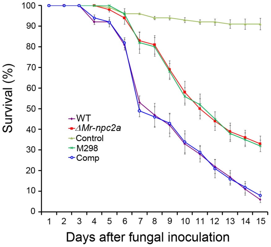
Wax worm larvae (G. mellonella) were treated with M. robertsii conidial suspensions (1×10⁷ spores/mL). WT: the wild type strain; ΔMr-npc2a: the mutant with the ORF of Mr-npc2a deleted; M298: the mutant with Mr-npc2a disrupted by T-DNA insertion; Comp: the complemented ΔMr-npc2a with a genomic clone of Mr-npc2a; control: insects treated with 0.05% Tween-80. The phenotype of ΔMr-npc2a on PDA (potato dextrose agar) plates was indistinguishable from the wild type strain, M298 and the complemented ΔMr-npc2a. Formation of infection structures (appressoria) by ΔMr-npc2a on the hindwings of Locusta migratoria manilensis (kindly provided by Dr. Wangpeng Shi at China Agricultural University) was not significantly different from the wild type strain, M298 and the complemented ΔMr-npc2a. The hyphal bodies [unicellular hyphal bodies (blastospores) and multicellular hyphal bodies] of ΔMr-npc2a, M298, the wild type strain and the complemented ΔMr-npc2a all began to appear in the hemolymph 36-hours after topical infection of G. mellonella, but the number of ΔMr-npc2a hyphal bodies (20±6.8 hyphal bodies/mL) and M298 (22±3.2 hyphal bodies/mL) was significantly less than the wild type (36 hyphal bodies ±9.3/mL) and the complemented ΔMr-npc2a (38 hyphal bodies ±9.5/mL). From 36 to 60 h post-inoculation, wild type hyphal bodies and the complemented ΔMr-npc2a hyphal bodies significantly outnumbered ΔMr-npc2a and M298 in the hemolymph. The number of the complemented ΔMr-npc2a hyphal bodies was not significantly different from the wild type strain at all time points (P>0.05). Likewise, the number of M298 hyphal bodies was not significantly different from ΔMr-npc2a at all time points (P>0.05) (Table S1). This set of data suggests that disruption of Mr-npc2a only impairs pathogenesis after M. robertsii enters the haemocoel.
All Galleria infected with the wild type strain or the complemented ΔMr-npc2a died as larvae, whereas nearly 40% of insects infected by ΔMr-npc2a or M298 survived. Similar numbers of conidia were produced on the cadavers of insects killed by the wild type strain, by the complemented ΔMr-npc2a, by ΔMr-npc2a and by M298 (P>0.05), so in terms of fecundity Mr-NPC2a enhanced fitness by 40%. Based on above data, M298 and ΔMr-npc2a are functionally indistinguishable, so only ΔMr-npc2a was used for following studies.
Metarhizium acquired the sterol carrier Mr-NPC2a from insect hosts by horizontal gene transfer
Genome analysis showed that there are 3 NPC2-like proteins in M. robertsii: MAA_03817 (Mr-NPC2a), MAA_10401 (Mr-NPC2b, Genbank accession no.: EFY94139) and MAA_03340 (Mr-NPC2c, Genbank accession no.: EFZ00744). All three Metarhizium NPC2-like proteins have conserved putative cholesterol/lipid binding sites, and six cysteine residues that form a hydrophobic core for sterol binding typical of Niemann-Pick type C2 (NPC2) sterol carriers. Mr-NPC2b shows 70% similarity to Mr-NPC2c (6e−53), but except for the aforementioned cholesterol/lipid binding sites and hydrophobic core, they show no significant similarity to Mr-NPC2a. We screened for homologs of Mr-NPC2a, Mr-NPC2b and Mr-NPC2c proteins using a protein based local alignment search tool (BLASTP, e-value cutoff 1e−05) against databases provided by NCBI (Genbank), JGI (Joint Genome Institute) and Broad Institute. No Mr-NPC2a homologs were identified when Mr-NPC2b or Mr-NPC2c were used as the query sequences; likewise no Mr-NPC2b and Mr-NPC2c homologs were identified when Mr-NPC2a was used as the query. Thus, BLASTP clearly differentiates between the evolutionary relationships of Mr-NPC2a and the related Mr-NPC2b and Mr-NPC2c. Homologs of Mr-NPC2b and Mr-NPC2c are widespread in the fungal kingdom being found in Ascomycota, Basidomycota, Zygomycota and Chytridiomycota. A phylogenetic analysis conducted with Mr-NPC2b, Mr-NPC2c and 30 other NPC2s showed that their phylogenetic relationship is consistent with previously established species phylogenies, demonstrating vertical inheritance (Fig. S3).
Mr-NPC2a homologs were identified in only two fungi (BLASTP, e-value cutoff 1e−05)–the ergot fungus Claviceps purpurea and Metarhizium acridum. All other Mr-NPC2a homologs were from insects and other animals including vertebrates. No homologs were found in plant, bacteria, archaea and viruses. A TBLASTn search against NCBI genomes (e-value cutoff 1e−05) only identified the same homologs as BLASTP. The origin of NPC2a was assessed using a variety of models and methods for phylogenetic reconstruction. The phylogenies consistently showed that Metarhizium and C. purpurea sequences exclusively clustered with insect sequences with high support (Fig. 2, Table S2). We then compared the topology of the obtained tree with those of alternative trees using eight tests including SH-test and AU-test provided by the program CONSEL [24]. The cluster of fungal NPC2as with vertebrate NPC2s was not statistically rejected, but these constrained trees were significantly less supported than the obtained tree. Furthermore, vertebrates lack close contact with Metarhizium and C. purpurea, whereas insects regularly encounter both fungi [17], [25]. Since sympatry is almost a prerequisite for HGT [26], the fungi are much more likely to have acquired NPC2a from insects than vertebrates. Details about the tree topology comparisons are presented in Table S3.
Fig. 2. Phylogenetic analysis of fungal NPC2a proteins and their homologs. 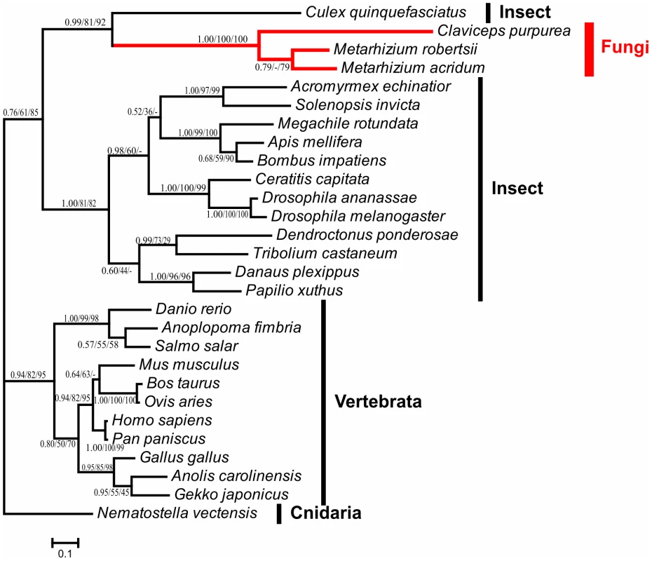
The Bayesian inference tree is shown unrooted. Numbers at nodes represent bayesian posterior probabilities (left) and bootstrap values of maximum likelihood (middle) and neighbor-joining (right) respectively. Hyphen (-) indicates no support value in corresponding method. The scale bar corresponds to the estimated number of amino acid substitutions per site. Branches of Clavicipitaceae (fungi) are indicated in red. The information about the sequences used in this analysis is presented in Table S2. Although it is the most likely that Metarhizium and C. purpurea acquired NPC2a from insects, the fungal NPC2as show low overall similarity to insect sequences (the lowest E value was 6e−15). However, NPC2 paralogs are remarkably divergent even in the same insect species, consistent with rapid diversification. For example, the fruitfly Drosophila melanogaster has 8 NPC2 proteins designated as NPC2a-NPC2h (Genbank accession numbers are NP_608637, NP_650331, NP_649976, NP_649975, NP_731439, NP_651219, NP_651823 and NP_651824, respectively), and the similarity of D. melanogaster NPC2a to other NPC2 proteins ranges from 3e−16 (NPC2b, 50% similarity) to as low as 3e−03 (NPC2g, 37% similarity). It is likely therefore that the insect ancestor of Mr-NPC2a could also have had low similarity to currently known insect NPC2 proteins.
We then looked at the genomic context of the npc2a genes in Metarhizium and C. purpurea to investigate how these fungi may have acquired npc2a gene from insects. The microsyntenies are almost the same in ∼40 kb M. robertsii and M. acridum DNA fragments containing the npc2a genes, indicating that the npc2a gene was acquired before the diversification of M. robertsii and M. acridum ∼33–43 MYA [22], and no significant genomic rearrangements have happened since. However, little microsynteny is retained between Metarhizium and other fungi including C. purpurea; only one M. robertsii gene (MAA_03818, Genbank accession no.: EFZ01222) adjacent to Mr-npc2a (MAA_03817) has a homolog (CPUR_07480, Genbank accession no.: CCE3355) adjacent to the C. purpurea npc2a gene (CPUR_07479, Genbank accession no.: CCE33554). CPUR_07480 and MAA_03818 have homologs in other fungi but not insects, showing that this gene is vertically inherited. CPUR_07480 and MAA_03818 have LITAF-like zinc ribbon domain, but their functions have not been characterized. Conversely, the microsynteny of the sequence around the C. purpurea npc2a gene (CPUR_07479) and CPUR_07480 is conserved among many fungi including Metarhizium (Fig. 3).
Fig. 3. Genomic context of the gene npc2a in M. robertsii, M. acridum, C. purpurea, and 16 other fungi (represented by Fusarium oxysporum and Cordyceps militaris). 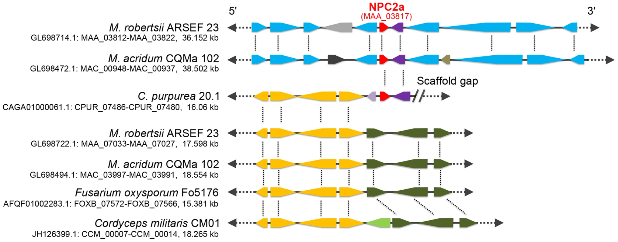
Homologs are shown in the same color. Red: npc2a gene. Purple: a gene that is adjacent to npc2a and is conserved among in M. robertsii, M. acridum and C. purpurea. Blue: genes are homologous between M. robertsii and M. acridum; Yellow: Genes that are co-linear in C. purpurea, M. robertsii, M. acridum, F. oxysporum and C. militaris. Dark olive: genes that are co-linear in M. robertsii, M. acridum, F. oxysporum and C. militaris. Note: a gap follows C. purpurea npc2a gene and the gene CPUR_07480. The lipid binding activity of Mr-NPC2a
Mr-NPC2a was expressed in Escherichia coli using the pET-15 system. The recombinant protein was purified to homogeneity and subjected to a lipid binding assay. Mr-NPC2a binds to cholesterol and bacterial lipid A that are absent in fungi, but did not bind to fungal ergosterol (Fig. 4), indicating that its binding specificity was similar to D. melanogaster NPC2 proteins that binds to cholesterol and bacterial lipid A [27], and distinct from yeast yNPC2p that binds to ergosterol and is involved in ergosterol homeostasis [28].
Fig. 4. Binding of recombinant Mr-NPC2a to diphosphoryl lipid A from E. coli, cholesterol and ergosterol. 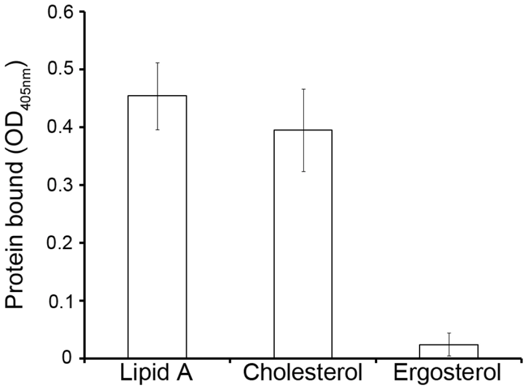
Purified Mr-NPC2a fusion protein was diluted to 500 mM and added to the ligand-coated plates for ELISA assay. Each bar represents the mean of 3 individual measurements±SEM. Expression pattern and cellular localization of Mr-NPC2a
We then investigated the biological function of Mr-NPC2a in M. robertsii. RT-PCR (reverse transcription PCR) did not detect Mr-npc2a transcripts in aerial hyphae, conidiating mycelium and conidia collected from PDA plates, hyphae and blastospores collected from SDB (Sabouraud dextrose broth), hyphae and blastospores grown in the in vitro prepared insect hemolymph, and appressoria on locust hindwings. However, Mr-npc2a was transcribed in the hyphal bodies collected from the hemolymph of living insects (Fig. 5A). The expression pattern of Mr-NPC2a was also investigated by observing GFP signal in three randomly selected transformants containing PMr-NPC2a:GFP. No differences in GFP signal at different developmental stages were observed between these transformants, so only data about one of the transformants is shown. GFP fluorescence was only observed in both unicellular (blastospores) and multicellular hyphal bodies after the fungus had penetrated the insect host (Manduca sexta larva) (Carolina, Burlington, NC) cuticle (Fig. 5B). Therefore, both RT-PCR analysis and GFP observation showed that Mr-NPC2a expresses exclusively in the hemolymph of living insects irrespective of whether the hyphal bodies were unicellular (blastospores) or multicellular. This pattern differs from other hemolymph specific promoters such as Mcl1 [29], as these are also active in in vitro prepared hemolymph.
Fig. 5. The expression pattern and cellular localization of Mr-NPC2a. 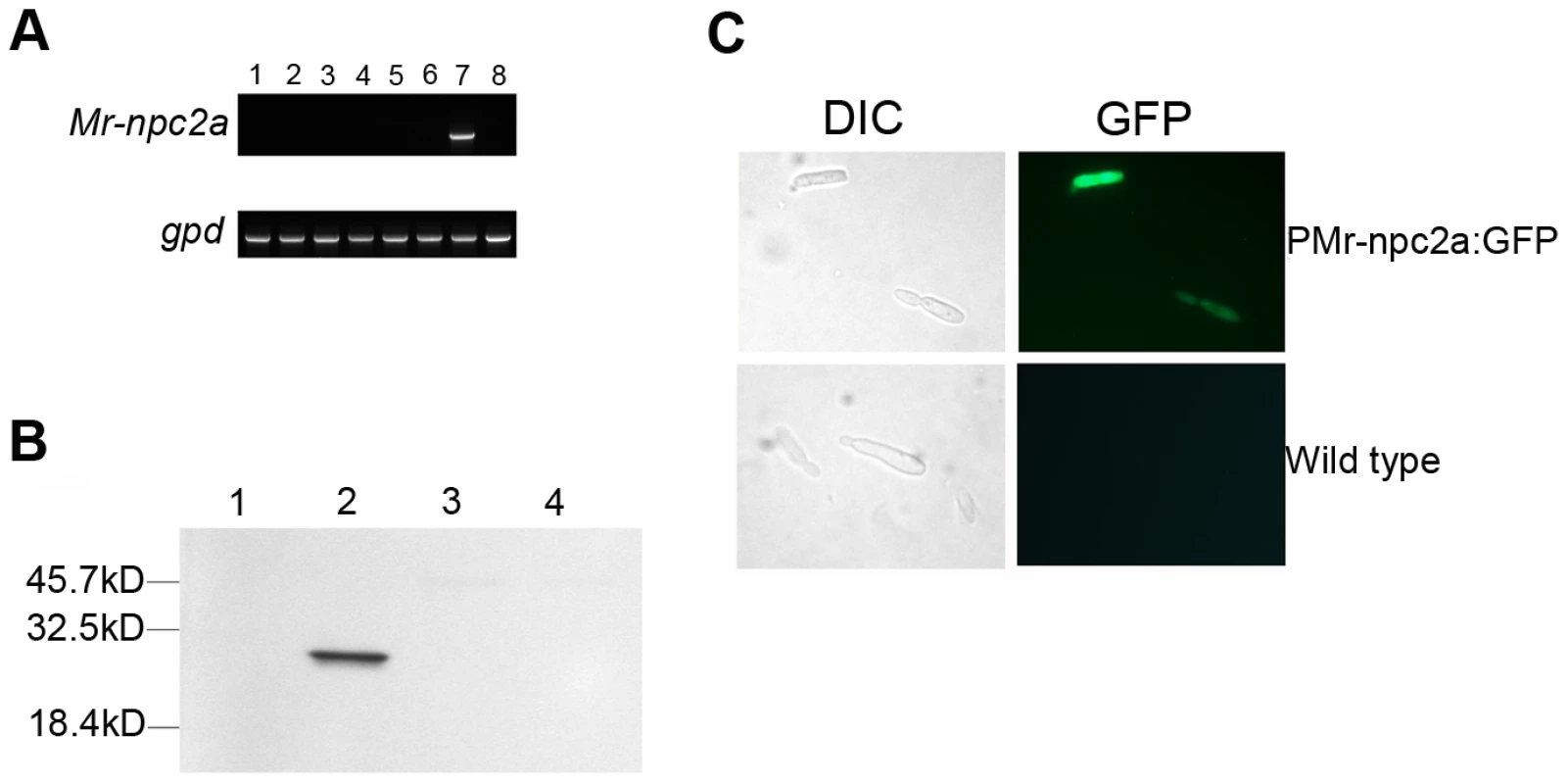
(A) RT-PCR analysis of Mr-npc2a expression in the wild type strain. Top row: the expression of Mr-npc2a in 1: aerial hyphae collected from a PDA plate; 2: conidiating mycelium collected from a PDA plate which has hyphae, conidiphores and conidia; 3: conidia collected from a PDA plate; 4: mycelium collected from SDB broth which comprises of hyphae and blastospores; 5: appressoria formed on locust hindwings; 6: hyphae and blastospores grown in the in vitro prepared hemolymph; 7: Unicellular hyphal bodies (blastospores) and multicellular hyphal bodies collected from the hemolymph of living insects which also contains insect hemocytes; 8: insect hemocytes of healthy insects. Bottom row: 1 to 7 represents the expression of the gene gpd in the same samples described in the top row; 8 is the expression of ribosomal protein S3 gene (Genbank accession no. U12708) in the hemocytes of M. sexta. (B) Analysis of expression pattern of Mr-npc2a by following GFP signal in a transformant where GFP expression was driven by the promoter region of Mr-npc2a. GFP fluorescence only occurred in hyphal bodies in the hemolymph of living M. sexta larvae. PMr-npc2a:GFP: a transformant with egfp driven by the promoter region of Mr-npc2a; wild type: the wild type strain of M. robertsii; DIC: differential interference contrast; GFP: GFP fluorescence. (C) Localization of Mr-NPC2a. M. robertsii transformants expressing the fusion protein Mr-NPC2a: GFP driven by the constitutive Pgpd promoter were constructed to investigate the localization of Mr-NPC2a. A representative transformant was grown in SDB for 36 h, and total proteins in mycelium and supernatant were separately prepared for Western blot analysis using rabbit anti-GFP antibody. Total protein was prepared from mycelium of (1) wild type, (2) mycelium of a transformant expressing Mr-NPC2a:GFP, (3) supernatant of the wild type strain, and (4) supernatant of the transformant expressing Mr-NPC2a:GFP. We tried to identify the hemolymph inducers of Mr-npc2a expression, but found sterols, an insect steroid hormone (20-Hydroxyecdysone), oxidative stress, hypoxia stress, osmotic stress did not trigger the expression of Mr-npc2a (Table S4).
In order to investigate the cellular distribution of Mr-NPC2a we placed the fusion protein Mr-NPC2a:GFP under control of the constitutive glyceraldehyde-3-phosphate dehydrogenase (gpd) promoter from Aspergillus nidulans [30]. No GFP was observed in the hyphae of the transformants, but Western blotting analysis detected Mr-NPC2a:GFP in cell lysates though not in liquid culture, indicating that Mr-NPC2a is an intracellular protein as reported for other NPC2s (Fig. 5C) [28]. There are other reports of failure in detecting GFP signal in fungal transformants expressing a hybrid gene, and the reason for the failure is not clear [31].
Mr-NPC2a is essential for membrane integrity of hyphal bodies
M. robertsii rapidly proliferates as hyphal bodies in host hemolymph. Having confirmed that Mr-NPC2a is a cholesterol carrier and specifically expressed in hemolymph, we investigated its role in hyphal body formation. Flipin staining confirmed a large reduction in sterol content of ΔMr-npc2a hyphal bodies relative to those of wild type and the complemented ΔMr-npc2a (Fig. 6A), indicating a role in maintaining sterols as the fungus proliferates. When cultured in a complex nutrient medium (PDA), ΔMr-npc2a, the wild-type and the complemented ΔMr-npc2a cells had similar levels of cell membrane sterols (Fig. S4).
Fig. 6. Filipin and sytox green staining of M. robertsii hyphal bodies collected from the hemolymph of living insects. 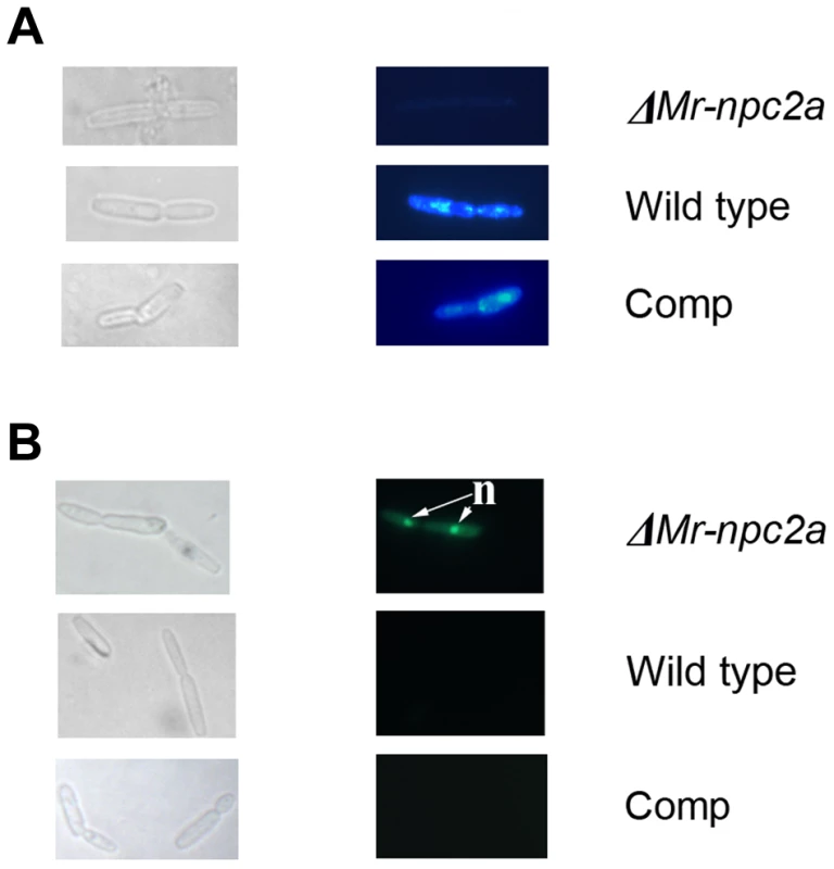
Left panels: differential interference contrast images of hyphal bodies; Right panels: fluorescence microscopy of the same hyphal bodies shown in the left panels. (A) Filipin staining of sterols in the cell membrane, (B) Sytox staining. ΔMr-npc2a: the mutant with the ORF of Mr-npc2a deleted; WT: the wild type strain of M. robertsii; Comp: the complemented ΔMr-npc2a with a genomic clone of Mr-npc2a; n = nuclei. The nucleic acid stain Sytox is unable to cross intact cell membranes and therefore only stains cells with poor membrane integrity [32]. We found that 7±2.7% of wild type hyphal bodies, 8±3.1% of the complemented ΔMr-npc2a and 55±6.5% of ΔMr-npc2a hyphal bodies were permeable to Sytox (Fig. 6B), confirming that the provision of sterols by Mr-NPC2a has an important role in maintaining the membrane integrity of hyphal bodies. This result is consistent with ΔMr-npc2a producing fewer hyphal bodies in hemolymph than the wild type strain.
Expression of Mr-NPC2a increased the virulence of the entomopathogenic fungus Beauveria bassiana
The genome of the well-studied insect-pathogenic fungus Beauveria bassiana (Genbank accession number: ADAH00000000) was searched for Mr-NPC2a homologs using BLASTP and TBLASTN (e-value cutoff 1e−05), but no significant hits were identified suggesting that this fungus lacks a Mr-NPC2a homolog. So, we decided to test whether transfer of Mr-NPC2a into B. bassiana can enhance its pathogenicity. Expression of Mr-NPC2a did not alter the growth, conidiation and germination of B. bassiana. Compared to the wild type strain, the LT50 (time taken to kill 50% of insects) of a transformant expressing Mr-NPC2a (Bb-Mr-NPC2a) was reduced by 22% (Fig. 7). Filipin staining did not detect obvious differences in sterol in the hyphal bodies of B. bassiana in hemolymph, but Sytox staining showed that the transformant expressing Mr-NPC2a had significantly fewer hyphal bodies (5±1.2%) with impaired cell membranes than did the wild type strain (10±1.8%).
Fig. 7. Kinetics of insect survivorship in bioassays with B. bassiana. 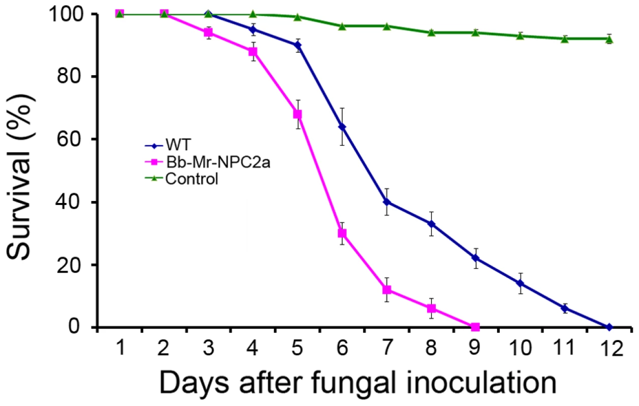
Wax worm larvae (G. mellonella) were inoculated with B. bassiana conidial suspensions (1×10⁷ spores/mL). Blue: the wild type B. bassiana; Purple: a B. bassiana transformant (Bb-Mr-NPC2a) expressing Mr-NPC2a; Green: control insects that were treated with 0.05% Tween-80. Discussion
The virulence of most fungal pathogens of plants and animals is a quantitative attribute, and thus many different genes and strategies are used simultaneously to cause disease [33]. Through evolution, fungi have acquired new virulence mechanisms, and increases in pathogen virulence have likely therefore evolved in a stepwise manner [34]. The important role of HGT in the evolution of fungal virulence is being illuminated by comparative genomics, but in only a few cases has the importance of the transferred genes been functionally demonstrated with gene knockouts, and no reports describe functional characterization of genes transferred from an animal to a eukaryotic pathogen. Lack of functional characterization is a major bottleneck in understanding the contribution of HGT to pathogen virulence. In this study, we describe how the insect pathogenic fungus Metarhizium may have acquired a sterol carrier gene Mr-npc2a from its insect hosts by HGT. This sterol carrier is involved in maintaining cell membrane sterols and thus membrane integrity of hyphal bodies in insect hemolymph. It thus helps Metarhizium adapt to the hemolymph and facilitates colonization of the host. A feature of Mr-npc2a is its specificity for the hemolymph of living insects. The promoter will thus be useful for driving expression of insecticidal proteins, and for manipulating gene expression in hemolymph, via antisense or overexpression constructs, to test whether candidate proteins play critical roles.
Ergosterol is an important constituent of fungal membrane lipids, similar to animal cholesterol, and modulates the fluidity, permeability and thickness of the membrane [35]. Under aerobic conditions, fungi synthesize ergosterol de novo and this process is oxygen dependent [36]. Under anaerobic conditions, ergosterol biosynthesis is suppressed and fungi need to take up sterols from the environment [37]. Metarhizium infects insects by direct penetration of the cuticle and entry into the haemocoel where the fungus kills insects with a combination of toxins and invasive growth by hyphal bodies [18]. However, the insect haemocoel is a hypoxic or even oxygen free environment, necessitating M. robertsii takes up sterols for the rapid proliferation of hyphal bodies. The insect hemolymph is the only environment where the fungal hyphal bodies can acquire sterols, and cholesterol is most likely to be exploited because it is the dominant insect sterol [38]. The bulk membrane function of the fungal ergosterol can be provided by structurally related sterols, including cholesterol [39]. Fungi can convert cholesterol into ergosterol or incorporate cholesterol directly into plasma membranes [28], [40].
Cholesterol is insoluble and insects use low density lipoproteins (LDL) to transport cholesterol through hemolymph to target tissues where they bind to LDL receptors. Metarhizium has a variety of genes with predicted functions that mimic those in insects including an LDL receptor (MAA_08841, Genbank accession no.: EFY95697) and close homologs of the S. cerevisiae ATP-binding cassette transporters AUS1 [M. robertsii's MAA_10338 (Genbank accession no.: EFY94212) and M. acridum's MAC_09600 (Genbank accession no.: EFY94889)] and PDR11 [M. robertsii's MAA_01883 (Genbank accession no.: EFY02301) and M. acridum's MAC_07971(Genbank accession no.: EFY96474)], required for cholesterol uptake in yeast [41].
The mechanisms of intracellular cholesterol transport are largely unknown, however, in mammals, cholesterol is exported to organelles by an NPC team that comprises an NPC1 (a lysosomal membrane protein) and NPC2 (a soluble protein in the lysosomal lumen). These mechanisms seem to be conserved in yeast and other fungi [28], [42]. M. robertsii has 1 NPC1-like protein (MAA_02001, Genbank accession no.: EFZ02419) as well as the 3 NPC2 proteins described in this study. Mr-NPC2b and Mr-NPC2c are typical fungal NPC2-like proteins, and their homolog in yeast (yNPC2p) is involved in ergosterol homeostasis [28]. The insect-like Mr-NPC2a is specifically expressed in insect hemolymph and is involved in maintaining cell membrane integrity only after the fungus enters the haemocoel. Although the disruption of Mr-npc2a greatly reduced the number of hyphal bodies in the hemolymph of living insects, i.e. suppressed growth, ΔMr-npc2a was still able to kill insects, suggesting that unidentified components can partially substitute for Mr-NPC2a when M. robertsii is in the hemolymph. The competition for cholesterol from Metarhizium might reduce the amount of sterols available to insects, which could alter the development of insects because insects cannot synthesize sterols and thus have dietary requirement for sterols. However, we did not see significant differences in development (pupation) between G. mellonella larva infected by ΔMr-npc2a and the wild type strain.
The existence of insect NPC2a homologs both in Metarhizium and the ergot fungus C. purpurea raises the question: when and how did these fungi acquire NPC2a from insects? There are four possible explanations. First, ancestral fungi had NPC2a proteins that were homologs of insect NPC2 proteins, but during evolution they were only retained by Metarhizium spp. and C. purpurea. This is unlikely as it would require independent loss of npc2a genes in all other fungal lineages. Secondly, Metarhizium and C. purpurea belong to the family Clavicipitaceae, and it is possible HGT occurred into the common ancestor of Metarhizium and C. purpurea. But microsynteny of sequence around C. purpurea npc2a is conserved with many fungi including Metarhizium. spp, whereas the microsynteny of the sequence around Metarhizium npc2a (Mr-npc2a) showed no conservation with other fungi including C. purpurea. So, if the ancestor of Metarhizium and C. purpurea acquired npc2a gene from an insect, this npc2a gene had moved from its ancient position to its current area during speciation of Metarhizium. spp. There is no evidence for Metarhizium npc2a (Mr-npc2a) having ever been within a transposon so genome rearrangement rather than transposition would be required for this move. Around Mr-npc2a (MAA_03817), only the gene MAA_03815 (Genbank accession no.: EFZ01219) is possibly involved in sterol metabolism which encodes C-4 methylsterol oxidase-like protein (Table S5), but M. acridum's C-4 methylsterol oxidase-like gene (MAC_09281, Genbank accession no.: EFY84691) does not cluster with its npc2a gene (MAC_00943 Genbank accession no.: EFY93160). Therefore, no obvious evolutionary force is seen that selected the genome rearrangement in Metarhizium genus. Thirdly, ancestors of C. purpurea and Metarhizium spp. may have independently acquired npc2a gene from ancestral insects. It is conceptually easy to understand an insect pathogenic fungus acquiring a gene from its host by HGT. In nature, C. purpurea also has physical contact with insects, as it produces ergot honeydew that attracts insects and these spread its conidia [25]. However, the gene adjacent to M. robertsii npc2a (Mr-npc2a) and that to C. purpurea npc2a are homologs with related genes in other fungi but not in insects. It is improbable that insect npc2a genes independently inserted beside the same gene in M. robertsii and C. purpurea. The final possibility is that an ancestor of C. purpurea or Metarhizium spp. acquired npc2a which was inserted beside the gene MAA_03818 in Metarhizium to become MAA_03817 (M. robertsii npc2a) or beside the gene CPUR_07480 in C. purpurea to become CPUR_07479 (C. purpurea npc2a). C. purpurea then obtained the two genes (the genes MAA_03818 and MAA_03817) from Metarhizium or vice versa. Ecological niche overlap allows HGT of gene clusters between fungi [8], making this the most likely possibility. The genetic distances between the generalist M. robertsii (can kill many kinds of insects) and the specialist M. acridum (specific for grasshoppers) is greater than that between most other species in the Metarhizium genus [43]. Since the ancestor of Metarhizium may have acquired npc2a gene from an insect before speciation and both genetically distant M. robertsii and M. acridum retain this gene, it is expected that most of other Metarhizium species still have this gene. Therefore, based on currently available information, more Metarhizium than Claviceps species have npc2a genes, so it is more likely that C. purpurea was the recipient. However, more Claviceps genomes will be needed to confirm the direction of HGT.
Materials and Methods
Fungal and bacterial strains
M. robertsii ARSEF2575 and B. bassiana ARSEF252 were cultured as previously described [18], [44]. Escherichia coli DH5α was used for plasmid construction. A. tumefaciens AGL1 was used for fungal transformation as described [45].
Gene cloning and disruption
The flanking sequences of T-DNA in mutants generated by the insertion of T-DNA into the genome of M. robertsii were cloned by Y-shaped adaptor-dependent extension as described [46]. The primers employed in this study and their usages are given in Table S6. PCR products were cloned into pGEM-T Easy (Promega, USA) for sequencing.
To construct the Mr-npc2a disruption plasmid, the 5′-end and 3′-end of Mr-npc2a were cloned by PCR and inserted into the XbaI and SpeI sites, respectively, of the plasmid pFBARGFP [18] to form the disruption plasmid pFBARGFP-dMr-npc2a. The disruption mutant (ΔMr-npc2a) was obtained utilizing A. tumefaciens-mediated fungal transformation. To complement ΔMr-npc2a, the genomic sequence of Mr-npc2a was cloned, inserted into the XbaI site of pSURGFP [43], and transformed into ΔMr-npc2a as described [47].
Phylogenetic analyses and tree topology tests
M. robertsii Mr-NPC2a protein sequence was used as the query for BLASTP searches against the NCBI non-redundant database (e-value cutoff 1e−05) to identify and retrieve its homologs. The selected homologous proteins of representative taxa were aligned using MUSCLE 3.7 [48], with a maximum number of iterations of 16. Poorly aligned positions and divergent regions of the alignment were removed using GBlocks0.91b [49], with half the gapped positions allowed, the minimum number of sequences for a conserved and a flank position set to 50% of the number of taxa plus one, the maximum of contiguous nonconserved positions set to 16, and the minimum length of a block set to 4. Phylogenetic analyses were performed with three approaches: maximum likelihood (ML), bayesian inference (BI) and distance-based neighbor-joining (NJ). ML phylogenies were constructed by PhyML 3.1 [50] using the best-fit evolutionary model as suggested by ProtTest 3.2 [51], in which case, a discrete gamma-distribution model with four rate-categories plus invariant positions was used. The gamma parameter and proportion of invariant sites were estimated from the data. Branch support values were obtained by 100 bootstrap pseudo-replicates. Bayesian reconstructions were performed with MrBayes 3.1.2 [52] under the WAG+Γ4+I model of amino acid substitution. The Markov chain Monte Carlo searches were run for 1,000,000 generations, sampling the Markov chains every 1000 generations; the first 250 trees were discarded as ‘burn-in’. In addition, we also employed distance-based neighbor-joining (NJ) trees, which were constructed by using the program Neighbor in MEGA5 [53]. Bootstrap support values were obtained by generating 1,000 pseudo-replicates.
We statistically tested the significance of the topology of the obtained tree (Fig. 2) by phylogenetic analysis in comparison with alternative trees in which fungal NPC2a proteins were constrained to the vertebrate clade to form monophyletic groups using the program CONSEL [24]. The value of site-wise likelihood of every constrained topology was calculated by PhyML [50].
Sterol binding assay of Mr-NPC2a
The DNA fragment encoding Mr-NPC2a (signal peptide excluded) was cloned with RT-PCR using total RNA extracted from hyphal bodies collected from the hemolymph of living insects as described below. The sequences of the primers are presented in Table S6. PCR product was purified and cloned into pGEM-T vector (Promega, Madison, WI, USA) for sequence verification. The DNA fragment was then subcloned into EcoRI/NotI restriction sites in the expression vector pET32a (EMD Millipore Corporation, Billerica, MA, USA) to form the pET32a-MrNPC2a plasmid that was then transformed into E. coli strain BL21 (DE3) (EMD Millipore Corporation, Billerica, MA, USA). Isopropyl-1-thio-β-D-galactopyranosid (1 mM) was used to induce recombinant protein expression. Recombinant Mr-NPC2a was purified with HisTALON Superflow Cartridges (Clontech, Mountain View, CA, USA), concentrated and dialyzed with Tris buffer (50 mM Tris-HCl, 50 mM NaCl, pH 8.0) at 4°C using Amicon Ultra centrifugal filter unit (EMD Millipore Corporation, Billerica, MA, USA). Control protein was expressed and purified from BL21 (DE3) cells transformed with pET32a empty vector with the same methods described above.
The ligand-binding assay was performed as described with some modifications [27]. Since the D. melangaster NPC2a can bind to bacterial Lipid A and cholesterol [27], both of them were used as ligands to compare binding specificity of Mr-NPC2a with that of the insect NPC2a. Ergosterol was also tested to look at the binding ability of Mr-NPC2a to the fungal sterol. Lipid A monophosphoryl from E. coli F583 (Rd mutant) and cholesterol were purchased from Sigma Aldrich (MO, USA), and ergosterol was from Santa Cruz Biotechnology (Texas, USA). Wells of flat-bottom 96-well plates (BD falcon, NJ, USA) were coated with 2 or 10 µg/well of each ligand. Control wells with no ligand were coated with the same amount of chloroform/water. The plates were then air dried overnight at room temperature, heated at 60°C for 30 min and then blocked with 200 µL/well of 1 mg/mL BSA in Tris buffer (50 mM Tris-HCl, 50 mM NaCl, pH 8.0) at 37°C for 2 h. Wells were washed with the Tris buffer (four times, each for 5 min) and 500 nM of purified Mr-NPC2a fusion protein or vector protein diluted in binding buffer (the Tris buffer with 0.1 mg/mL BSA) were added to each well of the coated plates (50 µL/well) and incubated at room temperature for 3 h. Plates were then washed and mouse monoclonal anti-polyHistidine antibody was added (100 µL/well) and incubated at 37°C for 2 h. Plates were washed again and alkaline phosphatase-conjugated goat anti-mouse IgG (Promega, 1∶2000 dilution in binding buffer) was added (100 µL/well) and incubated at 37°C for 2 h. Alkaline phosphatase substrate liquid (Sigma Aldrich MO, USA) was then added to each wells and incubated for 15 min at room temperature. Plates were shaken for 15 s and the absorbance at 405 nm of each well was determined using a plate reader (FilterMax F3, Molecular Devices, CA, USA). The absorbance from control wells was subtracted and specific binding was calculated by subtracting absorbance from vector protein from the total binding of NPC2 protein. Binding assays were repeated 3 times with 3 repeated wells.
Expression pattern of Mr-npc2a
The expression pattern of Mr-npc2a was investigated by RT-PCR and by following GFP fluorescence in transformants expressing GFP driven by the promoter region (2,203 bp) of Mr-npc2a. The promoter region of the Mr-npc2a and the ORF of egfp were both cloned by PCR using primers described in Table S6. The resultant egfp product was digested with EcoRV and XhoI, and inserted into the corresponding sites of pBARGPE1 [30] to form pGFP. The Mr-npc2a promoter was digested with EcoRI and SmaI, and inserted into EcoRI and EcoRV of pGFP to form PMr-NPC2a:GFP. The egfp cassette was then mobilized into Ppk2-bar [21] to form pPMr-NPC2a:GFP. This Ti plasmid was then transformed into the wild type M. robertsii mediated by A. tumefaciens [45]. For RT-PCR analysis, the gpd gene was used as a reference gene [54]. Primers for Mr-npc2a and gpd are presented in Table S6. Total RNA was isolated using the Plant RNeasy Kit (Qiagen USA). First-strand cDNA was synthesized using Revert Aid First Strand cDNA Synthesis Kit (Fermentas, USA). PCR was conducted using DreamTaq DNA polymerase (Thermo Scientific, USA).
Expression of Mr-npc2a was examined during saprophytic growth, infection and in several possible inducing conditions (Table S4). For saprophytic growth, the fungus was grown on PDA, aerial hyphae, conidiating mycelium and conidia were sampled as previously described [23] for GFP observation or RNA preparation. Blastospores were obtained by growing the fungus in nutrition rich medium SDB for 10 d and in the in vitro prepared hemolymph for 2 d. GFP fluorescence in blastospores and hyphae was examined directly under microscopy. Blastospores and hyphae were combined for RNA preparation.
The expression of Mr-npc2a during infection was examined in appressoria on insect cuticles (the hindwings of L. migratoria locusts) and hyphal bodies in the hemolymph of living insects. Appressoria were induced on locust hindwings as previously described [55]. To obtain hyphal bodies in insect hemolymph, 1×10⁵ conidia were injected into a 5th instar M. sexta larvae. After 20 h at 27°C, the insect was bled and GFP signal in the fungus in the hemolymph was checked by microscopy. The hemolymph with insect hemocytes and fungal hyphal bodies was subjected to RNA preparation. RNA of the hemolymph of healthy insects was prepared as a control for RT-PCR analysis.
In order to test the effect of sterols on the Mr-npc2a expression, the wild type strain (for RT-PCR analysis) or the transformant with PMr-npc2a:GFP (for GFP fluorescence observation) were grown in SDB for 24 h. The mycelium were then filtered, washed 3 times with sterile water and cultured in the minimal medium M100 [21] supplemented with the insect steroid hormone 20-Hydroxyecdysone (Sigma, USA), cholesterol (Sigma, USA) or ergosterol (Sigma, USA) (Table S4). To detect the effect of stresses on Mr-npc2a expression, the mycelium from the 24 h culture in SDB were cultured in the minimal medium M100 supplemented with different agents to generate stresses described in Table S4. For all treatments, GFP fluorescence in mycelium was examined and RNA was also prepared for RT-PCR.
Investigating localization of Mr-NPC2a
To investigate the localization of Mr-NPC2a in M. robertsii, we constructed the fusion protein Mr-NPC2a: GFP. The coding sequence of Mr-NPC2a (stop codon was excluded) was cloned by PCR, and inserted into the EcoRV site of the plasmid pGFP described above, resulting in pMr-NPC2a:GFP. Since Mr-NPC2a is only expressed in hyphal bodies produced in the hemolymph of living insects, it is hard to determine the localization of this protein when its native promoter is used to drive the expression of NPC2a:GFP. Therefore, the expression of the fusion gene was driven by the constitutive promoter Pgpd from A. nidulans in pGFP [30]. The Mr-NPC2a:GFP cassette was removed from pMr-NPC2a:GFP and inserted into Ppk2-bar to produce Ppk2-gpd:Mr-NPC2a:GFP that was then transformed into wild type M. robertsii mediated by A. tumefaciens to produce transformant Mr-NPC2a:GFP.
GFP observation and Western blot analysis were used to test the localization of Mr-NPC2a:GFP. The transformant Mr-NPC2a:GFP was inoculated in SDB broth (106 spores/mL) and grown for 36 h at 27°C with constant shaking (200 rpm). Mycelium was collected by filtration and ground into powder for protein preparation. The protein in the filtrant was precipitated with 80% (NH4)2SO4. The salt was then removed by centrifugation with Microcon filter with a 3 kDa cutoff (Millipore, USA). Total protein (15 µg) was subjected for Western blot analysis. Rabbit anti-GFP antibody (Proteintech Group Inc. USA) was used for Western blot analysis.
Expression of Mr-NPC2a in B. bassiana
The ORF of Mr-npc2a was cloned by PCR and inserted into BamHI and EcoRV sites downstream of the Pgpd promoter in the plasmid pBARGPE1 [30]. The Mr-npc2a expression cassette was then moved into pFBARGFP to form the expression plasmid pMr-NPC2a that was transformed into B. bassiana using A. tumefaciens.
Sytox staining of hyphal bodies collected from the hemolymph of living insects
Sytox Green nucleic acid staining is widely used to assess the integrity of eukaryotic and prokaryotic cell membranes [32]. Sytox Green can only enter cells if their cell membrane integrity is compromised, in which case it stains nucleic acids, resulting in a green fluorescence emission (absorption and emission maxima at 502 and 523 nm, respectively). To prepare hyphal bodies for Sytox staining, 50 µL of conidial suspension (107 conidia/mL) was injected into a fifth instar larva of M. sexta. After 20 h incubation at 27°C, the insect was bled and the hemolymph was collected into a 1.5 ml centrifuge tube that was then placed on ice. The hemolymph (100 µL) was then quickly mixed with 5 µL of sytox staining solution (10 µM) by pipetting up and down. The mixture was then immediately loaded on a glass slide and covered with a cover slip. This set was subsequently placed on wet paper towel in a small container and incubated at room temperature for 10 min. Stained nuclei were observed under microscopy.
Flipin staining
Hyphal bodies were prepared from the hemolymph of M. sexta as described above and stained with 5 µM Filipin (Sigma Aldrich, MO, USA) in ACES buffer as described [28].
Quantification of hyphal bodies in insect hemolymph
A fungal spore suspension (1×10⁷ spores/mL) was topically applied on the wax worm larvae (G. mellonella). At 12 h intervals after inoculation, ten larvae per treatment were surface sterilized with 1% bleach and individually bled as described [56]. Five microliter aliquots of blood from each larva were mixed with 95 µL of sterile water and spread onto Metarhizium selective medium for colony counts [56]. The number of colony forming units resulting from unicellular hyphal bodies (blastospores) or multicellular hyphal bodies is used to show the number of hyphal bodies.
Bioassay
Fungal virulence was assayed against wax worm (G.mellonella) as previously described [56]. The worms were maintained as manufacture's instruction (Pet Solutions, OH USA). The SPSS program was used to calculate the LT50 for each strain. There were 40 insects for each strain and the experiment was repeated 3 times.
Supporting Information
Zdroje
1. VoylesJ, YoungS, BergerL, CampbellC, VoylesWF, et al. (2009) Pathogenesis of chytridiomycosis, a cause of catastrophic amphibian declines. Science 326(5952): 582–585.
2. LorchJM, MeteyerCU, BehrMJ, BoylesJG, CryanPM, et al. (2011) Experimental infection of bats with Geomyces destructans causes white-nose syndrome. Nature 480(7377): 376–378.
3. SuhSO, NodaH, BlackwellM (2001) Insect symbiosis: derivation of yeast-like endosymbionts within an entomopathogenic filamentous lineage. Mol Biol Evol 18(6): 995–1000.
4. RicklefsRE, FallonSM (2002) Diversification and host switching in avian malaria parasites. Proc R Soc Lond B Biol Sci 269 : 885–892.
5. ArchieEA, LuikartG, EzenwaVO (2009) Infecting epidemiology with genetics: a new frontier in disease ecology. Trends Ecol Evol 24 : 21–30.
6. RaffaeleS, FarrerRA, CanoLM, StudholmeDJ, MacLeanD, et al. (2010) Genome evolution following host jumps in the Irish potato famine pathogen lineage. Science 330 : 1540–1543.
7. GilbertC, HernandezSS, Flores-BenabibJ, SmithEN, FeschotteC (2012) Rampant horizontal transfer of SPIN transposons in squamate reptiles. Mol Biol Evol 29 : 503–515.
8. SlotJC, RokasA (2011) Horizontal transfer of a large and highly toxic secondary metabolic gene cluster between fungi. Curr Biol 21(2): 134–139.
9. FriesenTL, StukenbrockEH, LiuZ, MeinhardtS, LingH, et al. (2006) Emergence of a new disease as a result of interspecific virulence gene transfer. Nat Genet 38 : 953–956.
10. RichardsTA, LeonardG, SoanesDM, TalbotNJ (2011) Gene transfer into the fungi. Fungal. Biol Rev 25 : 98–110.
11. SunBF, XiaoJH, HeS, LiuL, MurphyRW, et al. (2013) Multiple interkingdom horizontal gene transfers in Pyrenophora and closely related species and their contributions to phytopathogenic lifestyles. PLoS One 8(3): e60029.
12. MowerJP, StefanovicS, YoungGJ, PalmerJD (2004) Plant genetics: gene transfer from parasitic to host plants. Nature 432 : 165–166.
13. DavisCC, WurdackKJ (2004) Host-to-parasite gene transfer in flowering plants: phylogenetic evidence from Malpighiales. Science 305 : 676–678.
14. KeelingPJ, PalmerJD (2008) Horizontal gene transfer in eukaryotic evolution. Nat Rev Genet 9 : 605–618.
15. SelmanM, PombertJF, SolterL, FarinelliL, WeissLM, et al. (2011) Acquisition of an animal gene by microsporidian intracellular parasites. Curr Biol 21 : 576–577.
16. BarDZ (2011) Evidence of massive horizontal gene transfer between humans and Plasmodium vivax. Nature precedings doi:10.1038/npre.2011.5690.1
17. RobertsDW, St LegerRJ (2004) Metarhizium spp., cosmopolitan insect-pathogenic fungi: mycological aspects. Adv Appl Microbiol 54 : 1–70.
18. FangW, ScullyLR, ZhangL, PeiY, BidochkaMJ (2008) Implication of a regulator of G protein signalling (BbRGS1) in conidiation and conidial thermotolerance of the insect pathogenic fungus Beauveria bassiana. FEMS Microbiol Lett 279 : 146–156.
19. FangW, AzimzadehP, St LegerRJ (2012) Strain improvement of fungal insecticides for controlling insect pests and vector-borne diseases. Curr Opin Microbiol 15 : 232–238.
20. BehieSW, ZeliskoPM, BidochkaMJ (2012) Endophytic insect-parasitic fungi translocate nitrogen directly from insects to plants. Science 336 : 1576–1577.
21. FangW, St LegerRJ (2010) Mrt, a gene unique to fungi, encodes an oligosaccharide transporter and facilitates rhizosphere competency in Metarhizium robertsii. Plant Physiol 154 : 1549–1557.
22. XiaoG, YingSH, ZhengP, WangZL, ZhangS, et al. (2012) Genomic perspectives on the evolution of fungal entomopathogenicity in Beauveria bassiana. Sci Rep 2 : 483.
23. FangW, FernandesEK, RobertsDW, BidochkaMJ, St LegerRJ (2010) A laccase exclusively expressed by Metarhizium anisopliae during isotropic growth is involved in pigmentation, tolerance to abiotic stresses and virulence. Fungal Genet Biol 47 : 602–607.
24. ShimodairaH, HasegawaM (2001) CONSEL: for assessing the confidence of phylogenetic tree selection. Bioinformatics 17(12): 1246–1247.
25. ButlerMD, AldermanSC, HammondPC, BerryRE (2001) Association of Insects and Ergot (Claviceps purpurea) in Kentucky Bluegrass Seed Production Fields. J Econ Entomol 94 : 1471–1476.
26. DanchinEG, RossoMN, VieiraP, de Almeida-EnglerJ, CoutinhoPM, et al. (2010) Multiple lateral gene transfers and duplications have promoted plant parasitism ability in nematodes. Proc Natl Acad Sci USA 107(41): 17651–17656.
27. ShiXZ, ZhongX, YuXQ (2012) Drosophila melanogaster NPC2 proteins bind bacterial cell wall components and may function in immune signal pathways. Insect Biochem Mol Biol 42 : 545–556.
28. BergerAC, VanderfordTH, GernertKM, NicholsJW, FaundezV, et al. (2005) Saccharomyces cerevisiae Npc2p is a functionally conserved homologue of the human Niemann-Pick disease type C 2 protein, hNPC2. Eukaryot Cell 4 : 1851–1862.
29. WangC, St LegerRJ (2006) A collagenous protective coat enables Metarhizium anisopliae to evade insect immune responses. Proc Natl Acad Sci USA 103 : 6647–6652.
30. McCluskeyK (2003) The Fungal Genetics Stock Center: from molds to molecules. Adv Appl Microbiol 52 : 245–262.
31. ChenP, GaoR, ChenS, PuL, LiP, et al. (2012) A pericentrin-related protein homolog in Aspergillus nidulans plays important roles in nucleus positioning and cell polarity by affecting microtubule organization. Eukaryot Cell 11 : 1520–1530.
32. GerphagnonM, LatourD, ColombetJ, Sime-NgandoT (2013) A double staining method using SYTOX green and calcofluor white for studying fungal parasites of phytoplankton. Appl Environ Microbiol 79 : 3943–3951.
33. HorbachR, Navarro-QuesadaAR, KnoggeW, DeisingHB (2011) When and how to kill a plant cell: infection strategies of plant pathogenic fungi. J Plant Physiol 168 : 51–62.
34. GardinerDM, KazanK, MannersJM (2013) Cross-kingdom gene transfer facilitates the evolution of virulence in fungal pathogens. Plant Sci 210 : 151–158.
35. ZhangYQ, GamarraS, Garcia-EffronG, ParkS, PerlinDS, et al. (2010) Requirement for ergosterol in V-ATPase function underlies antifungal activity of azole drugs. PLoS Pathog 6: e1000939.
36. DupontS, LemetaisG, FerreiraT, CayotP, GervaisP, et al. (2012) Ergosterol biosynthesis: a fungal pathway for life on land? Evolution 66 : 2961–2968.
37. AndreasenAA, StierTJ (1953) Anaerobic nutrition of Saccharomyces cerevisiae. I. Ergosterol requirement for growth in a defined medium. J Cell Physiol 41 : 23–36.
38. JingX, GrebenokRJ, BehmerST (2013) Sterol/steroid metabolism and absorption in a generalist and specialist caterpillar: Effects of dietary sterol/steroid structure, mixture and ratio. Insect Biochem Mol Biol 43 : 580–587.
39. JacquierN, SchneiterR (2012) Mechanisms of sterol uptake and transport in yeast. J Steroid Biochem Mol Biol 129 : 70–78.
40. XiongQ, HassanSA, WilsonWK, HanXY, MayGS, et al. (2005) Cholesterol import by Aspergillus fumigatus and its influence on antifungal potency of sterol biosynthesis inhibitors. Antimicrob Agents Chemother 49 : 518–524.
41. AlimardaniP, RegnacqM, Moreau-VauzelleC, FerreiraT, RossignolT, et al. (2004) SUT1-promoted sterol uptake involves the ABC transporter Aus1 and the mannoprotein Dan1 whose synergistic action is sufficient for this process. Biochem J 381 : 195–202.
42. BergerAC, HansonPK, Wylie NicholsJ, CorbettAH (2005) A yeast model system for functional analysis of the Niemann-Pick type C protein 1 homolog, Ncr1p. Traffic 6 : 907–917.
43. BischoffJF, RehnerSA, HumberRA (2009) A multilocus phylogeny of the Metarhizium anisopliae lineage. Mycologia 101 : 512–530.
44. FangW, PeiY, BidochkaMJ (2007) A regulator of a G protein signaling (RGS) gene, cag8, from the insect-pathogenic fungus Metarhizium anisopliae is involved in conidiation, virulence and hydrophobin synthesis. Microbiology 153 : 1017–1025.
45. FangW, PeiY, BidochkaMJ (2006) Transformation of Metarhizium anisopliae mediated by Agrobacterium tumefaciens. Can J Microbiol 52 : 623–626.
46. FangW, LengB, XiaoY, JinK, MaJ, et al. (2005) Cloning of Beauveria bassiana chitinase gene Bbchit1 and its application to improve fungal strain virulence. Appl Environ Microbiol 71 : 363–370.
47. FangW, St LegerRJ (2012) Enhanced UV resistance and improved killing of malaria mosquitoes by photolyase transgenic entomopathogenic fungi. PLoS One 7: e43069.
48. EdgarRC (2004) MUSCLE: multiple sequence alignment with high accuracy and high throughput. Nucleic Acids Res 32 : 1792–1797.
49. CastresanaJ (2000) Selection of conserved blocks from multiple alignments for their use in phylogenetic analysis. Mol Biol Evol 17 : 540–552.
50. GuindonS, GascuelO (2003) A simple, fast, and accurate algorithm to estimate large phylogenies by maximum likelihood. Syst Biol 52 : 696–704.
51. AbascalF, ZardoyaR, PosadaD (2005) ProtTest: selection of best-fit models of protein evolution. Bioinformatics 21 : 2104–2105.
52. HuelsenbeckJP, RonquistF (2001) MR BAYES: Bayesian inference of phylogenetic trees. Bioinformatics 17 : 754–755.
53. TamuraK, PetersonD, PetersonN, StecherG, NeiM, et al. (2011) MEGA5: Molecular evolutionary genetics analysis using maximum likelihood, evolutionary distance, and maximum parsimony methods. Mol Biol Evol 28 : 2731–2739.
54. FangW, BidochkaMJ (2006) Expression of genes involved in germination, conidiogenesis and pathogenesis in Metarhizium anisopliae using quantitative real-time RT-PCR. Mycol Res 110 : 1165–1171.
55. FangW, Pava-ripollM, WangS, St.LegerRJ (2009) Protein kinase A regulates production of virulence determinants by the entomopathogenic fungus, Metarhizium anisopliae. Fungal Genet Biol 46 : 277–85.
56. FangW, FengJ, FanY, ZhangY, BidochkaMJ, et al. (2009) Expressing a fusion protein with protease and chitinase activities increases the virulence of the insect pathogen Beauveria bassiana. J Invertebr Pathol 102 : 155–159.
Štítky
Hygiena a epidemiologie Infekční lékařství Laboratoř
Článek Affinity Proteomics Reveals Elevated Muscle Proteins in Plasma of Children with Cerebral MalariaČlánek The Transcriptional Activator LdtR from ‘ Liberibacter asiaticus’ Mediates Osmotic Stress ToleranceČlánek Complement-Related Proteins Control the Flavivirus Infection of by Inducing Antimicrobial PeptidesČlánek Fungal Chitin Dampens Inflammation through IL-10 Induction Mediated by NOD2 and TLR9 ActivationČlánek Parasite Fate and Involvement of Infected Cells in the Induction of CD4 and CD8 T Cell Responses to
Článek vyšel v časopisePLOS Pathogens
Nejčtenější tento týden
2014 Číslo 4- Stillova choroba: vzácné a závažné systémové onemocnění
- Perorální antivirotika jako vysoce efektivní nástroj prevence hospitalizací kvůli COVID-19 − otázky a odpovědi pro praxi
- Diagnostika virových hepatitid v kostce – zorientujte se (nejen) v sérologii
- Jak souvisí postcovidový syndrom s poškozením mozku?
- Familiární středomořská horečka
-
Všechny články tohoto čísla
- , , , Genetic Variability: Cryptic Biological Species or Clonal Near-Clades?
- Early Mortality Syndrome Outbreaks: A Microbial Management Issue in Shrimp Farming?
- Wormholes in Host Defense: How Helminths Manipulate Host Tissues to Survive and Reproduce
- Plastic Proteins and Monkey Blocks: How Lentiviruses Evolved to Replicate in the Presence of Primate Restriction Factors
- The 2010 Cholera Outbreak in Haiti: How Science Solved a Controversy
- Affinity Proteomics Reveals Elevated Muscle Proteins in Plasma of Children with Cerebral Malaria
- Noncanonical Role for the Host Vps4 AAA+ ATPase ESCRT Protein in the Formation of Replicase
- Efficient Parvovirus Replication Requires CRL4-Targeted Depletion of p21 to Prevent Its Inhibitory Interaction with PCNA
- Host-to-Pathogen Gene Transfer Facilitated Infection of Insects by a Pathogenic Fungus
- The Transcriptional Activator LdtR from ‘ Liberibacter asiaticus’ Mediates Osmotic Stress Tolerance
- Coxsackievirus B Exits the Host Cell in Shed Microvesicles Displaying Autophagosomal Markers
- TCR Affinity Associated with Functional Differences between Dominant and Subdominant SIV Epitope-Specific CD8 T Cells in Rhesus Monkeys
- Coxsackievirus-Induced miR-21 Disrupts Cardiomyocyte Interactions via the Downregulation of Intercalated Disk Components
- Ligands of MDA5 and RIG-I in Measles Virus-Infected Cells
- Kind Discrimination and Competitive Exclusion Mediated by Contact-Dependent Growth Inhibition Systems Shape Biofilm Community Structure
- Structural Differences Explain Diverse Functions of Actins
- HSCARG Negatively Regulates the Cellular Antiviral RIG-I Like Receptor Signaling Pathway by Inhibiting TRAF3 Ubiquitination Recruiting OTUB1
- Vaginitis: When Opportunism Knocks, the Host Responds
- Complement-Related Proteins Control the Flavivirus Infection of by Inducing Antimicrobial Peptides
- Fungal Chitin Dampens Inflammation through IL-10 Induction Mediated by NOD2 and TLR9 Activation
- Microbial Pathogens Trigger Host DNA Double-Strand Breaks Whose Abundance Is Reduced by Plant Defense Responses
- Alveolar Macrophages Are Essential for Protection from Respiratory Failure and Associated Morbidity following Influenza Virus Infection
- An Interaction between Glutathione and the Capsid Is Required for the Morphogenesis of C-Cluster Enteroviruses
- Concerted Spatio-Temporal Dynamics of Imported DNA and ComE DNA Uptake Protein during Gonococcal Transformation
- Potent Dengue Virus Neutralization by a Therapeutic Antibody with Low Monovalent Affinity Requires Bivalent Engagement
- Regulation of Human T-Lymphotropic Virus Type I Latency and Reactivation by HBZ and Rex
- Functionally Redundant RXLR Effectors from Act at Different Steps to Suppress Early flg22-Triggered Immunity
- The Pathogenic Mechanism of the Virulence Factor, Mycolactone, Depends on Blockade of Protein Translocation into the ER
- Role of Calmodulin-Calmodulin Kinase II, cAMP/Protein Kinase A and ERK 1/2 on -Induced Apoptosis of Head Kidney Macrophages
- An Overview of Respiratory Syncytial Virus
- First Experimental Model of Enhanced Dengue Disease Severity through Maternally Acquired Heterotypic Dengue Antibodies
- Binding of Glutathione to Enterovirus Capsids Is Essential for Virion Morphogenesis
- IFITM3 Restricts Influenza A Virus Entry by Blocking the Formation of Fusion Pores following Virus-Endosome Hemifusion
- Parasite Fate and Involvement of Infected Cells in the Induction of CD4 and CD8 T Cell Responses to
- Deficient IFN Signaling by Myeloid Cells Leads to MAVS-Dependent Virus-Induced Sepsis
- Pernicious Pathogens or Expedient Elements of Inheritance: The Significance of Yeast Prions
- The HMW1C-Like Glycosyltransferases—An Enzyme Family with a Sweet Tooth for Simple Sugars
- The Expanding Functions of Cellular Helicases: The Tombusvirus RNA Replication Enhancer Co-opts the Plant eIF4AIII-Like AtRH2 and the DDX5-Like AtRH5 DEAD-Box RNA Helicases to Promote Viral Asymmetric RNA Replication
- Mining Herbaria for Plant Pathogen Genomes: Back to the Future
- Inferring Influenza Infection Attack Rate from Seroprevalence Data
- A Human Lung Xenograft Mouse Model of Nipah Virus Infection
- Mast Cells Expedite Control of Pulmonary Murine Cytomegalovirus Infection by Enhancing the Recruitment of Protective CD8 T Cells to the Lungs
- Cytosolic Peroxidases Protect the Lysosome of Bloodstream African Trypanosomes from Iron-Mediated Membrane Damage
- Abortive T Follicular Helper Development Is Associated with a Defective Humoral Response in -Infected Macaques
- JC Polyomavirus Infection Is Strongly Controlled by Human Leucocyte Antigen Class II Variants
- Cationic Antimicrobial Peptides Promote Microbial Mutagenesis and Pathoadaptation in Chronic Infections
- Estimating the Fitness Advantage Conferred by Permissive Neuraminidase Mutations in Recent Oseltamivir-Resistant A(H1N1)pdm09 Influenza Viruses
- Progressive Accumulation of Activated ERK2 within Highly Stable ORF45-Containing Nuclear Complexes Promotes Lytic Gammaherpesvirus Infection
- Caspase-1-Like Regulation of the proPO-System and Role of ppA and Caspase-1-Like Cleaved Peptides from proPO in Innate Immunity
- Is Required for High Efficiency Viral Replication
- Modified Vaccinia Virus Ankara Triggers Type I IFN Production in Murine Conventional Dendritic Cells via a cGAS/STING-Mediated Cytosolic DNA-Sensing Pathway
- Evidence That Bank Vole PrP Is a Universal Acceptor for Prions
- Rapid Response to Selection, Competitive Release and Increased Transmission Potential of Artesunate-Selected Malaria Parasites
- Inactivation of Genes for Antigenic Variation in the Relapsing Fever Spirochete Reduces Infectivity in Mice and Transmission by Ticks
- Exposure-Dependent Control of Malaria-Induced Inflammation in Children
- A Neutralizing Anti-gH/gL Monoclonal Antibody Is Protective in the Guinea Pig Model of Congenital CMV Infection
- The Apical Complex Provides a Regulated Gateway for Secretion of Invasion Factors in
- A Highly Conserved Haplotype Directs Resistance to Toxoplasmosis and Its Associated Caspase-1 Dependent Killing of Parasite and Host Macrophage
- A Quantitative High-Resolution Genetic Profile Rapidly Identifies Sequence Determinants of Hepatitis C Viral Fitness and Drug Sensitivity
- Histone Deacetylase Inhibitor Romidepsin Induces HIV Expression in CD4 T Cells from Patients on Suppressive Antiretroviral Therapy at Concentrations Achieved by Clinical Dosing
- PLOS Pathogens
- Archiv čísel
- Aktuální číslo
- Informace o časopisu
Nejčtenější v tomto čísle- The 2010 Cholera Outbreak in Haiti: How Science Solved a Controversy
- , , , Genetic Variability: Cryptic Biological Species or Clonal Near-Clades?
- Efficient Parvovirus Replication Requires CRL4-Targeted Depletion of p21 to Prevent Its Inhibitory Interaction with PCNA
- An Overview of Respiratory Syncytial Virus
Kurzy
Zvyšte si kvalifikaci online z pohodlí domova
Autoři: prof. MUDr. Vladimír Palička, CSc., Dr.h.c., doc. MUDr. Václav Vyskočil, Ph.D., MUDr. Petr Kasalický, CSc., MUDr. Jan Rosa, Ing. Pavel Havlík, Ing. Jan Adam, Hana Hejnová, DiS., Jana Křenková
Autoři: MUDr. Irena Krčmová, CSc.
Autoři: MDDr. Eleonóra Ivančová, PhD., MHA
Autoři: prof. MUDr. Eva Kubala Havrdová, DrSc.
Všechny kurzyPřihlášení#ADS_BOTTOM_SCRIPTS#Zapomenuté hesloZadejte e-mailovou adresu, se kterou jste vytvářel(a) účet, budou Vám na ni zaslány informace k nastavení nového hesla.
- Vzdělávání


