
Prenatal diagnosis of agenesis of the portal system type II Abernethy malformation associated with trisomy 21
Authors:
A. C. Mendes Ribeiro 1
 ; M. Araújo 1
; A. de Nazaré Miziara 2
; L. R. A. S. de Figueiredo 1
; Y. R. de Abreu 1
; Edward Araujo Júnior 3,4
; A. B. Peixoto 1,5
Authors place of work:
Department of Gynecology and Obstetrics, Federal University of Triângulo Mineiro (UFTM), Uberaba – MG, Brazil
1; Discipline of Cardiology, Federal University of Triângulo Mineiro (UFTM), Uberaba – MG, Brazil
2; Department of Obstetrics, Paulista School of Medicine – Federal University of São Paulo (EPM-UNIFESP), São Paulo – SP, Brazil
3; Discipline of Woman Health, Municipal University of São Caetano do Sul (USCS), São Caetano do Sul – SP, Brazil
4; Service of Gynecology and Obstetrics, Mário Palmério University Hospital, University of Uberaba (UNIUBE), Uberaba – MG, Brazil
5
Published in the journal:
Ceska Gynekol 2026; 91(5): 395-400
Category:
Kazuistika
doi:
https://doi.org/10.48095/cccg2026395
Summary
Congenital agenesis of the portal vein (CAPV) is a rare vascular anomaly characterized by absence of the portal vein, resulting in abnormal shunting of splenic and mesenteric blood directly into the systemic circulation without adequate hepatic perfusion. This case report describes a fetus diagnosed prenatally with CAPV associated with trisomy 21 (Down syndrome), confirmed postnatally. Detailed ultrasonographic and Doppler findings revealed a porto-caval shunt and vascular abnormalities, while genetic analysis confirmed the chromosomal anomaly. The newborn presented with moderate pulmonary hypertension and mild congenital heart defects, requiring multidisciplinary management. This report highlights the importance of early prenatal diagnosis through imaging, enabling appropriate obstetric planning and postnatal care. The association between CAPV and trisomy 21, although rare, underscores the need for genetic investigation in fetuses with vascular malformations. Follow-up demonstrated persistence of the porto-caval shunt – type II Abernethy malformation, reinforcing the pathological nature of the condition and necessity for specialized monitoring. This case contributes to the understanding of the clinical spectrum and embryological origins of CAPV and its syndromic associations.
Keywords:
Down syndrome – prenatal diagnosis – portal vein agenesis – congenital vascular anomalies – fetal malformations
Introduction
Congenital agenesis of the portal vein (CAPV) – Abernethy malformation is a rare vascular anomaly characterized by the partial or complete absence of the portal vein, causing diversion of splenic and mesenteric blood directly into the systemic circulation, without adequate hepatic perfusion [1]. This condition is divided into type I, in which the liver does not receive portal blood due to a complete shunt and is characterized by the absence of intrahepatic portal veins, and Type II, in which the liver is perfused with portal blood in the presence of a partial shunt, usually to the inferior vena cava. This alteration results from failures in the remodeling of the vitelline veins between the 3rd and 8th weeks of gestation, a critical period for the formation of the portal-hepatic system [2].
CAPV – Abernethy malformation leads to a wide spectrum of anatomical and clinical manifestations, ranging from asymptomatic forms to severe conditions such as hepatic encephalopathy, pulmonary hypertension, and benign or malignant liver tumors. CAPV may present in isolation or be associated with other anomalies, especially cardiovascular and genetic ones, such as trisomy 21 [3]. Prenatal diagnosis is challenging and depends on the identification, by Doppler ultrasonography, of the absence of the portal tract and abnormal drainage of the umbilical and mesenteric veins [4]. Intrauterine detection has significant implications, as it allows planning of delivery, anticipation of possible hemodynamic repercussions, and establishing multidisciplinary postnatal follow-up [1]. This case report is relevant for documenting the association between agenesis of the portal-hepatic system – Abernethy malformation type II – and Down syndrome, a condition rarely described in the literature. The analysis of ultrasonographic and genetic findings contributes to the early recognition of complex vascular anomalies and reinforces the importance of detailed prenatal diagnosis in the management of rare fetal malformations [1].
We describe a case of CAPV – Abernethy malformation with trisomy 21with prenatal and postnatal follow-upfindings.
Case report
A 32 years-old, primigravida, with regular prenatal care and a history of hypothyroidism managed with levothyroxine. The pregnancy was dated based on the last menstrual period and confirmed by ultrasound examination with 14 weeks of gestational age (GA). Throughout prenatal care, six consultations were conducted, with laboratory and serological tests within normal limits, including immunity to CMV and rubella, and negative serologies for HIV, venereal disease research laboratory, hepatitis, and human T-lymphotropic virus. Thyroid function was adequately controlled following therapeutic adjustment.
The 2nd trimester scan performed at 22 weeks demonstrated a fetus in cephalic presentation, estimated weight at the 55th percentile, aberrant right subclavian artery, and femur length below the 5th percentile for GA, without other apparent structural abnormalities. In the context of minor markers of aneuploidy, the risk of trisomy 21 during the 2nd trimester was assessed, resulting in an adjusted risk of 1 : 96. Fetal karyotyping was subsequently offered to the patient. However, she declined the karyotyping, despite receiving counseling, due to concerns about potential fetal loss and a determination to proceed with the pregnancy irrespective of the test outcome.
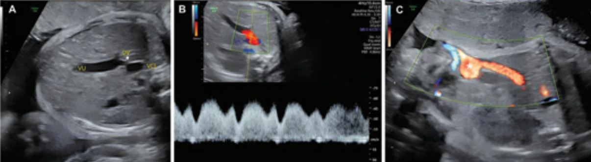
The patient was referred to a tertiary care center, where fetal echocardiography at 25 weeks identified a perimembranous inlet ventricular septal defect, ectasia of the inferior vena cava (Fig. 1) and umbilical vein, and suspicion of agenesis of the ductus venosus.

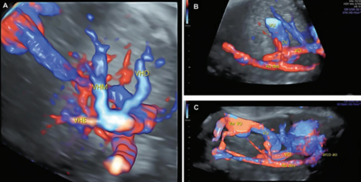
In a subsequent ultrasonographic evaluation at 30 weeks and 2 days, the cardiac findings were confirmed, along with umbilical vein varices, abnormal course of the ductus venosus (Fig. 2), and agenesis of the portal vein (Fig. 3).
At the 37th week, Doppler ultrasound showed appropriate fetal growth, umbilical vein varices, venous dilation, and agenesis of the portal vein, with normal pulsatility indexes (umbilical and middle cerebral arteries) and no signs of blood flow centralization or fetal growth restriction. The approach was expectant, with obstetric and cardiological monitoring.
At 37 weeks and 5 days, the patient presented premature rupture of ovular membranes and moderate abdominal pain, without vaginal bleeding. Clinical examination and cardiotocography indicated good fetal vitality. She was admitted for delivery, and started on crystalline penicillin for prophylaxis of perinatal infection. After failed induction of labor due to cervical dystocia, a cesarean section was performed without complications.
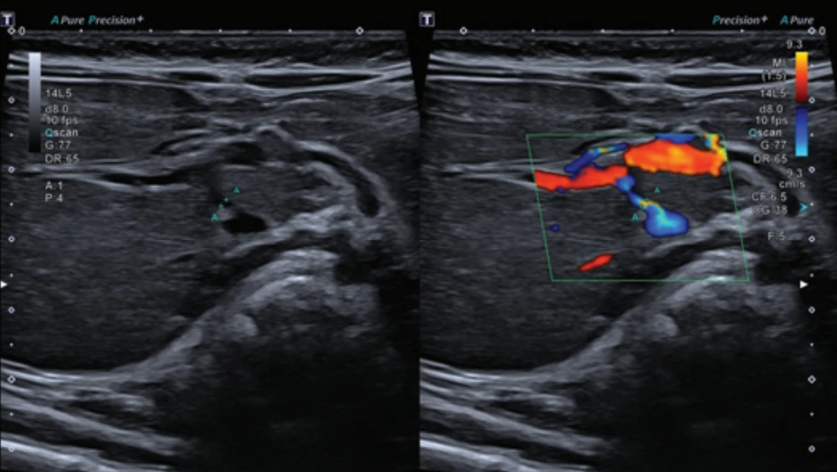
The female newborn presented good vitality at birth (Apgar scores 8 and 8 at the 1st and 5th minute, resp.), weighed 3,265 g, and did not require resuscitation. In the immediate neonatal period, she had episodes of central cyanosis and moderate pulmonary hypertension, which were treated with inhaled nitric oxide and milrinone, with good clinical response. Transthoracic echocardiogram revealed a patent foramen ovale, persistent ductus arteriosus, enlargement of the right chambers, thickening of the right ventricle, moderate tricuspid insufficiency, and significant pulmonary hypertension. Abdominal ultrasound with color Doppler showed a portocaval shunt with communication between the right portal vein and the inferior vena cava, a hypoplastic portal vein, and portosystemic flow, with no signs of thrombosis (Fig. 4). During hospitalization, neonatal conjunctivitis and congenital cataract in the left eye were diagnosed, with scheduled follow-up in ophthalmology. After clinical stabilization, the patient was discharged with multidisciplinary follow-up in pediatric, cardiology, gastroenterology, ophthalmology, and genetics teams.
The postnatal karyotype confirmed trisomy 21, supporting the prenatal syndromic suspicion. The patient underwent genetic counseling after receiving the newborn’s karyotype results. During outpatient follow-up, Doppler ultrasound showed persistence of the porto-caval shunt, portal vein with a diameter at the lower limit of normal, and preserved hepatic flow, with no additional structural alterations.
Discussion
The case presented illustrates the complexity of diagnosing and managing congenital heart diseases associated with hepatic vascular anomalies, such as agenesis of the portal vein and abnormal course of the ductus venosus, rare conditions that can lead to significant hemodynamic repercussions. Prenatal identification of these alterations through fetal echocardiography and Doppler ultrasound allows for close monitoring of fetal development, contributing to decisions regarding obstetric management and preparation of the neonatal care team. The association with trisomy 21 further underscores the need for a multidisciplinary approach, considering possible comorbidities, such as pulmonary hypertension and tricuspid insufficiency, which require specific interventions in the neonatal period.
In this case, the persistence of a connection between the right portal vein and the inferior vena cava observed at 90 days of life rules out the possibility of a transient physiological finding, since the ductus venosus, a structure typical of fetal life, normally closes both functionally and anatomically within the first weeks after birth, forming the ligamentum venosum [5]. Therefore, identification of this shunt characterizes a congenital pathological vascular alteration, compatible with a type II congenital portosystemic shunt (type II Abernethy malformation), as the portal vein is present, though of smaller caliber, and part of the portal flow continues to perfuse the liver. This is a rare condition resulting from failures in the remodeling of the vitelline veins during embryogenesis [6].
Doppler ultrasound findings such as visualization of an abnormal vessel connecting the right portal vein directly to the inferior vena cava or hepatic vein; absence or reduced caliber of intrahepatic portal branches; direction of portal flow towards the systemic circulation; elevated velocities in the shunt; and turbulence at the site of communication with a continuous, minimally pulsatile flow pattern all correspond to widely accepted diagnostic criteria for congenital portosystemic shunts. Doppler stands out as a fundamental initial tool for both diagnosis and follow-up, as it is the exam that allows dynamic assessment of flow and hepatic repercussions. Angio-computed tomography or magnetic resonance angiography can be performed for anatomical confirmation [5].
Clinically, the evolution of the newborn with moderate pulmonary hypertension in the immediate neonatal period is an extrahepatic manifestation of the portosystemic shunt. This pulmonary repercussion is a consequence of increased systemic blood flow and circulation of vasoactive mediators not metabolized by the liver, leading to pulmonary circulatory overload. The good response to clinical treatment and subsequent hemodynamic stability reinforce the importance of early recognition of this complication and specialized management [5,7]. Furthermore, the coexistence of mild congenital heart disease and trisomy 21 suggests a syndromic context that may imply a correlation between genetic syndromes and portosystemic anomalies. Although rare, this association highlights the need for genetic investigation in the presence of unusual fetal vascular findings [7].
For management, the type II congenital portosystemic shunt allowed for the conservative approach taken, in contrast to a type I portosystemic shunt, in which there is a total absence of the intrahepatic portal vein and no portal flow perfusing the liver. This anatomical configuration can lead to more severe repercussions, evolving to the need for endovascular or surgical treatment, or even as an early indication for liver transplantation [6].
Conclusion
In summary, this report highlights the importance of regular prenatal care, emphasizing the relevance of imaging exams, especially ultrasound, for the identification of fetal vascular abnormalities such as agenesis of the portal venous system. Early diagnoses allow for proper planning of obstetric and neonatal care, as well as enabling the diagnosis and management of postnatal repercussions. The persistence of portosystemic shunt beyond the neonatal period reinforces the pathological nature of the condition and the need for specialized follow-up. Although rare, the association with trisomy 21 broadens the understanding of the clinical spectrum of these anomalies and underscores the need for a multidisciplinary approach to optimize patient prognosis and safety.
Zdroje
1. Carneiro DN, Rossi I, Oliveira NT et al. Congenital intra-hepatic porto-systemic shunts diagnosed during intrauterine life: systematic review. J Clin Ultrasound 2023; 51 (5): 803–811. doi: 10.1002/jcu.23341.
2. Mistinova J, Valacsai F, Varga I. Congenital absence of the portal vein – case report and a review of literature. Clin Anat 2010; 23 (7): 750–758. doi: 10.1002/ca.21007.
3. Tannuri U, Galvão F, Leal AJ et al. Congenital absence of the portal vein: a complex disease with multiple manifestations and types of treatment. Eur J Pediatr Surg 2011; 21 (4): 269–272. doi: 10.1055/s-0031-1275675.
4. Rossi I, Peixoto AB, de Souza MA et al. Intrauterine diagnosis of portosystemic shunt associated with trisomy 21. J Obstet Gynaecol Can 2023; 45 (10): 101784. doi: 10.1016/j.jogc.2021.06.011.
5. Hlavata T, Kaldararova M, Klauco F et al. Congenital absence of the portal vein as a rare cause of portopulmonary hypertension – a case study series. Medicina (Kaunas) 2022; 58 (10): 1484. doi: 10.3390/medicina58101484.
6. Laverdure N, Lallier M, Dubois J et al. Congenital absence of the portal vein: define the portosystemic shunt, avoid liver transplantation. Can Liver J 2021; 4 (3): 322–327. doi: 10.3138/canlivj-2020-0011.
7. Kong B, Yan X, Gui Y et al. Prenatal sonographic characteristics and postnatal outcomes of congenital portosystemic shunt diagnosed during the fetal period: a systematic review. Orphanet J Rare Dis 2025; 20 (1): 257. doi: 10.1186/s13023-025-03811-3.
Štítky
Dětská gynekologie Gynekologie a porodnictví Reprodukční medicínaČlánek vyšel v časopise
Česká gynekologie
2026 Číslo 5
- I „pouhé“ doporučení znamená velkou pomoc. Nasměrujte své pacienty pod křídla Dobrých andělů
- Neuromodulace v terapii hyperaktivního močového měchýře – výsledky klinických studií a možnost náboru do studie
- Inosin pranobex v léčbě chřipky a dalších respiračních infekcí virové etiologie
- Dlouhodobá ketodieta může poškozovat naše orgány
- Stresová inkontinence moči se týká žen i mužů. Jak vypadají aktuální možnosti léčby?
Nejčtenější v tomto čísle
- Methylation test versus dual staining in the triage of HR-HPV positive women – pilot data from cervical cancer screening
- Transverse B-Lynch suture – specific modality of a uterine compression suture
- Hysteroscopic septum resection and reproductive outcomes – a retrospective cohort study
- Female ejaculation and squirting – definition, classification and differential diagnosis