
Biological and Structural Characterization of a Host-Adapting Amino Acid in Influenza Virus
Two amino acids (lysine at position 627 or asparagine at position 701) in the polymerase subunit PB2 protein are considered critical for the adaptation of avian influenza A viruses to mammals. However, the recently emerged pandemic H1N1 viruses lack these amino acids. Here, we report that a basic amino acid at position 591 of PB2 can compensate for the lack of lysine at position 627 and confers efficient viral replication to pandemic H1N1 viruses in mammals. Moreover, a basic amino acid at position 591 of PB2 substantially increased the lethality of an avian H5N1 virus in mice. We also present the X-ray crystallographic structure of the C-terminus of a pandemic H1N1 virus PB2 protein. Arginine at position 591 fills the cleft found in H5N1 PB2 proteins in this area, resulting in differences in surface shape and charge for H1N1 PB2 proteins. These differences may affect the protein's interaction with viral and/or cellular factors, and hence its ability to support virus replication in mammals.
Published in the journal:
. PLoS Pathog 6(8): e32767. doi:10.1371/journal.ppat.1001034
Category:
Research Article
doi:
https://doi.org/10.1371/journal.ppat.1001034
Summary
Two amino acids (lysine at position 627 or asparagine at position 701) in the polymerase subunit PB2 protein are considered critical for the adaptation of avian influenza A viruses to mammals. However, the recently emerged pandemic H1N1 viruses lack these amino acids. Here, we report that a basic amino acid at position 591 of PB2 can compensate for the lack of lysine at position 627 and confers efficient viral replication to pandemic H1N1 viruses in mammals. Moreover, a basic amino acid at position 591 of PB2 substantially increased the lethality of an avian H5N1 virus in mice. We also present the X-ray crystallographic structure of the C-terminus of a pandemic H1N1 virus PB2 protein. Arginine at position 591 fills the cleft found in H5N1 PB2 proteins in this area, resulting in differences in surface shape and charge for H1N1 PB2 proteins. These differences may affect the protein's interaction with viral and/or cellular factors, and hence its ability to support virus replication in mammals.
Introduction
Influenza viruses pose an ongoing threat to human health, as underscored by the current H1N1 influenza pandemic and the sporadic transmission of highly pathogenic avian H5N1 influenza viruses to humans [1], [2]. Viral determinants of virulence and transmissibility are still poorly understood, although lysine at position 627 of the polymerase subunit PB2 (PB2-627K) is now known to be important for avian influenza virus adaptation to mammals [3], [4]. Most avian influenza viruses (with the exception of the Qinghai Lake-lineage of H5N1 viruses) possess glutamic acid at this position (PB2-627E), whereas most human influenza viruses (with the notable exception of the current pandemic H1N1 viruses) have lysine (PB2-627K). In addition, replacement of the aspartic acid at position 701 of PB2 (PB2-701D) found in most avian influenza viruses with asparagine (PB2-701N) conferred high pathogenicity to an H5N1 influenza virus in mice [5]. These mammalian-type amino acids (i.e., PB2-627K and PB2-701N) are found in some H5N1 (http://www.flu.lanl.gov) or H7N7 [6] influenza viruses isolated from humans, are selected during replication of H5N1 viruses in humans [7], and facilitate virus transmission in ferret [8] and guinea pig models [9]. Collectively, these results have led to the concept that PB2-627K or PB2-701N are critical for efficient influenza virus replication in mammalian species. Nevertheless, the pandemic H1N1 viruses and some H5N1 influenza viruses isolated from humans do not possess these amino acids. Here, we sought to identify additional amino acid changes that facilitate virus adaptation in mammalian species.
Results/Discussion
Lysine at position 591 of the PB2 protein confers efficient replication to an H5N1 influenza virus in mammals
An H5N1 influenza virus isolated from an infected person in Indonesia in 2005 (A/Indonesia/UT3006/05, UT3006) replicated more efficiently than an avian H5N1 virus (A/chicken/Indonesia/UT3091/05 virus; CkUT3091) in normal human bronchioepithelial (NHBE) cells (Fig. 1), despite having avian-type amino acids at PB2-627 and PB2-701. In addition, the mouse lethal dose 50 (MLD50) of UT3006 is 180 plaque-forming units (PFU), indicating appreciable virulence in mice. Reverse genetics approaches demonstrated a critical role of the UT3006 PB2 segment in facilitating more efficient growth of UT3006-CkUT3091 reassortants in NHBE cells (Fig. 1A). Although the UT3006 and CkUT3091 PB2 proteins both encode the avian-type amino acids at positions 627 and 701, they differ by nine other amino acids (62-R/K, 117-T/I, 288-R/Q, 344-M/V, 524-T/I, 526-K/R, 591-Q/K, 676-K/T, 756-T/M; where the first amino acid indicates the residue found in CkUT3091 and the second indicates that found in UT3006). Five of these changes (at positions 62, 117, 524, 526, and 591) are not commonly found in avian influenza viruses, suggesting that they may play a role in mammalian adaptation. Introduction of the UT3006 sequence at these five sites appreciably increased the replicative ability of CkUT3091 in NHBE cells (CkUT3091-5aa, Fig. 1B), although virus titers did not reach the level of UT3006 virus. The respective changes were tested individually for their ability to facilitate avian influenza virus replication in human cells and introduction of the PB2-Q591K change appreciably enhanced the growth properties of CkUT3091 in NHBE cells (Fig. 1B). In contrast, the CkUT3091 variant possessing the PB2-T117I mutant was comparable in its growth to the parental CkUT3091 virus (Fig. 1B). The mutants containing PB2-R62K, PB2-T524I, or PB2-K526R changes were not viable, suggesting that these residues require compensatory changes at other sites. These findings suggest that, in the absence of PB2-627K or 701N, PB2-591K is a key amino acid for efficient influenza virus replication in human cells. Hence, replacement of the highly conserved glutamine at position 591 with lysine may facilitate mammalian adaptation in the context of an avian-type PB2 protein.

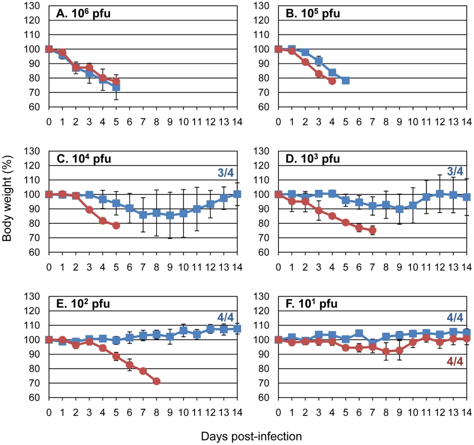
Next, we tested CkUT3091 and its mutant possessing the PB2-Q591K substitution (CkUT3091-PB2-Q591K) for their virulence in mice. All mice infected intranasally with high doses of virus [106 or 105 PFU of virus] lost more than 25% of their pre-infection weight and had to be euthanized on day 4 or 5 post-infection (Fig. 2A and B). At lower infection doses (102–104 PFU), most animals infected with CkUT3091 virus survived (Figs. 2C–E), while all mice infected with CkUT3091-PB2-Q591K had to be euthanized. At the lowest doses tested (101 PFU), infected animals did not experience significant weight loss and survived (Fig. 2F). The MLD50 was 104.3 PFU for CkUT3091 and 50 PFU for the mutant possessing PB2-Q591K (CkUT3091-PB2-Q591K). These findings demonstrated that the glutamine-to-lysine mutation at position 591 of PB2 increased the virulence of an avian H5N1 influenza virus in mice.
Lysine at position 591 of the PB2 protein is selected during H5N1 virus replication in ostrich cells
We previously reported that the human-adapting amino acids PB2-627K and PB2-701N are not only selected during replication of avian influenza viruses in mammalian cells, but also during replication of avian H5N1 viruses in cultured primary ostrich cells or ostriches [10]. Although not fully understood, the selection processes in ostriches thus appear to mimic those in mammalian cells. Since the significance of PB2-591 had not been recognized at the time, we focused our previous analysis on PB2-627 and PB2-701. Here, we therefore reexamined all amino acid changes acquired during replication of A/duck/Vietnam/5001/04 (H5N1) or A/duck/Vietnam/NCVD-18/03 (H5N1) in ostrich cells in vitro or in vivo. PB2-591K was not detected after replication of A/duck/Vietnam/NCVD-18/03 in an infected ostrich, but 12 of 73 PB2 molecular clones derived from viruses isolated from the trachea of an ostrich infected with A/duck/Vietnam/5001/04 (originally possessing PB2-591Q) had acquired the PB2-591K substitution (Table 1). Interestingly, five passages of A/duck/Vietnam/5001/04 in ostrich embryonic cells also yielded one clone (out of 16) with a PB2-Q591R mutation, as found in pandemic H1N1 viruses (see below). Comparable to mammalian cells, the replication of avian H5N1 influenza viruses in ostriches thus resulted in the selection of a basic amino acid at position 591; it is interesting to note that both PB2-591K (as found in some mammalian-adapted H5N1 viruses) and PB2-591R (as found in pandemic H1N1 viruses) were detected. The observed differences in adaptation to ostriches between the two duck viruses may result from differences in their genetic composition. The selection of a basic amino acid at PB2-591 in ostrich cells that seem to mimic the selective pressure in mammals provided further support for the role of PB2-591 in avian influenza virus adaptation to mammalian species.

All 12 ostrich PB2 clones possessing the PB2-591K mutation encoded PB2-627E (i.e., the wild-type, avian-type amino acid; Table 1). Conversely, the remaining 61 clones acquired the PB2-627K (mammalian-type) mutation but encoded the wild-type amino acid at position 591 (PB2-591Q) (Table 1). These findings from our in vivo selection study are consistent with three published ostrich sequences (Supplementary Figure S1) that show the PB2-591Q/627K or PB2-591K/627E combinations, but not mutations at both positions (i.e., PB2-591K/627K). Hence, PB2-591K appears to be selected in combination with PB2-627E, but not in combination with PB2-627K, suggesting that PB2-591K may functionally compensate for the lack of a positive charge at position 627.
Role of PB2 amino acids 591, 627, and 701 in pandemic H1N1 viruses
Our data indicated that a basic amino acid at PB2-591 can compensate for PB2-627K in mammalian adaptation; interestingly, these two residues are positioned near each other on the surface of PB2 [11], [12]. These findings offered an explanation for the efficient replication of the newly emerged pandemic H1N1 viruses in humans: although these viruses lack mammalian-type amino acids at position 627 and 701, they encode a basic amino acid (i.e., arginine) at position 591 (PB2-591R). In fact, a recent study demonstrated that PB2-590S and PB2-591R are important for efficient polymerase activity in an in vitro assay, and for efficient replication of a mixed human/avian influenza virus (with the polymerase and nucleoproteins of avian virus origin) in human cells [13]. Importantly, the additional introduction of PB2-627K did not increase the replicative ability of the mutant virus further [13], consistent with our finding that the combination of basic amino acids at positions 591 and 627 is not selected for in vivo. However, these studies were not carried out with authentic pandemic H1N1 virus and therefore leave in question the biological significance of PB2-591R for the efficient replication of pandemic H1N1 viruses in humans.
To test the significance of specific PB2 amino acid changes in the background of authentic pandemic H1N1 viruses in animal models, we first generated the following PB2 protein variants that were all based on A/California/04/09 (Cal04), an early pandemic H1N1 virus that we previously characterized in vitro and in several animal models [14]: (i) wild-type PB2, (ii) PB2-627K (mammalian-type) instead of PB2-627E (avian-type), (iii) PB2-701N (mammalian-type) instead of PB2-701D (avian-type), or (iv) PB2-591Q (consensus amino acid at this position) instead of PB2-591R (found in pandemic H1N1 viruses). These variants were assessed for their polymerase activity in minireplicon assays, essentially as described [15]. Replacement of PB2-591R with PB2-591Q reduced the polymerase activity (Supplementary Figure S2), further indicating a critical role of PB2-591R in efficient pandemic H1N1 virus replication. Replacement of PB2-627E with PB2-627K, or of PB2-701D with PB2-701N, had mild or no effects. Herfst et al. [16] recently reported increased polymerase activity in minireplicon assays for pandemic H1N1 variants possessing the PB2-627K or PB2-701N mutations, but used different pandemic H1N1 virus isolates.
Next, the PB2 mutations described above were introduced into the genetic background of Cal04 virus, using reverse genetics. Growth curves in Madin-Darby canine kidney (MDCK) cells revealed no significant differences (Supplementary Figure S3), as has been reported by others for the PB2-627K and PB2-701N variants [16], [17]. Intranasal inoculation of BALB/c mice with 104 or 105 PFU of virus resulted in increased weight loss for the PB2-627K and PB2-701N variants (Supplementary Figure S4); however, virus titers in the lungs and nasal turbinates on days 3 and 6 post-infection were comparable for all variants tested (Supplementary Table S1). Similar findings (i.e., weight loss in mice, but no increase in virus titers) were recently reported by others for pandemic H1N1 viruses possessing PB2-627K or PB2-701N [16], [17]. The effects of mutations in PB2 on pandemic H1N1 viruses were further tested in competitive transmission studies in ferrets, an established animal model in influenza virus research; in addition, pandemic H1N1 viruses are known to efficiently transmit in ferrets [14], [18], [19]. Wild-type Cal04 (PB2-591R/627E/701D) and its PB2 variants (PB2-591R/627K/701D, PB2-591R/627E/701N, or PB2-591Q/627E/701D) were mixed at a 1∶1 ratio based on plaque-forming units; however, deep sequencing revealed a slight excess of wild-type Cal04 PB2 vRNA over mutant PB2 vRNAs (Fig. 3B–D). Three ferrets (one per cage) were infected intranasally with virus mix. One day later, one ferret each was placed in a cage adjacent to an infected ferret (‘contact’ ferret) to monitor virus transmission by aerosols. At the indicated time points, nasal wash samples were assessed for the relative ratios of the two viruses (Figs. 3B–D; and Supplementary Table S2). In both infected and contact ferrets, mutations at position PB2-627 (PB2-591R/627K/701D) or PB2-701 (PB2-591R/627E/701N) did not provide a replicative advantage to the respective viruses when compared to wild-type Cal04 virus (PB2-591R/627E/701D). This is consistent with recent findings that E627K or D701N mutations did not affect the virulence or transmissibility of pandemic H1N1 viruses in ferrets [16]. However, in competition studies between wild-type Cal04 virus and a mutant virus possessing PB2-591Q (PB2-591Q/627E/701D), the relative amounts of Cal04 virus increased over time in infected animals (Fig. 3D), indicating that in the background of PB2-627E/701D, PB2-591R provides a replicative advantage over PB2-591Q (the amino acid commonly found at this position). Interestingly, two out of three contact animals showed enrichment of the PB2-591Q mutant virus on day 3 after co-housing; thus, although further studies with larger numbers of animals are needed, we speculate that PB2-591Q might enhance transmission, but not replication in the new host. Collectively, our findings supported the hypothesis that PB2 possessing 591R supports efficient viral replication in mammals, so that there is no strong selective pressure to acquire the mammalian-type amino acids at position 627 and 701.

Based on our experimental data and the evaluation of published influenza virus sequences, we suggest the following scenario: efficient replication in mammalian species can be conferred by PB2-591Q/627K [found in some avian H5N1 viruses that acquired the ability to replicate in mammalian cells [7] (http://www.flu.lanl.gov), and found in human influenza viruses], PB2-591K/627E [found in some avian H5N1 viruses that acquired the ability to replicate in mammalian cells, (http://www.flu.lanl.gov), and identified in this study], or PB2-591R/627E (found in pandemic H1N1 viruses). However, the combination of PB2-591R/K and PB2-627K does not seem to further enhance replication in mammalian cells (Figs. 3B and C). Currently, it is not clear why PB2-591R is found in pandemic H1N1 viruses, while PB2-591K is selected in avian H5N1 viruses. In addition, PB2-701N also confers to H5N1 viruses the ability to replicate in mammalian cells in the absence of PB2-627E [5]. Moreover, other amino acids changes in PB2 may confer efficient replication in mammalian species as well (for example, [20], [21]).
Crystal structure of the C-terminal portion of PB2 from a 2009 H1N1 pandemic virus
Previous X-ray crystallographic studies have demonstrated the close proximity of residues 591 and 627 on the surface of PB2 [11], [12]. This has lead to the suggestion (based on modeling) that PB2-591R neutralizes the PB2-627E in pandemic H1N1 viruses, and thereby partially restores the positively charged surface of the 627 domain of PB2 [13]. To address this critical question, we present here the high-resolution crystal structure of the C-terminal domain of PB2 from a 2009 H1N1 pandemic virus (A/Mexico/InDRE4487/2009) (Fig. 4A); the PB2 proteins of A/Mexico/InDRE4487/2009 and Cal04 are identical in the region crystallized; i.e., amino acid residues 538–741. For comparison, we also present the crystal structure of the C-terminal portion of an H5N1 PB2 protein encoding PB2-627K (A/Vietnam/1203/04; Fig. 4B).

The PB2 protein of the pandemic H1N1 virus shows an altered polar surface when compared to the avian H5N1 PB2-627K protein. Interestingly, our structure of the pandemic H1N1 PB2 protein reveals that PB2-591R fills the cleft found in H5N1 PB2 proteins in this area (compare Fig. 4A and 4B). In particular, the side chains of R591 and S590 in the structure of H1N1 PB2 occupy what would otherwise be the positively charged cleft in the H5N1 PB2. PB2-591R is located 4 Ångstrom from PB2-627E, with the side chain of R591 rotated away from PB2-627E and participating in hydrogen-bonded and water-mediated hydrogen bond networks with the main chain carbonyl backbone between residues A587 and T588 (Fig. 4C), even though the two residues have space to assume rotomers that could form a neutralizing salt bridge (see Fig. 4C). The R591 residue in H1N1 PB2 thus affects both the shape and charges on the surface of the protein, which may affect its interaction with other viral and/or host factors.
A previous study [22] suggested that an inhibitory host factor may suppress the activity of avian-type polymerases (possessing PB2-627E), but not that of human-type polymerases (possessing PB2-627K). In this scenario, the positively charged residue (PB2-591R) that rotates away from PB2-627E may prevent the inhibitory host factor from interacting with PB2 by either charged repulsion or steric hindrance by filling the distinct cleft on the surface of the H5N1 PB2. Alternatively, the positive charge associated with PB2-627K or PB2-591R may facilitate interaction with a stimulatory host factor. Thus, efficient replication in mammalian cells may require a PB2 structure that prevents the action of the inhibitory host factor, or facilitates the action of the stimulatory host factor. Our finding that PB2-591Q/627K (found in human viruses) and PB2-591R/627E (found in pandemic H1N1 viruses) adopt different conformations (i.e. a positively charged, ‘cleft’ vs. a positively-charged residue that fills the cleft and is located next to a negatively-charged patch) probably favors the former possibility, since it is less likely that they would both enhance interaction with the same host protein, but the positive charge introduced by either change could easily disrupt interaction with an inhibitory host protein.
A serine at position 590 of PB2 is found in pandemic H1N1 viruses (but not consistently in older human H1N1 isolates), and this residue (PB2-590S) has been suggested to play a role in the enhanced replicative ability of pandemic H1N1 viruses in mammals [13]. Our results that a basic amino acid at PB2-591 provided a replicative advantage in mammals to avian H5N1 viruses (which possess a glycine at PB2-590), argue against a critical role of PB2-590S in the adaptation of H5N1 influenza viruses to mammals. Our H1N1 structure shows that while the side chain of PB2-590S is in close proximity (<4 Ångstroms) to 627E, the hydroxyl moiety points away from the glutamatic acid and does not participate in hydrogen bonds or polar interactions with the side chains of 591R or 627E. Furthermore, the side chain atoms of PB2-590S also protrude into the positively charged cleft and may provide a steric platform that reduces the conformational flexibility of neighboring residue 627E. These findings suggest that PB2-590S constrains any side chain at position 627. This constraint may be critical for PB2-627K which may need conformational freedom to reach its binding partner. The constraint by PB2-590S on PB2-627E may be less critical, and may in fact help to shield the negative charge.
Our study identifies a new marker (PB2-591R or K) for influenza virus adaptation in mammals that compensates for the lack of PB2-627K. This finding provides an explanation for the efficient replication of pandemic H1N1 viruses (which possess PB2-591R) in mammals. Our X-ray crystal structure of the C-terminal portion of a pandemic H1N1 PB2 protein reveals changes in surface shape and charge created by PB2-591R which may prevent an inhibitory host factor as suggested by Mehle & Doudna [22] from binding to PB2, hence allowing efficient influenza virus replication. Although a few human H1N1 isolates have now been found to possess PB2-627K (http://www.promedmail.org/pls/otn/f?p=2400 : 1001 : 19224::NO::F2400_P1001_BACK_PAGE,F2400_P1001_PUB_MAIL_ID:1010,79432), this mutation did not appear to increase the severity of disease, and does not seem to spread in human populations, consistent with our conclusion that the PB2-627K mutation does not provide a significant replicative advantage to pandemic H1N1 viruses. Based on findings with H5N1 influenza viruses, it was feared that the introduction of PB2-627K into pandemic H1N1 viruses could increase the pathogenicity of the pandemic viruses. However, our data and recent findings by others [13] indicate that PB2-627K does not provide a replicative advantage in the background of a PB2 protein possessing a basic amino acid at PB2-591. From a public health perspective, the notion that the introduction of PB2-627K into pandemic H1N1 viruses is rare and unlikely to create a more pathogenic variant is thus reassuring.
Materials and Methods
Viruses and cells
Human embryonic kidney (293 or 293T) cells were maintained in Dulbecco's modified essential medium (DMEM) containing 10% fetal calf serum and antibiotics. Madin-Darby canine kidney (MDCK) cells were maintained in Eagle's minimal essential medium (MEM) containing 5% newborn calf serum. Normal human bronchioepithelial cells (NHBE) were obtained from Lonza (Walkersville, MD) and maintained in serum-free and hormone-supplemented growth medium according to the manufacturer's instructions. All cells were incubated at 37°C with 5% CO2.
All influenza viruses used in this study were amplified in MDCK cells. All viruses were stored at −80°C until their use in experiments. The titers of stock viruses were determined by plaque assays in MDCK cells (for H1N1 viruses) or NHBE cells (for H5N1 viruses). All experiments with H5N1 viruses were performed in enhanced biosafety level 3 (BSL3) containment laboratories at the University of Wisconsin-Madison, which are USDA approved for such use by the CDC and the U.S. Department of Agriculture, or in BSL3 containment laboratories at the University of Tokyo (Tokyo, Japan), which are approved for such use by the Ministry of Agriculture, Forestry and Fisheries, Japan.
Construction of plasmids and generation of wild-type and mutant influenza viruses
The cDNAs of the A/chicken/Indonesia/UT3091/05 (H5N1; CkUT3091), A/Indonesia/UT3006/05 (H5N1; UT3006), and A/California/04/09 (H1N1; Cal04) viruses were synthesized by reverse transcription as previously described [23], [24]. Viruses were amplified and titrated as described above. All viruses were sequenced to ensure the absence of unwanted mutations.
Mutations in the PB2 genes of CkUT3091 or Cal04 viruses were generated by PCR amplification of the respective RNA polymerase I PB2 construct with primers possessing the desired mutations (primer sequences will be provided upon request). All constructs were sequenced to ensure the absence of unwanted mutations.
Minireplicon assays
To assess the viral polymerase activity, we performed luciferase activity-based minireplicon assay as described previously [15] using the following plasmids; expression plasmids for Cal04-PB2, -PB1, -PA, and -NP (0.2 µg each), pPolI-WNA-Fluc (0.02 µg), which express NA gene encoding firefly luciferase gene, and pGL4.74[hRluc/TK] (Promega, 0.02 µg). The luciferase activity in the plasmid-transfected 293 cells was measured by Dual-Luciferase Reporter Assay System (Promega) at 24 h post-transfection.
Growth curves in NHBE or MDCK cells
NHBE cells (Lonza) were infected with the indicated viruses at a multiplicity of infection (m.o.i) of 0.0005. One hour later, the viral inoculum was removed and replaced with bronchial epithelial growth medium (SAGM; Cambrex) containing bovine pituitary extract (BPE; 30 µg/ml), hydrocortisone (0.5 µg/ml), human epidermal growth factor (hEGF; 0.5 ng/ml), epinephrine (0.5 µg/ml), transferrin (10 µg/ml), insulin (5 µg/ml), triiodothyronine (6.5 ng/ml), bovine serum albumin – fatty-acid free (BSA-FAF; 50 µg/ml), retinoic acid (RA; 0.1 ng/ml), gentamycin (30 µg/ml) and amphotericin B (15 ng/ml). Infected cells were incubated at 37°C and 5% CO2. At the indicated times post infection, the virus titers in the cell culture supernatant were determined by plaque assays in NHBE cells. For the determination of viral growth kinetics in MDCK cells, cells were infected at an m.o.i. of 0.001; at the indicated time points, virus titers were determined by plaque assays.
Virulence in mice
Six-week-old female BALB/c mice (Japan SLC Inc., Shizuoka, Japan; The Jackson Laboratory, Bar Harbor, Maine, USA) were used in this study. Baseline body weights were measured prior to infection. Four mice per group were anesthetized with isoflurane and intranasally inoculated with 101–106 PFU (50 µl) of CkUT3001 or CkUT3091-PB2-Q591K, or with 103 or 106 PFU of Cal04 possessing wild-type or mutant PB2 protein. Body weight and survival were monitored daily for 14 days; mice with body weight loss of more than 25% of pre-infection values were euthanized. For virus titration in organs, mice were infected intranasally with 105 PFU of virus and euthanized on days 3 and 6 postinfection.
Ferret competition-transmission study
We used six-to-seven-month-old female ferrets (Triple F Farms, Sayre, PA), which were serologically negative by hemagglutination inhibition (HI) assay for currently circulating human influenza viruses (including pandemic H1N1 viruses). The animals were housed in adjacent transmission cages that prevent direct and indirect contact between animals but allow spread of influenza virus through the air. Three ferrets were intranasally inoculated with 106 plaque-forming units (pfu; in 500 µl) of the mixture of Cal04 and mutant virus at a 1∶1 ratio (0.5×10⁶ pfu of Cal04 plus 0.5×10⁶ pfu of mutant virus). One day after infection, three naive ferrets were each placed in a cage adjacent to an inoculated ferret (contact ferrets). Hence, we tested three pairs of infected/contact ferrets for each virus. Nasal washes from ferrets were collected on days 1, 3, 5, and 7 post-infection or co-housing and titrated for virus titers in MDCK cells. Samples with greater than 1×10² pfu/ml of virus were subjected to sequence analysis as described below. All animal experiments were carried out in accordance with institutional guidelines.
Sequence analysis of viruses obtained from infected or contact ferrets
Viral RNA was extracted from nasal wash samples and twice from the inoculum (to verify the 1∶1 ratio of the two test viruses). The cDNAs were synthesized as previously described [23]. A panel of PCR primer pairs was used to generate 410 base pair amplicons of the PB2 region covering 591Q, 627K, and 701N of A/California/04/2009. All primers contained Roche/454 Titanium Amplicon adapter sequences and distinctive multiplier identifier (MID) tags engineered at their 5′ termini. After gel purification and normalization to 108 molecules/µl, emulsion PCR was performed on four pools containing 14 MID-tagged amplicons each.
The resulting pools were pyrosequenced in individual regions of a 70×75 PicoTiterPlate with a 16-well gasket on a Roche/454 GS FLX instrument following protocols provided by the manufacturer (www.454.com). An average of 2221 sequence reads was obtained for each sample (range: 705–5102). All sequences from a single time point were aligned to the PB2 sequence of A/California/04/09 (Roche/454 AVA software). The frequencies of wild-type and mutant amino acids at residues 591, 627, and 701 were tabulated from the aligned sequences. To confirm the pyrosequencing results, independent PB2 sequences from 37 of the 42 samples were generated using alternative sets of PCR primers and pyrosequenced using the Roche/454 FLX chemistry.
X-ray crystal structure determination of PB2 C-terminal domains
Codon engineered synthetic genes for the PB2 gene of influenza A virus strains A/Vietnam/1203/04 (H5N1) (AAT73550) and A/Mexico/InDRE4487/09 (H1N1) (ACQ73384) were designed in Gene Composer (Emerald BioSystems, Bainbridge Island, WA) using a combined E. coli and baculovirus codon usage table with a threshold setting of 0.2% and synthesized (DNA 2.0, Menlo Park, CA) [25], [26]. The C-terminal amino acid residues 538–741 from both strains were cloned into an engineered pET vector using the Polymerase Incomplete Primer Extension (PIPE) cloning technology [27]. Cloning, expression, and purification followed reported protocols from the Seattle Structural Genomics Center for Infectious Disease [28]. Constructs were expressed in vectors containing an N-terminal 6×His Smt fusion protein [29]. Site specific removal of the 11kDA 6×His Smt fusion partner was accomplished with 0.2 µg Ulp1 protease/mg of recombinant target [29], [30]. Final protein buffer composition following size exclusion chromatography (Superdex 75, GE Healthcare, Piscataway, NJ) was 25 mM Hepes/NaOH pH 7.2, 500 mM NaCl, 2 mM DTT, 5% glycerol. Protein crystallization trials were conducted by sitting drop vapor diffusion at 16°C in 96-well crystallization plates. Crystallization conditions for H5N1-PB2(538–741) were 5% octyl β-D-1-thioglucopyranoside (SBOG), 100 mM N-cyclohexyl-3-aminopropanesulfonic acid (CAPS) pH 10.5, 1.2 M sodium phosphate, 0.2 M lithium phosphate, 0.8 M potassium phosphate at protein concentration of 8 mg/ml. Crystallization conditions for H1N1-PB2(538–741) were 20% w/v PEG 3350, 200 mM ammonium citrate at protein concentration of 24 mg/ml. All crystal drop sizes used were 0.4 µl crystallant plus 0.4 µl protein. Crystals were harvested by transfer into drops containing crystallant plus either 25% glycerol or ethylene diol as cryoprotectant and flash frozen in liquid nitrogen. X-ray data were collected on a Rigaku (Rigaku Americas, The Woodlands, Texas) FR-E+ X-ray generator equipped with a Saturn-944 CCD detector. One degree images were collected for 30 seconds and scaled with HKL2000 [31]. Structures were determined by molecular replacement with the coordinates from PDBID 3CW4 using CCP4/PHASER [32]. Iterative refinement and modeling of coordinates was conducted using REFMAC [33] and COOT [34]. Final crystallographic refinement statistics are presented in Table 2. Structure figures were generated using PYMOL [35].

Ethics
Our experiments in mice, ostriches, and ferrets followed the University of Tokyo's Regulations for Animal Care and Use and the University of Wisconsin-Madison's Animal Care and Use Protocol. They were approved by the Animal Experiment Committee of the Institute of Medical Science, the University of Tokyo (approval number: 19–29) and the Animal Care and Use Committee of the University of Wisconsin-Madison (protocol number V00806), which acknowledged and accepted both the legal and ethical responsibility for the animals, as specified in the Fundamental Guidelines for Proper Conduct of Animal Experiment and Related Activities in Academic Research Institutions under the jurisdiction of the Ministry of Education, Culture, Sports, Science and Technology, 2006 (Japan) and in the Animal Welfare Act and associated Animal Welfare Regulations and Public Health Service Policy (USA).
Supporting Information
Zdroje
1. NeumannG
NodaT
KawaokaY
2009 Emergence and pandemic potential of swine-origin H1N1 influenza virus. Nature 459 931 939
2. YenHL
AldridgeJR
BoonAC
IlyushinaNA
SalomonR
2009 Changes in H5N1 influenza virus hemagglutinin receptor binding domain affect systemic spread. Proc Natl Acad Sci U S A 106 286 291
3. HattaM
GaoP
HalfmannP
KawaokaY
2001 Molecular basis for high virulence of Hong Kong H5N1 influenza A viruses. Science 293 1840 1842
4. SubbaraoEK
LondonW
MurphyBR
1993 A single amino acid in the PB2 gene of influenza A virus is a determinant of host range. J Virol 67 1761 1764
5. LiZ
ChenH
JiaoP
DengG
TianG
2005 Molecular basis of replication of duck H5N1 influenza viruses in a mammalian mouse model. J Virol 79 12058 12064
6. FouchierRA
SchneebergerPM
RozendaalFW
BroekmanJM
KeminkSA
2004 Avian influenza A virus (H7N7) associated with human conjunctivitis and a fatal case of acute respiratory distress syndrome. Proc Natl Acad Sci U S A 101 1356 1361
7. LeQM
Sakai-TagawaY
OzawaM
ItoM
KawaokaY
2009 Selection of H5N1 influenza virus PB2 during replication in humans. J Virol 83 5278 5281
8. Van HoevenN
PappasC
BelserJA
MainesTR
ZengH
2009 Human HA and polymerase subunit PB2 proteins confer transmission of an avian influenza virus through the air. Proc Natl Acad Sci U S A 106 3366 3371
9. SteelJ
LowenAC
MubarekaS
PaleseP
2009 Transmission of influenza virus in a mammalian host is increased by PB2 amino acids 627K or 627E/701N. PLoS Pathog 5 e1000252
10. ShinyaK
MakinoA
OzawaM
KimJH
Sakai-TagawaY
2009 Ostrich involvement in the selection of H5N1 influenza virus possessing mammalian-type amino acids in the PB2 protein. J Virol 83 13015 13018
11. KuzuharaT
KiseD
YoshidaH
HoritaT
MurazakiY
2009 Structural basis of the influenza A virus RNA polymerase PB2 RNA-binding domain containing the pathogenicity-determinant lysine 627 residue. J Biol Chem 284 6855 6860
12. TarendeauF
CrepinT
GuilligayD
RuigrokRW
CusackS
2008 Host determinant residue lysine 627 lies on the surface of a discrete, folded domain of influenza virus polymerase PB2 subunit. PLoS Pathog 4 e1000136
13. MehleA
DoudnaJA
2009 Adaptive strategies of the influenza virus polymerase for replication in humans. Proc Natl Acad Sci U S A
14. ItohY
ShinyaK
KisoM
WatanabeT
SakodaY
2009 In vitro and in vivo characterization of new swine-origin H1N1 influenza viruses. Nature 460 1021 1025
15. OzawaM
FujiiK
MuramotoY
YamadaS
YamayoshiS
2007 Contributions of two nuclear localization signals of influenza A virus nucleoprotein to viral replication. J Virol 81 30 41
16. HerfstS
ChutinimitkulS
YeJ
de WitE
MunsterVJ
2010 Introduction of virulence markers in PB2 of pandemic swine-origin influenza virus does not result in enhanced virulence or transmission. J Virol 84 3752 3758
17. ZhuH
WangJ
WangP
SongW
ZhengZ
2010 Substitution of lysine at 627 position in PB2 protein does not change virulence of the 2009 pandemic H1N1 virus in mice. Virology 401 1 5
18. MainesTR
JayaramanA
BelserJA
WadfordDA
PappasC
2009 Transmission and pathogenesis of swine-origin 2009 A(H1N1) influenza viruses in ferrets and mice. Science 325 484 487
19. MunsterVJ
de WitE
van den BrandJM
HerfstS
SchrauwenEJ
2009 Pathogenesis and transmission of swine-origin 2009 A(H1N1) influenza virus in ferrets. Science 325 481 483
20. BrownEG
BaillyJE
1999 Genetic analysis of mouse-adapted influenza A virus identifies roles for the NA, PB1, and PB2 genes in virulence. Virus Res 61 63 76
21. YaoY
MingayLJ
McCauleyJW
BarclayWS
2001 Sequences in influenza A virus PB2 protein that determine productive infection for an avian influenza virus in mouse and human cell lines. J Virol 75 5410 5415
22. MehleA
DoudnaJA
2008 An inhibitory activity in human cells restricts the function of an avian-like influenza virus polymerase. Cell Host Microbe 4 111 122
23. KatzJM
WangM
WebsterRG
1990 Direct sequencing of the HA gene of influenza (H3N2) virus in original clinical samples reveals sequence identity with mammalian cell-grown virus. J Virol 64 1808 1811
24. NeumannG
WatanabeT
ItoH
WatanabeS
GotoH
1999 Generation of influenza A viruses entirely from cloned cDNAs. Proc Natl Acad Sci U S A 96 9345 9350
25. LorimerD
RaymondA
WalchliJ
MixonM
WallaceE
2009 Gene Composer: Database software for protein construct design, codon engineering, and gene synthesis. BMC Biotechnol 9 36
26. RaymondA
LovellS
LorimerD
WalchliJ
MixonM
2009 Combined protein construct and synthetic gene engineering for heterologous protein expression and crystallization using Gene Composer. BMC Biotechnol 9 37
27. KlockHE
KoesmaEJ
KnuthMW
LesleySA
2008 Combining the polymerase incomplete primer extension method for cloning and mutagenesis with microscreening to accelerate structural genomics efforts. Proteins 1 982 994
28. MylerPJ
StacyR
StewartL
StakerB
Van VoorhisWC
2009 The Seattle Structural Genomics Center for Infectious Disease (SSGCID). Infect Disord Drug Targets
29. MossessovaE
LimaCD
2000 Ulp1-SUMO crystal structure and genetic analysis reveal conserved interactions and a regulatory element essential for cell growth in yeast. Mol Cell 5 865 876
30. LiSJ
HochstrasserM
1999 A new protease required for cell-cycle progression in yeast. Nature 398 246 251
31. OtwinowskiZ
MinorW
1997 Processing of X-ray Diffraction Data Collected in Oscillation Mode Methonds in Enzymology 276 307 326
32. Collaborative Computational Project N 1994 The CCP4 Suite: Programs for Protein Crystallography. Acta Cryst D D50 760 763
33. MurshudovaGN
VaginAA
DodsonEJ
1997 Refinement of macromolecular structures by the maximum-likelihood method. Acta Crystallographica Section D-Biological Crystallography D53 240 255
34. EmsleyP
CowtanK
2004 Coot: model - building tools for molecular graphics. Acta Crystallographica Section D-Biological Crystallography 60 2126 2132
35. DeLanoWL
2002 The PyMOL User's Manual: DeLano Scientific, Palo Alto, CA, USA
Štítky
Hygiena a epidemiologie Infekční lékařství LaboratořČlánek vyšel v časopise
PLOS Pathogens
2010 Číslo 8
- Parazitičtí červi v terapii Crohnovy choroby a dalších zánětlivých autoimunitních onemocnění
- Vakcíny proti klíšťové encefalitidě
- Kdy je nejlepší očkovat
- Možné vedlejší účinky očkování
- Imunogenita vakcín
Nejčtenější v tomto čísle
- The Transcription Factor Rbf1 Is the Master Regulator for -Mating Type Controlled Pathogenic Development in
- PKC Signaling Regulates Drug Resistance of the Fungal Pathogen via Circuitry Comprised of Mkc1, Calcineurin, and Hsp90
- Contribution of Coagulases towards Disease and Protective Immunity
- Early Severe Inflammatory Responses to Uropathogenic Predispose to Chronic and Recurrent Urinary Tract Infection
Zvyšte si kvalifikaci online z pohodlí domova
Mazová zátka a její řešení
nový kurzVšechny kurzy