
Evaluation of the effect of magnesium sulfate on maternal cerebral hemodynamics in severe preeclampsia using an ophthalmic artery Doppler
Authors:
K. V. Zamprogno 1
 ; C. A. de Oliveira 1
; Luis Guillermo Coca Velarde 1
; L. M. Teixeira 2
; Edward Araujo Júnior 2,3
; R. A. M. De Sá 1
Authors place of work:
Department of Obstetrics and Gynecology, Fluminense Federal University (UFF), Niterói – RJ, Brazil
1; Discipline of Woman Health, Municipal University of São Caetano do Sul (USCS), São Caetano do Sul – SP, Brazil
2; Department of Obstetrics, Paulista School of Medicine, Federal University of São Paulo (EPM-UNIFESP), São Paulo – SP, Brazil
3
Published in the journal:
Ceska Gynekol 2026; 91(5): 384-389
Category:
Původní práce
doi:
https://doi.org/10.48095/cccg2026384
Summary
Objective: To describe the Doppler indices of the ophthalmic artery (OAD) in women with severe preeclampsia (PE) and singleton pregnancies before and after the use of magnesium sulfate. Methods: A prospective cross-sectional study was conducted including singleton pregnancies, absence of labor, and criteria for initiation of magnesium sulfate for maternal neuroprotection, defined by the presence of severe hypertension and signs suggestive of imminent eclampsia. The right OAD was evaluated before initiation of magnesium sulfate infusion and at 10, 30, and 60 minutes after the drug was started. The pulsatility index (PI), resistance index (RI), and peak ratio (PR) were assessed. Systolic blood pressure (SBP), diastolic blood pressure (DBP), and heart rate were measured at all OAD assessment time points. Results: Eighteen pregnant women were evaluated, with a median maternal age of 28.5 years and a median gestational age of 33 weeks at the time of OAD assessment. There was an increase in RI and PI values without statistical significance and a statistically significant decrease in PR after magnesium sulfate administration. SBP and DBP showed statistically significant reductions during the evaluations. No statistically significant change in heart rate was observed. Conclusion: The decrease in PR reflected increased impedance to flow in the ophthalmic artery, and consequently indicated reduced cerebral perfusion after magnesium sulfate administration.
Keywords:
preeclampsia – Doppler – magnesium sulfate – ophthalmic artery
Introduction
Preeclampsia (PE) is a hypertensive disorder that develops during pregnancy after 20 weeks of gestation and may progress to eclampsia if untreated [1]. PE complicates 5–10% of pregnancies and is the leading cause of maternal mortality worldwide [2–4]. Acute cerebral complications of PE, such as eclampsia, intracranial hemorrhage, and cerebral edema, account for at least 75% of deaths in these patients [5–7]. PE is also associated with significant short and long-term maternal morbidity, and neurological sequelae account for much of this morbidity, including decreased visual acuity, permanent neurological deficits after stroke, and late cognitive impairment [8].
Despite its importance, data regarding alterations in cerebral circulation in pregnant women with preeclampsia remain scarce and conflicting [9,10]. The most widely accepted theory currently suggests that severe hypertension in a compromised cerebral endothelium exceeds the limits of cerebral blood flow autoregulation, leading to forced vasodilation, increased hydrostatic pressure, hyperperfusion, and subsequent cerebral edema. This phenomenon has received considerable attention in recent years and is described as posterior reversible encephalopathy syndrome (PRES) [2–4,10].
Magnesium sulfate has been widely used as a prophylactic agent for eclampsia, significantly reducing seizures compared with placebo. This makes magnesium sulfate an important ally in preventing maternal death caused by hypertension [11–13]. However, the mechanism of action of magnesium sulfate in providing cerebral protection in patients with severe PE remains undefined. Given its vasodilatory effect, it has been suggested that magnesium sulfate induces cerebral and retinal vasodilation, a theory that does not fully explain its protective effect in severe PE, since neurological involvement in PE/eclampsia fulfills the criteria for PRES, a condition associated with vasogenic cerebral edema. Although still uncertain, studies suggest that the pathophysiology of PRES is related to elevated cerebral perfusion pressure leading to impaired cerebral autoregulation, resulting in vasodilation and cerebral edema, with endothelial injury – also present in PE – being the primary pathophysiological substrate of PRES [14–16].
Ophthalmic artery Doppler (OAD) is a noninvasive examination used to study vascular flow in the cerebral territory [4,5], providing important data regarding cerebral circulation. Due to embryological, anatomical, and functional similarities between small intracranial vessels and orbital arteries, Doppler assessment of orbital vessels has been used to infer intracranial circulation [17]. Although several studies have used OAD in the evaluation of hypertensive pregnant women, no studies have addressed the behavior of ophthalmic artery flow following magnesium sulfate administration for the prevention of cerebral injury in severe PE.
The objective of this study was to evaluate OAD parameters in women with severe PE and singleton pregnancies before and after magnesium sulfate administration, and to correlate these parameters with the recognized protective effect of magnesium sulfate on cerebral arterial blood flow.
Methods
A prospective cross-sectional study was conducted between June 2017 and June 2019 at Antonio Pedro University Hospital, Fluminense Federal University, Niterói, Rio de Janeiro, Brazil. The study was approved by the Ethics Committee of the Federal University of Rio de Janeiro (No. 1,125,688), and all participants signed informed consent. Inclusion criteria were singleton pregnancies, absence of labor, and criteria for initiation of magnesium sulfate for maternal neuroprotection in women with severe
PE and/or signs of imminent eclampsia:
1. Severe hypertension – systolic blood pressure ≥ 160 mmHg and/or diastolic blood pressure ≥ 110 mmHg confirmed after 15 minutes of rest.
2. Maternal symptoms suggestive of imminent eclampsia risk – persistent or atypical headache, visual disturbances, persistent right upper quadrant or upper abdominal pain.
Exclusion criteria were:
1. Smoking, due to its influence on vascular endothelium, including endothelial activity blockade and reduced systemic inflammatory activity.
2. Eclampsia.
3. Associated comorbidities, including chronic hypertension.
4. Serum creatinine > 1 mg/dL, due to renal excretion of magnesium sulfate.
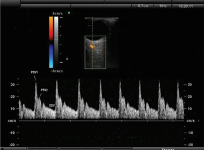
Magnesium sulfate was administered intravenously, with a loading dose of 4 g over 20 minutes followed by a maintenance dose of 1 g/hour according to the Zuspan regimen [18] and Sibai et al. [19]. All patients had right ophthalmic artery flow evaluated at four time points: before infusion and at 10, 30, and 60 minutes after initiation. SBP, DBP, and heart rate were measured at all OAD assessment times. The right ophthalmic artery was chosen due to better examiner positioning, facilitating technique and examination performance. Studies show no difference between right and left ophthalmic artery assessments [20]. Intravenous hydralazine 5 mg was administered every 20 minutes in all patients until SBP was < 160 mmHg and/or DBP < 110 mmHg. For OAD evaluation, a drop of gel was applied over the closed eyelids beneath the eyebrows. A linear transducer was gently placed to avoid pressure artifacts and oriented transversely. Cranial and caudal movements were performed to identify vessels. The ophthalmic artery was identified deep in the temporal retrobulbar region, crossing anterior to the optic nerve and proceeding medially in its superficial portion. Sample volume was adjusted to 2–3 mm and positioned nasally and superiorly to the optic nerve, beside the hypoechoic band corresponding to the nerve, approximately 10–15 mm from the posterior scleral wall. After insonation of the ophthalmic artery, verification was conducted to assure the insonation angle remained below 20 degrees. Measurements were taken after obtaining six consecutive waveforms of similar morphology and size [21]. Each exam lasted approximately 5 minutes. A linear transducer (7.0–12.5 MHz) from Voluson S8 (General Electric Healthcare, Zipf, Austria) and Sonoace X8 (Samsung Corp., Seoul, South Korea) devices was used. Examinations at 10, 30, and 60 minutes after magnesium sulfate administration were performed at bedside. The following OAD parameters were assessed: resistance index (RI), pulsatility index (PI), and peak ratio (PR) (Fig. 1).
Data were analyzed using R software version 3.3.2, available at www.r-project.org. All parameters were expressed as median and quartiles. Wilcoxon signed-rank test was used to compare indices before and after magnesium sulfate administration, as well as SBP, DBP, and heart rate values. A P-value ≤ 0.05 was considered statistically significant.
Results
Eighteen patients agreed to participate in the study, and all underwent OAD assessment before and after administration of the magnesium sulfate loading and maintenance doses (10, 30, and 60 minutes). Of the 18 patients, 4 (22.2%) were not using antihypertensive medi-cation at the time of examination, whereas 14 (77.7%) were receiving methyldopa, and among these, 2 (14.2%) were also using nifedipine. The median maternal age and gestational age at the time of OAD assessment were 28.5 years (IQR 25.5–34.7) and 33 weeks (IQR 31–36), resp.
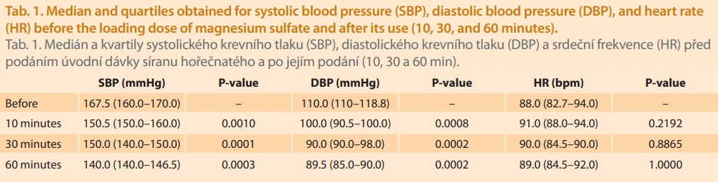
Tab. 1 shows the median and quartiles for SBP, DBP, and heart rate before the magnesium sulfate loading dose and after administration (10, 30, and 60 minutes). There was a statistically significant reduction in SBP and DBP; however, no statistically significant change in heart rate was observed.
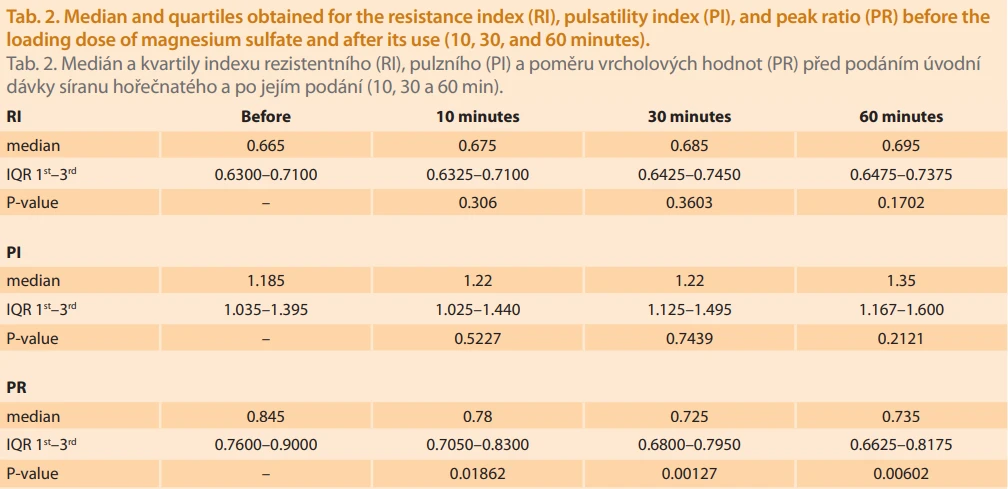
Tab. 2 shows the median and quartiles for RI, PI, and PR before the magnesium sulfate loading dose and after administration (10, 30, and 60 minutes). RI and PI values increased without statistical significance, and PR decreased significantly after magnesium sulfate administration.
Discussion
Acute cerebral complications of PE account for at least 75% of PE-related deaths, with eclampsia being its most severe complication [5,8,9]. Magnesium sulfate is indicated for central nervous system protection, preventing seizures in women with severe PE, and its incorporation into clinical practice has been associated with a significant reduction in maternal mortality [11,12]. Since publication of the MAGPIE trial in 2002, which evaluated 10,110 women with PE and concluded that magnesium sulfate reduces the risk of developing eclampsia, its use has increased worldwide for eclampsia prevention in women with PE [13]. In 2006, a subsequent publication of the MAGPIE study confirmed its safety after 2 years, demonstrating that the reduction in eclampsia risk following magnesium sulfate prophylaxis was not associated with excess mortality or disability at 2-year follow-up [22]. Nevertheless, the mechanism of action of magnesium sulfate in providing cerebral protection in pregnant women with severe PE remains unclear. The most accepted theory explaining the etiology of eclamptic seizures suggests that severe hypertension exceeds the limits of cerebral blood flow autoregulation, leading to hyperperfusion and cerebral edema [9,23]. Failure of autoregulation, together with endothelial dysfunction, appears to be part of the pathophysiology of posterior reversible encephalopathy syndrome (PRES), characterized by visual disturbances, headache, seizures, severe hypertension, and altered mental status. PE and eclampsia have been described as the most common causes of PRES [24]. Identification of cerebral hyperperfusion in patients with PE has been described as a marker of cerebral hemorrhage risk and a feature of disease severity [5]. Therefore, the vasodilatory effect of magnesium sulfate alone would not justify its role in preventing eclampsia in patients with severe PE. Signs of vasodilation in the ophthalmic and central retinal arteries associated with orbital hyperperfusion in PE patients have previously been described by several authors [4,25–27].
PR appears to be the most important index related to PE severity and was initially proposed by Nakatsuka et al. [28]. PR analyzes the behavior of the flow velocity waveform during systole, better quantifying vessel-specific flow changes in vessels with a dicrotic waveform. In patients with PE, the ophthalmic artery flow velocity waveform shows a greater elevation of the second systolic peak following the first notch, representing local hyperperfusion (lower flow impedance). The higher the velocity peak after the first notch, the higher the PR (approaching 1) [5]. Increased PR values in severe PE suggest reduced flow impedance in orbital circulation and, therefore, hyperflow. Although the mechanism underlying elevation of this index remains unclear, PR has been proposed as the most sensitive indicator of orbital vascular changes associated with cerebral hyperperfusion [2,4,8].
In the present study, a decrease in PR values was observed after magnesium sulfate administration, demonstrating increased impedance to flow in the ophthalmic artery and suggesting reduced cerebral perfusion in patients with severe PE after magnesium sulfate use. Belfort et al. [11] studied the effect of magnesium sulfate on cerebral hemodynamics in PE by performing middle cerebral artery Doppler in 15 PE patients with SBP < 160 mmHg and DBP < 110 mmHg before and after magnesium sulfate administration. No other vasoactive medication was used. The authors concluded that magnesium sulfate reduced cerebral perfusion pressure in patients with elevated baseline cerebral perfusion pressure but not in those with normal baseline perfusion pressure. This finding may be important for the prevention of hypertensive encephalopathy. Until 2017, in a pilot study, no research had addressed the mechanism of action of magnesium sulfate in preventing eclamptic seizures in patients with severe PE. That study already demonstrated a trend toward increased impedance to flow in the ophthalmic artery with consequent reduction in cerebral perfusion after magnesium sulfate administration in patients with severe PE [7]. These data could explain its protective effect by preventing cerebral injury and acute seizures. In the present study, this trend was maintained in a larger, though still limited, sample size, suggesting that this may be the mechanism by which magnesium sulfate prevents seizures in patients with severe PE. Since all studied patients met criteria for initiation of antihypertensive therapy for blood pressure reduction and there was a significant decrease in SBP and DBP after magnesium sulfate administration, it is possible that PR reduction was influenced by blood pressure reduction rather than magnesium sulfate itself. Hydralazine, the antihypertensive medication indicated for blood pressure control in hypertensive emergencies during pregnancy, is a vasodilator that acts on vascular smooth muscle, promoting muscle relaxation with consequent vasodilation and reduction of peripheral vascular resistance [29]. Evaluation of ocular flow in patients with signs of imminent eclampsia and SBP below 160 mmHg and/or DBP below 110 mmHg – therefore without criteria for acute antihypertensive therapy – could better elucidate the isolated effect of magnesium sulfate on cerebral circulation
Conclusion
The decrease in PR reflects increased impedance to flow in the ophthalmic artery and consequently indicates reduced cerebral perfusion after magnesium sulfate administration. This reduction may explain the mechanism by which magnesium sulfate protects against cerebral injury in severe PE, preventing acute seizures in these patients, since the most accepted current theory suggests that severe PE involves impairment of cerebral blood flow autoregulation, leading to hyperperfusion responsible for cerebral damage.
Zdroje
1. Olatunji RB, Adekanmi AJ, Obajimi MO et al. Maternal ophthalmic artery Doppler velocimetry in pre-eclampsia in Southwestern Nigeria. Int J Womens Health 2015; 7 : 723–734. doi: 10.2147/IJWH.S86314.
2. Sibai B, Dekker G, Kupferminc M. Pre-eclampsia. Lancet 2005; 365 (9461): 785–799. doi: 10.1016/S0140-6736 (05) 17987-2.
3. Magee LA, Helewa M, Rey E. RETIRED: diag-nosis, evaluation, and management of the hypertensive disorders of pregnancy. J Obstet Gynaecol Can 2008; 206 (3 Suppl): S1–S2. doi: 10.1016/S1701-2163 (16) 32776-1.
4. Diniz AL, Moron AF, Santos MC et al. Ophthalmic artery Doppler as a measure of severe pre-eclampsia. Int J Gynaecol Obstet 2008; 100 (3): 216–220. doi: 10.1016/j.ijgo.2007.07.013.
5. de Oliveira CA, de Sá RA, Velarde LG et al. Changes in ophthalmic artery Doppler indices in hypertensive disorders during pregnancy. J Ultrasound Med 2013; 32 (4): 609–616. doi: 10.7863/jum.2013.32.4.609.
6. Zeeman GG. Neurologic complications of preeclampsia. Semin Perinatol 2009; 33 (3): 166–172. doi: 10.1053/j.semperi.2009.02.003.
7. Oliveira CA, de Sá RA, Zamprogno KV et al. Magnesium sulfate and ophthalmic artery Doppler velocimetry in patients with severe preeclampsia: a case series. J Med Case Rep 2017; 11 (1): 326. doi: 10.1186/s13256-017-1490-1.
8. Barbosa AS, Pereira AK, Reis ZS et al. Ophthalmic artery-resistive index and evidence of overperfusion-related encephalopathy in severe preeclampsia. Hypertension 2010; 55 (1): 189–193. doi: 10.1161/HYPERTENSIONAHA.109.143586.
9. Bartynski WS. Posterior reversible encephalopathy syndrome. Part 2: controversies surrounding pathophysiology of vasogenic edema. AJNR Am J Neuroradiol 2008; 29 (6): 1043–1049. doi: 10.3174/ajnr.A0929.
10. da Silva FC, de Sá RA, Velarde LG et al. Doppler sonography of maternal cerebral arteries in pregnancy: side-to-side differences. Gynecol Obstet Invest 2011; 72 : 25–31. doi: 10.1159/00 0322218.
11. Belfort M, Allred J, Dildy G. Magnesium sulfate decreases cerebral perfusion pressure in preeclampsia. Hypertens Pregnancy 2008; 27 (4): 315–327. doi: 10.1080/10641950801955683.
12. Belfort MA, Clark SL, Sibai B. Cerebral Hemodynamics in preeclampsia: cerebral perfusion and the rationale for an alternative to magnesium sulfate. Obstet Gynecol Surv 2006; 61 (10): 655–665. doi: 10.1097/01.ogx.0000 238670.29492.84.
13. Altman D, Carroli G, Duley L et al. Do women with pre-eclampsia, and their babies, benefit from magnesium sulphate? The Magpie Trial: a randomized placebo-controlled trial. Lancet 2002; 359 (9321): 1877–1890. doi: 10.1016/s01 40-6736 (02) 08778-0.
14. Belfort MA, Saade GR, Moise KJ. The effect of magnesium sulfate on maternal retinal blood flow in preeclampsia: a randomized placebo-controlled study. Am J Obstet Gynecol 1992; 167 (6): 48–53. doi: 10.1016/0002-9378 (92) 91737-u.
15. Naidu S, Payne AJ, Moodley J et al. Randomized study assessing the effect of phenytoin and magnesium sulfate on maternal cerebral circulation in eclampsia using transcranial Doppler ultrasound. Br J Obstet Gynaecol 1996; 103 (2): 111–116. doi: 10.1111/j.1471-0528.1996.tb09660.x.
16. Hatab MR, Zeeman GG, Twickler DM. The effect of magnesium sulfate on large cerebral artery blood flow in severe preeclampsia. J Matern Fetal Neonatal Med 2005; 17 (3): 187–192. doi: 10.1080/14767050500073050.
17. Ayaz T, Akansel G, Hayirlioglu A et al. Ophthalmic artery color Doppler ultrasonography in mild-to-moderate preeclampsia. Eur J Radiol 2003; 46 (3): 244–249. doi: 10.1016/s0720-048 x (02) 00194-8.
18. Zuspan FP. Treatment of severe preeclampsia and eclampsia. Clin Obstet Gynecol 1966; 9 (4): 954–972. doi: 10.1097/00003081-196612000-00010.
19. Sibai BM, Lipshitz J, Anderson GD et al. Reassessment of intravenous MgSO4 therapy in preeclampsia-eclampsia. Obstet Gynecol 1981; 57 (2): 199–202.
20. de Oliveira CA, de Sá RA, Velarde LG et al. Doppler velocimetry of the ophthalmic artery in normal pregnancy: reference values. J Ultrasound Med 2009; 28 (5): 563–569. doi: 10.7863/jum.2009.28.5.563.
21. Diniz AL, Moron AF, Santos MC et al. Color dopplervelocimetry of orbital vessels: technique and normal vascular anatomy. Radiol Bras 2004; 37 (4): 287–290. doi: 10.1590/S0100-39842 24000400013.
22. Magpie Trial Follow-Up Study Collaborative Group. The Magpie Trial: a randomised trial comparing magnesium sulphate with placebo for pre-eclampsia. Outcome for women at 2 years. BJOG 2007; 114 (3): 300–309. doi: 10.1111/j.1471-0528.2006.01166.x.
23. Sibai BM. Hypertensive disorders of pregnancy: the United States perspective. Curr Opin Obstet Gynecol 2008; 20 (2): 102–106. doi: 10.1097/GCO.0b013e3282f73380.
24. Sesar A, Cavar I, Sesar AP et al. Transient cortical blindness in posterior reversible encephalopathy syndrome after postpartum eclampsia. Taiwan J Ophthalmol 2018; 8 (2): 111–114. doi: 10.4103/tjo.tjo_5_18.
25. Ohno Y, Kawai M, Wakahara Y et al. Ophthalmic artery velocimetry in normotensive and preeclamptic women with or without photophobia. Obstet Gynecol 1999; 94 (3): 361–363. doi: 10.1016/s0029-7844 (99) 00294-x.
26. Ayaz T, Akansel G, Hayirlioglu A et al. Ophthalmic artery color Doppler ultrasonography in mild-to-moderate preeclampsia. Eur J Radiol 2003; 46 (3): 244–249. doi: 10.1016/s07 20-048x (02) 00194-8.
27. Diniz AL, Moron AF, Santos MC et al. Ophthalmic artery Doppler in differential diagnosis of hypertensive disorders in pregnancy. Ultrasound Obstet Gynecol 2006; 28 (4): 385. doi: 10.1002/uog.2951.
28. Nakatsuka M, Takata M, Tada K et al. Effect of a nitric oxide donor on the ophthalmic artery flow velocity waveform in preeclampsia women. J Ultrasound Med 2002; 21 (3): 309–313. doi: 10.7863/jum.2002.21.3.309.
29. Kohlmann O Jr, Gus M, Ribeiro AB et al. Drug therapy. J Bras Nefrol 2010; 32 (Suppl 1): 29–43. doi: 10.1590/s0101-28002010000500008.
Štítky
Dětská gynekologie Gynekologie a porodnictví Reprodukční medicínaČlánek vyšel v časopise
Česká gynekologie
2026 Číslo 5
- I „pouhé“ doporučení znamená velkou pomoc. Nasměrujte své pacienty pod křídla Dobrých andělů
- Neuromodulace v terapii hyperaktivního močového měchýře – výsledky klinických studií a možnost náboru do studie
- Inosin pranobex v léčbě chřipky a dalších respiračních infekcí virové etiologie
- Dlouhodobá ketodieta může poškozovat naše orgány
- Stresová inkontinence moči se týká žen i mužů. Jak vypadají aktuální možnosti léčby?
Nejčtenější v tomto čísle
- Methylation test versus dual staining in the triage of HR-HPV positive women – pilot data from cervical cancer screening
- Transverse B-Lynch suture – specific modality of a uterine compression suture
- Hysteroscopic septum resection and reproductive outcomes – a retrospective cohort study
- Female ejaculation and squirting – definition, classification and differential diagnosis