
Do adipose-derived mesenchymal stem cells improve reproductive potential in patientswith inadequate ovarian response?
Authors:
M. C. Çolakoğlu 1
 ; Jule Eriç Horasanlı 2
; Fatih Akkuş 3
; Jule Eriç Horasanlı 2
; Fatih Akkuş 3
Authors place of work:
Department of Obstetrics and Gynecology, Faculty of Medicine, Ankara Medipol University, Ankara, Turkey
1; Department of Obstetrics and Gynecology, Faculty of Medicine, Necmettin Erbakan University, Konya, Turkey
2; Department of Obstetrics and Gynecology, Perinatology Clinic, Kütahya City Hospital, Kütahya, Turkey
3
Published in the journal:
Ceska Gynekol 2026; 91(4): 290-298
Category:
Původní práce
doi:
https://doi.org/10.48095/cccg2026290
Summary
Objective: To evaluate whether intraovarian infusion of autologous adipose-derived mesenchymal stem cells (MSCs) improves ovarian reserve parameters and oocyte retrieval outcomes in women with poor ovarian response. Materials and methods: This retrospective pre-post cohort study included 29 women aged 25–40 years who met the European Society of Human Reproduction and Embryology (ESHRE) criteria for poor ovarian response and had experienced at least one previous Assisted Reproductive Technology (ART) cycle without oocyte retrieval. A total of 1 × 10⁶ autologous adipose-derived MSCs were laparoscopically infused into four cortical quadrants of each ovary. Serum anti-Müllerian hormone (AMH) levels were measured at baseline and 2 months after treatment. Controlled ovarian stimulation using a gonadotropin-releasing hormone (GnRH) antagonist protocol was initiated two months post-infusion, followed by oocyte retrieval. Paired comparisons were performed using the Wilcoxon signed-rank test. Multivariable linear regression evaluated factors associated with change in AMH (DAMH), and binomial logistic regression assessed predictors of successful oocyte retrieval. Results: Serum AMH levels increased significantly following MSC therapy. Median AMH rose from 0.01 ng/mL (0.01–0.02) to 0.02 ng/mL (0.01–0.04) (P = 0.001). No baseline demographic or clinical variables were independently associated with DAMH (all P > 0.05). DAMH was the only variable significantly associated with successful oocyte retrieval (likelihood ratio c² = 6.54, P = 0.011). Higher DAMH values corresponded to progressively increased predicted probabilities of oocyte retrieval. Conclusion: Intraovarian administration of autologous adipose-derived MSCs was associated with modest but significant improvements in AMH levels and oocyte retrieval probability. These exploratory findings warrant confirmation in larger prospective controlled studies with definitive reproductive endpoints.
Keywords:
mesenchymal stem cells – In vitro fertilization – anti-Müllerian hormone – poor ovarian response
Introduction
Premature ovarian insufficiency (POI), formerly known as “ovarian failure”, involves an abnormally early decline in normal ovarian function, occurring long before the expected timeline for healthy women. A marked reduction in the remaining ovarian follicles and the resulting decrease in ovarian sex hormones are key features of POI. Consequently, affected women experience subfertility and estrogen deficiency many years sometimes even decades before the typical age of menopause [1]. The characteristic triad of POI includes irregular or missing menstrual periods, increased levels of serum gonadotropins, and decreased serum estrogen levels. The ESHRE (European Society of Human Reproduction and Embryology) standard states that a diagnosis of poor ovarian response (POR) requires two of the following:
- advanced maternal age (≥ 40 years) or any other POR risk factor;
- a history of a prior cycle in which three oocytes were obtained using conventional stimulation;
- an abnormal ovarian reserve test (i.e., antral follicle count (AFC), < 5–7 follicles or anti-Müllerian hormone (AMH), < 0.5–1.1 ng/mL) [2].
POI may be the result of a woman’s limited ovarian reserve or the number of primordial follicles at the time, findings consistent with this condition, such as a low antral follicle count (< 5), small ovarian volumes, and endometrial thinness (< 4 mm), are observed on transvaginal ultrasonography. A complex spectrum of diseases with genetic, immunological, or toxic causes can trigger POI by accelerating the loss of the oocyte complement. Autoimmune ovarian damage, infectious causes, iatrogenic causes, and lifestyle factors are also recognized as etiologies in patients with POI. According to earlier research, 10–12% of individuals with POI have chromosomal abnormalities, most of which are related to the X chromosome Only a tiny percentage are linked to autosomal chromosome problems [3].
POI, which is caused by early ovarian follicle depletion and follicle destruction, frequently coexists with infertility. Only 5–10% of women of childbearing age conceive spontaneously and give birth after being diagnosed with POI. Approximately 1% of women of reproductive age are affected [4,5]. Adoption or oocyte donation are currently thought to be the best options, but people with POI are very interested in having genetic children. While ovulation induction produced an overall pregnancy rate of 6.3%, controlled studies comparing gonadotropin-releasing hormone agonist (GnRH-a) suppression to placebo indicated no statistically significant difference in pregnancy rates. Traditional infertility treatments have demonstrated limited efficacy [6].
Regenerative methods include strategies such as ovarian tissue transplantation, platelet-rich plasma injection, and drug stimulation of dormant follicles. Among these methods, mesenchymal stem cell (MSCs) therapy holds particular promise due to its potential to remodel the ovarian microenvironment through paracrine signaling, angiogenic factor secretion, anti-apoptotic cytokines, and exosomal microRNAs (e. g., miR-21, miR--17-92) [7]. Growth factors produced by these cells, insulin-like growth factor 1 (IGF-1), transforming growth factor b (TGF-b), fibroblast growth factors 1 and 2 (FGF1 and FGF2), and epidermal growth factor (EGF), have previously been reported to be associated with ovarian follicle formation [8]. Because of the ability for self-renewal and pluripotency, stem cells have the potential to regenerate, which makes them an attractive treatment option. According to experimental evidence, mesenchymal stem cells may use angiogenic and anti-apoptotic mechanisms to restore ovarian function [9].
Although women with POR have a markedly diminished ovarian reserve, residual dormant primordial follicles may still be present in some patients and could potentially be activated by regenerative therapies. Supporting this concept, Herraiz et al. demonstrated that stem cell therapy improved ovarian function in a mouse model of POI. Spontaneous pregnancies were achieved in the ovarian failure mouse model after infusion of human bone marrow-derived mesenchymal stem cells (BMSCs). In this study, when human BMSCs were infused into mice xenografted with human ovarian cortex from POR patients, the injected cells localized near vessels and granulosa cells, promoting follicular growth [10,11]. These findings provide a biological rationale for investigating MSC therapy as a regenerative approach in women with POR, although clinical evidence remains limited.
In recent years, the basal layer of the endometrium has been shown to contain clonogenic stromal cells that proliferate regularly and dynamically, making the endometrium one of the preferred sources for researchers to access stem cells. The self-renewal capacity, transdifferentiation capacity, and expression pattern of various cell surface antigens of these cells contain biological characteristics similar to stromal-like MSCs [12].
Therefore, in the context of legal restrictions on oocyte donation in our country and the limited effectiveness of conventional fertility treatments for women with severely diminished ovarian reserve, the present study was designed as a retrospective pre–post cohort analysis to evaluate the biochemical and functional ovarian outcomes following autologous mesenchymal stem cell therapy in women with premature ovarian insufficiency, as described in the subsequent Materials and methods section.
Materials and methods
Study design and participants
This retrospective pre-post cohort was approved by the NEU (2025-5867). The study was conducted between 2020 and 2024 with twenty-nine low-responder patients aged 25–40 years who met the ESHRE POR criteria.
Inclusion criteria
Inclusion criteria were: written informed consent; women aged 25–40 years with poor ovarian response (POR) diagnosis; serum anti-Müllerian hormone (AMH) < 1.1 ng/mL; antral follicle count (AFC) < 4 in both ovaries; no oocyte retrieval despite at least one previous conventional in vitro fertilization and embryo transfer (IVF-ET) or intracytoplasmic sperm injection and embryo transfer (ICSI-ET) cycle; no known history of autoimmune disorders.
Exclusion criteria
History of hydrosalpinx or past pelvic abscess, Müllerian anomaly, those whose partners have severe male infertility (azospermia), prior ovarian surgery, active cancer, and autoimmune diseases requiring immunosuppression were excluded from the study.
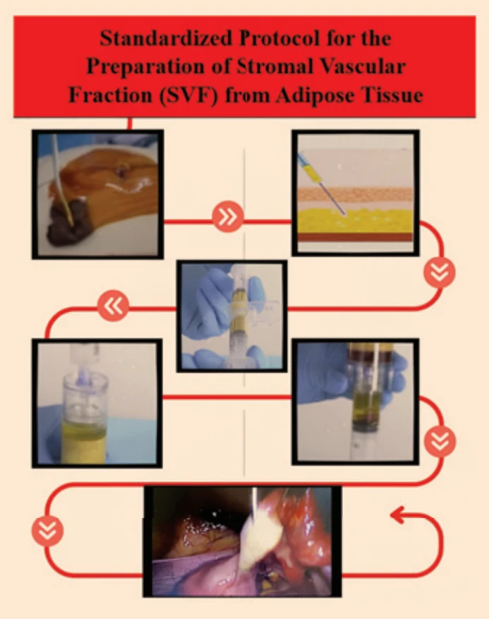
Fat aspiration was performed from the patient’s abdominal region. Patients with an antral follicle count of < 5 on transvaginal ultrasonography were included in the study. Exclusion criteria included prior ovarian surgery, active cancer, and autoimmune diseases requiring immunosuppression.
Preparation of adipose-derived stem cell culture
Adipose tissue samples were mechanically minced into small fragments before processing. This step was omitted when adipose-derived stem cells (ASCs) were obtained from lipoaspirates.
After this period, the culture medium is removed and the ASCs are left attached to the plate bottom. Iliac-crest aspirates (60 mL) were processed with Ficoll to isolate mononuclear cells. MSCs were culture-expanded to passage 1 and confirmed by surface markers (CD73+/CD90+/CD105+/HLA-DR–) with > 95% viability.
Intervention
A total of 1 × 10⁶ MSCs were infused into the four cortical quadrants of each ovary via laparoscopic surgery performed under general anesthesia. All patients were discharged the same day without complications. Following controlled ovarian stimulation (COS), the patients underwent oocyte retrieval two months after the injection (Fig. 1).
Follow-up schedule
To evaluate the safety and efficacy of MSCs injections in the participants included in the study, their medical history, vital signs, blood chemistry, physical examination reports, vaginal ultrasonography, and ovarian function tests were reviewed at baseline and at a 2-month follow-up.
Ovarian stimulation in participants
Ovarian stimulation in participants was performed using a GnRH antagonist protocol. After injection of recombinant human FSH (GONAL-f; Merck Serono, Germany, 300 IU) and Human Menopausal Gonadotropin (HMG) (Menotropins; Karma, Germany,150 IU), ovarian follicle size and assessment were evaluated by vaginal ultrasound. When the primary follicle reached 14 mm, GnRH antagonists (Cetrotide; Merck Serono, Germany) were administered, and their effects were monitored by vaginal ultrasound (Honda 2000 – 5 MHz, Japan). 0.25 mg was administered subcutaneously daily until the follicle size reached at least 18 mm. Thirty-six hours after intramuscular administration of 10,000 IU HCG (CETROTIDE 250 mcg (Merck Serono, Germany), egg retrieval was performed under vaginal ultrasound guidance. Embryo transfer (ET) was performed on the 3rd–5th day following egg retrieval. If the endometrium was unsuitable for transfer, embryos were frozen and transferred during a frozen embryo transfer cycle. Prior to ET, 400 mg PROGESTAN 200 mg (Koçak Farma, Turkey) was administered vaginally twice daily and continued.
Statistical analysis
We used IBM SPSS Statistics for Windows, version 26.0 (IBM Corp., Armonk, NY, USA) and JASP software (version 0.19.3; University of Amsterdam, The Netherlands) to do the statistical analyses. The analyses were performed on a Windows operating system. We used histograms and the Shapiro-Wilk test to check if continuous variables were normal. For variables that were normally distributed, descriptive statistics were shown as mean ± standard deviation (SD). For variables that were not normally distributed, they were shown as median (Min.–Max.). We used frequencies and percentages to sum up categorical variables. Wilcoxon signed-rank test was used to compare serum anti-Müllerian hormone (AMH) levels before and after stem cell therapy because AMH levels did not follow a normal distribution. We used a multivariable linear regression analysis to look at the factors that are linked to changes in AMH levels (DAMH). The model incorporated demographic characteristics, hormonal parameters, and pertinent clinical variables grounded in clinical significance. The results of the regression were given as regression coefficients (b) with standard errors (SE) and p values. Binomial logistic regression was used to examine the factors that increase the likelihood of obtaining egg cells after stem cell therapy. Due to the small sample size and the existence of extreme coefficient estimates, predictor significance was assessed using likelihood ratio tests instead of odds ratios. Log-odds coefficients, likelihood ratio Chi-square (c²) statistics, degrees of freedom, and p values were used to report the results. We didn’t show odds ratios because they could be misinterpreted because of the small sample size and the way the variables were scaled. We made predicted probability plots to show how changes in AMH levels (DAMH) are related to the chance of getting a positive oocyte retrieval. We used two-tailed tests for all of our statistical analyses, and we set the level of statistical significance at P < 0.05.
Results
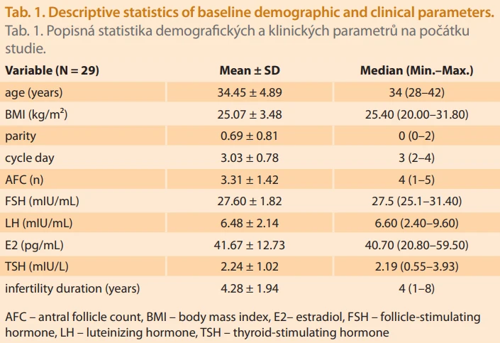
The final analysis included 29 patients. Tab. 1 shows the basic demographic and clinical information about the study group. The average age of the patients was 34.45 years, and the average body mass index (BMI) was 25.07 kg/m². At the start, most patients had a low ovarian reserve, with a median antral follicle count (AFC) of 4 (range, 1–5). The median duration of infertility was 4 years (range, 1–8), indicating a cohort with prolonged infertility.
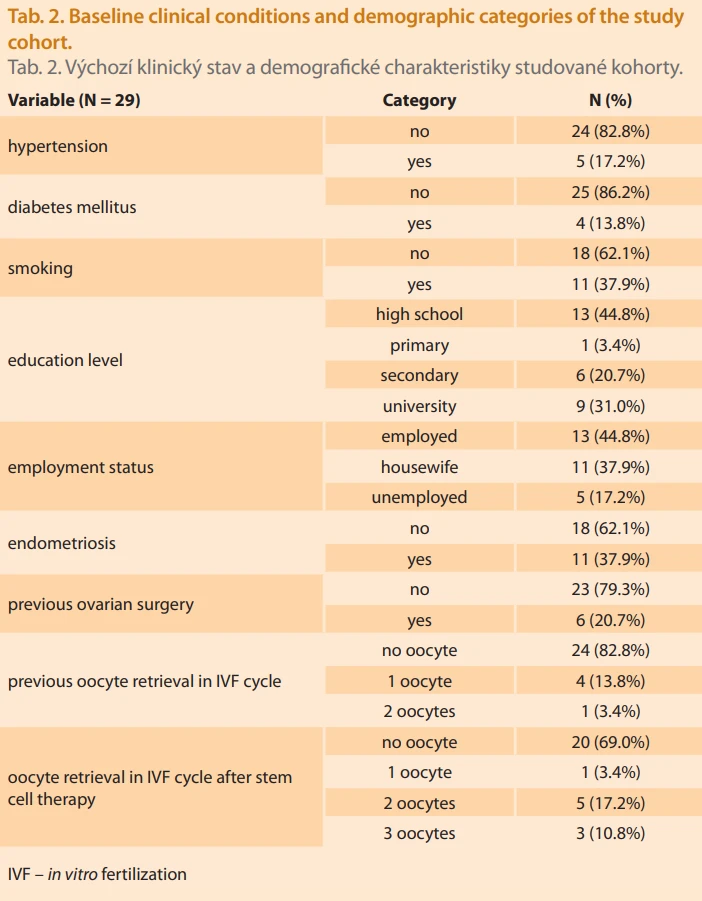
Tab. 2 shows the baseline clinical conditions and categorical demographic variables. Seventeen percent of patients had high blood pressure, and thirteen percent had diabetes. 37.9% of the group said they smoked. Endometriosis was diagnosed in 37.9% of patients, and 20.7% had a history of prior ovarian surgery. In terms of IVF outcomes, 82.8% of patients did not have any oocytes retrieved before stem cell therapy. After treatment, this number dropped to 69.0%. Notably, the number of patients obtaining one or more oocytes increased after treatment, indicating a clinically significant enhancement in ovarian response.
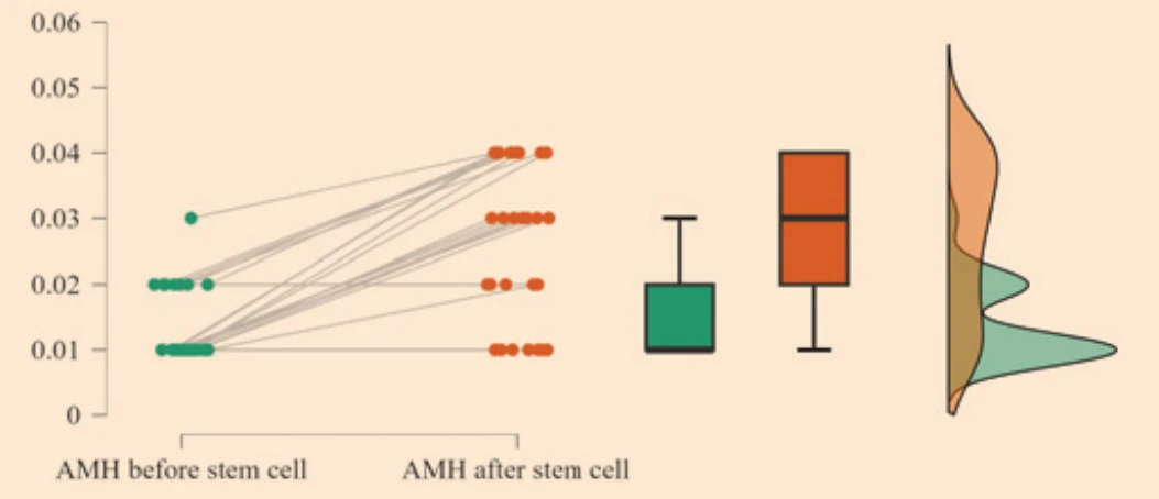
Tab. 3 shows the difference in serum AMH levels before and after stem cell therapy. The average AMH levels went up from 0.0138 ± 0.0056 before treatment to 0.0266 ± 0.0123 after treatment. This is about twice as much. This change was statistically significant (P = 0.001). Fig. 2 shows the individual AMH levels of each person before and after treatment. It also shows that the overall AMH levels in the group went up after stem cell therapy.

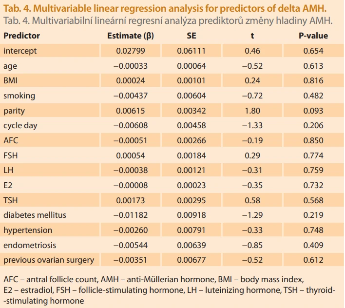
To investigate potential determinants of AMH enhancement, factors correlated with variations in AMH levels (DAMH) were assessed through multivariable linear regression (Tab. 4). None of the demographic, hormonal, or clinical variables analyzed – including age, BMI, smoking status, AFC, baseline gonadotropin levels, thyroid function, or comorbid conditions – demonstrated an independent association with DAMH (all P > 0.05). There was a tendency for parity to be positively related to DAMH, but this was not statistically significant (b = 0.00615, P = 0.093).
We used binomial logistic regression (Tab. 5) to look into what factors make it more likely that oocyte retrieval will be successful after stem cell therapy. Using likelihood ratio testing, DAMH was the only variable that was strongly linked to positive oocyte retrieval (c² = 6.54, df = 1, P = 0.011). Conversely, age, BMI, AFC, and the existence of diabetes mellitus, hypertension, or endometriosis did not show a significant correlation with oocyte retrieval outcomes.
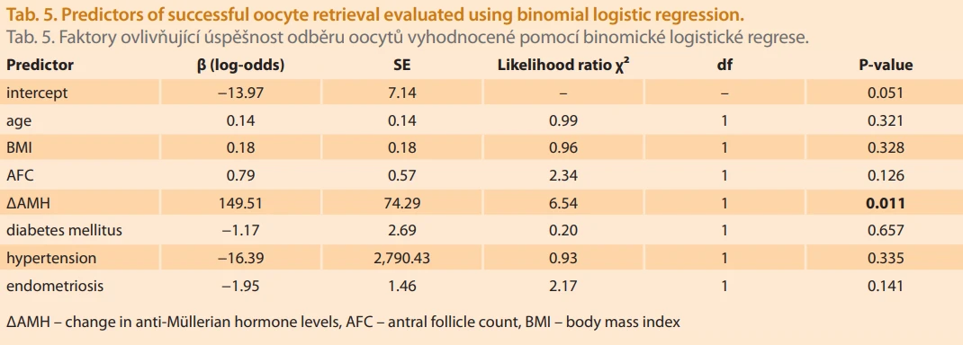
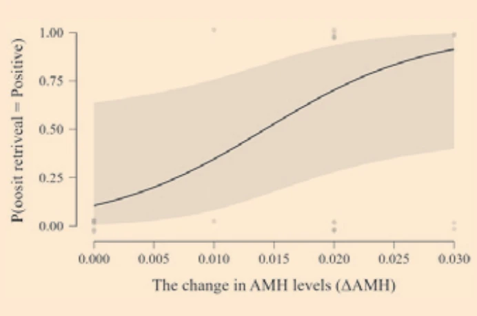
Fig. 3 shows how DAMH is related to the chance of getting a positive oocyte retrieval. Higher DAMH values were linked to a steady and gradual increase in the predicted likelihood of successful oocyte retrieval. This means that bigger improvements in AMH levels after treatment led to better IVF results.
Discussion
This study investigated the changes in AMH levels and the likelihood of oocyte retrieval following autologous adipose-derived MSCs therapy in women diagnosed with premature ovarian failure (POI) undergoing IVF. The key findings highlight a statistically significant increase in serum AMH levels and a modest but clinically significant improvement in oocyte retrieval outcomes following MSCs treatment. Furthermore, it was found that change in AMH (DAMH), rather than demographic or clinical variables in patients, was the only variable significantly inked to oocyte retrieval success. However, while these results suggest that MSCs treatment may have a potential biological effect on ovarian function, they should be interpreted cautiously due to various methodological limitations.
Research on adult stem cells has concentrated on mesenchymal and hematopoietic adult stem cells. Mesenchymal stem cells, relative to tissue cellular, have a very high development ability. On the other hand, they have the potential to differentiate themselves when they are cultivated in vitro to other cells, including osteocytes, adipocytes, and chondrocytes [13].
Damous et al. showed that adipose tissue–derived stem cell therapy enhanced ovarian graft quality by increasing vascular endothelial growth factor-A (VEGF-A) gene expression and blood vessel density in ovarian tissue, leading to earlier restoration of function in newly transplanted ovaries of adult female rats [14]. Hirakawa et al. investigated the effects of adipose tissue-derived MSCs on ovarian function in aged mice. In addition to evaluating their impact on ovulation rate, fertilization rate, and blastocyst formation, the authors measured ovarian follicle counts and serum AMH levels. The MSCs exhibited characteristic mesenchymal stem cell properties and were shown to distribute to various tissues, including the ovarian stroma. Following transplantation, oocyte numbers and ovulation increased, accompanied by elevated AMH levels. Genetic analyses further demonstrated improvements in oocyte quality, as well as increased fertilization and blastocyst formation rates [15].
Cacciottola et al., in one of the recent reviews, sought to address the question of whether MSCs infusion or localized MSCs therapy can contribute to the repair of injured ovaries. Upon reviewing the available literature, they observed that various types of MSCs have been tested with the aim of improving fertility. The studies predominantly comprised several animal experiments and a limited number of human trials. The authors concluded that, in addition to encouraging follicular survival and growth, MSCs may reverse chemotherapy-induced ovarian damage and improve the survival of the follicular pool by promoting revascularization of ovarian tissue [16]. In another experimental study conducted by the same group of researchers, immunodeficient mice were transplanted with adipose tissue–derived stem cells and human ovarian tissue, and blood samples were collected monthly for six months. Four months after transplantation, a significant increase in plasma AMH and estradiol levels was observed in the MSCs treated group. The long-term profit of preserving ovarian reserve and promoting follicular development have been demonstrated. In our study, the primary focus was on improvement in AMH levels and an increase in oocyte yield. Accordingly, a statistically significant response was observed in both parameters. These findings may be interpreted as evidence that MSCs support follicular survival and growth.
One of the primary limitations of this study is its retrospective pre–post design without a parallel control group. Although spontaneous recovery of ovarian function in POI is considered rare, it is well recognized that a small subset of patients may exhibit intermittent follicular activity and temporal hormonal fluctuations. In the absence of a control arm, it is not possible to definitively exclude the contribution of regression to the mean, natural hormonal variability, or spontaneous follicular activation to the observed increase in AMH levels. Therefore, establishing a clear causal relationship between MSCs administration and ovarian response may not be possible.
Second, the small sample size substantially limits the statistical power and the precision of effect estimates. The clinical significance of the increase in AMH levels should also be interpreted with caution. Despite a statistically significant increase in post-treatment AMH levels, these levels were still below the criteria usually associated with normal ovarian reserve. Therefore, this increase should not be interpreted as a restoration of normal ovarian function, but rather as partial activation or improved survival of a limited number of residual follicles. This distinction is especially crucial for managing clinical expectations about MSC-based therapies and for patient counseling. Another important limitation concerns the nature of the outcome measures. This study primarily focused on intermediate and surrogate endpoints, such as AMH levels and oocyte retrieval, and did not assess definitive reproductive outcomes, including embryo development, clinical pregnancy, or live birth. Although oocyte retrieval represents a critical step for patients seeking genetically related pregnancies, it does not, on its own, guarantee reproductive success. The absence of long-term reproductive outcomes therefore limits inferences regarding the true clinical benefit of MSC therapy.
The biological heterogeneity of POI represents another factor that complicates interpretation. POI encompasses a broad etiological spectrum, including genetic, autoimmune, iatrogenic, and idiopathic causes, and these subgroups may respond differently to regenerative therapies. Although several clinical variables were incorporated into the regression models, the study lacked sufficient statistical power to perform meaningful subgroup analyses. Consequently, it remains unclear whether specific patient subgroups derive greater or lesser benefit from MSC therapy.
In addition, this study does not provide direct evidence regarding the mechanisms by which MSC therapy may influence ovarian function. While previous experimental studies have suggested that MSCs exert their effects predominantly through paracrine, angiogenic, and anti-apoptotic pathways, no molecular, histological, or imaging data were obtained in the present study to support these mechanisms. Therefore, mechanistic interpretations should be regarded as hypothesis-based and derived from preclinical literature rather than direct evidence.
Despite these limitations, the study has several notable strengths. The cohort represents a well-defined POI population with profoundly diminished ovarian reserve and very limited therapeutic options. The consistent temporal association between MSC treatment and improvements in AMH levels and oocyte retrieval, along with the strong association between DAMH and the probability of oocyte retrieval, suggests the presence of a signal warranting further investigation, rather than immediate translation into clinical practice.
In conclusion, these findings should be considered exploratory and hypothesis-generating. MSC therapy may be associated with partial biochemical and functional ovarian responses in selected patients with POI; however, definitive conclusions regarding efficacy, durability, and clinical benefit cannot yet be drawn.
Conclusion
In summary, this study provides preliminary evidence that autologous mesenchymal stem cell therapy may be associated with modest biochemical and functional ovarian responses in women with POI. The observed increase in serum AMH levels and its association with oocyte retrieval probability suggest a potential, albeit limited, impact on residual ovarian activity rather than restoration of normal ovarian function. Larger, prospective, controlled studies incorporating mechanistic assessments and definitive reproductive endpoints are required to clarify the clinical relevance, durability, and safety of stem cell-based interventions in this challenging patient population.
Submitted/Doručeno: 8. 2. 2026
Accepted/Přijato: 10. 2. 2026
Assoc. Prof. Fatih Akkuş, MD
Department of Obstetrics and Gynecology
Division of Perinatology
Kütahya City Hospital
Evliya Çelebi, Eken Paşa Cd. No: 19
43100 Kütahya
Turkey
fakkus1987@gmail.com
Zdroje
1. Visser JA, Schipper I, Laven JS et al. Anti-Müllerian hormone: an ovarian reserve marker in primary ovarian insufficiency. Nat Rev Endocrinol 2012; 8 (6): 331–341. doi: 10.1038/nrendo. 2011.224.
2. Ferraretti A, La Marca A, Fauser BC et al. ESHRE consensus on the definition of ‘poor response’ to ovarian stimulation for in vitro fertilization: the Bologna criteria. Hum Reprod 2011; 26 (7): 1616–1624. doi: 10.1093/humrep/der092.
3. Kalantari H, Madani T, Moradi SZ et al. Cytogenetic analysis of 179 Iranian women with premature ovarian failure. Gynecol Endocrinol 2013; 29 (6): 588–591. doi: 10.3109/09513590.2013.788625.
4. Nelson LM. Clinical practice. Primary ovarian insufficiency. N Engl J Med 2009; 360 (6): 606–614. doi: 10.1056/NEJMcp0808697.
5. Luborsky J, Meyer P, Sowers MF et al. Premature menopause in a multi‐ethnic population study of the menopause transition. Hum Reprod 2003; 18 (1): 199–206. doi: 10.1093/humrep/deg005.
6. van Kasteren YM, Schoemaker J. Premature ovarian failure: a systematic review on therapeutic interventions to restore ovarian function and achieve pregnancy. Hum Reprod Update 1999; 5 (5): 483–492. doi: 10.1093/humupd/5.5.483.
7. Kim HK, Kim TJ. Current status and future prospects of stem cell therapy for infertile patients with premature ovarian insufficiency. Biomolecules 2024; 14 (2): 242. doi: 10.3390/biom14020242.
8. Fàbregues F, Ferreri J, Méndez M et al. In vitro follicular activation and stem cell therapy as anovel treatment strategies in diminished ovarian reserve and primary ovarian insufficiency. Front Endocrinol (Lausanne) 2021; 11 : 617704. doi: 10.3389/fendo.2020.617704.
9. Petrou P, Kassis I, Levin N et al. Beneficial effects of autologous mesenchymal stem cell transplantation in active progressive multiple sclerosis. Brain 2020; 143 (12): 3574–3588. doi: 10.1093/brain/awaa333.
10. Herraiz S, Buigues A, Diaz-Garcia C et al. Bone marrow derived stem cells promote vascularization and follicular growth in xenografted human ovarian cortex. Canada, Montreal: SRI 63rd Annual Scientific Meeting 2016, Society for Reproductive Investigation.
11. Sanchez-Mata A, Gonzalez-Muñoz E. Understanding menstrual blood-derived stromal/stem cells: definition and properties. Are we rushing into their therapeutic applications? IScience 2021; 24 (12): 103501. doi: 10.1016/j.isci.2021.103501.
12. Zafardoust S, Kazemnejad S, Fathi-Kazerooni M et al. The effects of intraovarian injection of autologous menstrual blood-derived mesenchymal stromal cells on pregnancy outcomes in women with poor ovarian response. Stem Cell Res Ther 2023; 14 (1): 332. doi: 10.1186/s13287-023-03568-1.
13. Zhao YX, Chen SR, Su PP et al. Using mesenchymal stem cells to treat female infertility: an update on female reproductive diseases. Stem Cells Int 2019; 2019 : 9071720. doi: 10.1155/2019/9071720.
14. Damous LL, Nakamuta JS, Saturi de Carvalho AE et al. Does adipose tissue-derived stem cell therapy improve graft quality in freshly grafted ovaries? Reprod Biol Endocrinol 2015; 13 (1): 108. doi: 10.1186/s12958-015-0104-2.
15. Hirakawa T, Yotsumoto F, Imi S et al. Adipose-derived mesenchymal stem cell transplantation improves ovarian function and oocyte quality in aged mice. Anticancer Res 2024; 44 (8): 3577–3586. doi: 10.21873/anticanres.17180.
16. Cacciottola L, Vitale F, Donnez J et al. Use of mesenchymal stem cells to enhance or restore fertility potential: a systematic review of available experimental strategies. Hum Reprod Open 2023; 2023 (4): hoad040. doi: 10.1093/hropen/hoad040.
Štítky
Dětská gynekologie Gynekologie a porodnictví Reprodukční medicínaČlánek vyšel v časopise
Česká gynekologie
2026 Číslo 4
- Vědecké poznatky rozptylující obavy spojené s používáním nitroděložní antikoncepce i u mladých žen
- Používání intrauterinních tělísek uvolňujících měď a riziko tubární infertility u nuligravidních žen
- Účinnost dlouhodobě působící reverzibilní antikoncepce
- Antikoncepční pilulka přestává být nejpřitažlivější metodou antikoncepce
- Nitroděložní systém s levonorgestrelem by mohl snižovat riziko adenokarcinomu endometria
Nejčtenější v tomto čísle
- Srovnání diagnostické přesnosti ambulantní hysteroskopie a trojrozměrného transvaginálního ultrazvukového vyšetření s histopatologickým vyšetřením u žen s abnormálním děložním krvácením
- 150 let porodnice u Apolináře v Praze – historie jedné z nejdéle nepřetržitě fungujících porodnic na světě
- Personalizovaný přístup k léčbě sterility u pacientky s polyendokrinním metabolickým ovariálním syndromem a chronickou pankreatitidou
- Ultrazvukové vyšetrenie počas pôrodu – prospektívna pilotná komparatívna štúdia presnosti vaginálneho a ultrasonografického vyšetrenia
Zvyšte si kvalifikaci online z pohodlí domova
Mazová zátka a její řešení
nový kurzVšechny kurzy