
Measurement of vaginal pressure profile during horseback riding and its effect on pelvic floor function in a woman with stress urinary incontinence: a case report
Meranie vaginálneho tlakového profilu počas jazdy na koni a jeho vplyv na funkciu panvového dna u ženy s únikom moču: prípadová štúdia
Únik moču pri fyzickej aktivite sa vyskytuje najmenej raz týždenne u jednej tretiny dospelých žien. Ďalšie štúdie preukázali, že jazda na koni zlepšuje funkčný stav svalov panvového dna v pokoji aj pri fyzickej aktivite. Zvyšuje aj pokojový tonus svalov panvového dna. Doposiaľ nebola realizovaná žiadna štúdia, v ktorej by bol hodnotený vaginálny tlakový profil pomocou Femfitu® počas rôznych typov jazdy na koni. Nebol hodnotený vplyv jazdenia na funkciu panvového dna u ženy s miernymi príznakmi stresovej inkontinencie moču (SUI – stress urinary incontinence). Táto prípadová štúdia opisuje 46-ročnú ženu so SUI, ktorá sa venovala rekreačnej jazde na koni počas 12 týždňov. Boli pozorované významné rozdiely medzi cvalom a všetkými ostatnými typmi jazdy, pričom najvyššie hodnoty boli zaznamenané počas cvalu. Vyšetrenie funkcie panvového dna bolo realizované (pomocou manometra, 2D/3D USG) . Bolo zistené zvýšenie maximálnej vôľovej kontrakcie z 27,5 (1,4) na 45,7 (2,2) cm H2O a jej trvania z 5 (0,2) na 8 (0,4) s. Počas kontrakcie bolo zaznamenané zmenšenie urogenitálneho hiatu (z 16,5 (0,8) na 15,0 (0,7) cm2) a počas Valsalvovho manévra z 19,6 (0,9) na 18,8 (0,9) cm2. Jazda na koni s aktiváciou svalov panvového dna pozitívne zlepšila funkciu svalov panvového dna a znížila príznaky úniku moču. Bolo by prospešné vykonať podobné merania na väčšej skupine jazdkýň.
Klíčová slova:
stresová inkontinencia moču – tréning svalov panvového dna – vaginálny tlakový profil – jazda na koni
Authors:
M. Hagovská 1; A. Buková 2; J. Svihra 3
Authors place of work:
Department of Physiatry, Balneology and Medical Rehabilitation, Faculty of Medicine, PJ Safarik University, Kosice, Slovak Republic
1; Institute of Physical Education and Sport, PJ Safarik University, Kosice, Slovak Republic
2; Department of Urology, Jessenius Faculty of Medicine, Comenius University Bratislava, Martin, Slovak Republic
3
Published in the journal:
Rehabil. fyz. Lék., 105, 2025, No. 2, pp. 88-94.
Category:
Kazuistika
doi:
https://doi.org/10.48095/ccrhfl202588
Summary
Stress urinary incontinence (SUI), the complaint of involuntary leakage during effort or exertion, occurs at least weekly in one third of adult women. Additional studies have demonstrated that horseback riding promotes pelvic floor training in women with stress incontinence, leading to increased resting pelvic floor tension and stronger tension during active pelvic floor muscle contraction. No study to date has measured the vaginal pressure profile using FemFit® during different types of horseback riding or assessed its effects on pelvic floor function in a woman with mild symptoms of SUI. This case report describes a 46-year-old woman with SUI who practiced recreational horseback riding over a period of 12 weeks. Significant differences were observed between the gallop and all other types of riding, with the highest values recorded during the gallop. Examination of pelvic floor muscle function (via manometer, 2D/3D ultrasound using volume contrast imaging software) revealed an increase in maximal voluntary contraction from 27.5 (1.4) to 45.7 (2.2) cm H2O and its duration from 5 (0.2) to 8 (0.4) s. A decrease in the hiatal area was noted during contraction (from 16.5 (0.8) to 15.0 (0.7) cm2) and during the Valsalva maneuver from 19.6 (0.9) to 18.8 (0.9) cm2. Horseback riding with pelvic floor muscle activation positively improved pelvic floor muscle function and reduced urinary leakage symptoms. It would be beneficial to perform similar measurements on larger groups of female horse riders.
Keywords:
stress urinary incontinence – pelvic floor muscle training – vaginal pressure profile – horse riding

Introduction
Urinary incontinence (UI) is an involuntary, objectively demonstrable leakage of urine, which is unhygienic and a social problem. The most common types of UI are stress, urgency, and mixed urinary incontinences [1]. Additional studies have demonstrated that horseback riding promotes pelvic floor training in women with stress incontinence, leading to increased resting pelvic floor tension and stronger tension during active pelvic floor muscle contraction.
In recent years, interest in horseback riding as a leisure and recreational activity for women has increased. Horseback riding is a physical activity with moderate exertion, classified at 3–6 metabolic equivalents, with aerobic costs rising as the horse’s speed increases [2]. Riding positively influences gross and fine motor skills, optimizes whole-body muscle tone, improves posture, and activates the deep stabilization system. It also has significant benefits for the static, anticipatory, and dynamic components of balance [3–6].
The slowest movement of a horse is known as horse walking. During horse walking, the rider’s body experiences a swinging motion, and the horse’s average stride speed is 100 m/min [7]. The trot is a faster movement than horse walking. During a trot, a horse typically reaches a speed of 290 m/min. The gallop is a fast form of horse movement. It typically occurs at a speed of 300–600 m/min [7].
Electromyography (EMG) biofeedback studies using a vaginal probe have shown that pelvic floor tension in women increases as the horse’s speed increases. The highest tension was recorded during a gallop in the two-point position; slightly lower tension was observed in the sitting position, and the tension returned to resting state when the horse stopped [8,9]. Based on these findings, it has been hypothesized that horseback riding positively affects the prevention of pelvic floor dysfunction in women [10,11].
No study to date has measured the vaginal pressure profile using FemFit® during different types of horseback riding or assessed its effects on pelvic floor function in a woman with mild symptoms of SUI [12].
The primary aim of this case study was to measure changes in the vaginal pressure profile using FemFit® during horseback riding. The secondary aim was to quantify changes in pelvic floor function following the horseback riding programme in a woman with mild symptoms of SUI.
Case presentation
A 46-year-old woman with mild symptoms of SUI began practicing recreational horseback riding. She weighed 53 kg, was 160 cm tall, and had two prior vaginal deliveries. She reported experiencing mild SUI symptoms for 5 years without worsening.
he experienced urinary leakage, on average, twice a week while walking. The amount of urine leakage was minimal. The impact on her quality of life was assessed as a visual analogue scale score of 4. The International Consultation on Incontinence Questionnaire-Urinary Incontinence Short Form (ICIQ-UI SF) score was 1 + 0 + 4 = 5.
The woman had a sedentary occupation. She exhibited a sagging posture when sitting and standing. Other kinesiological findings were within normal limits. As part of her leisure routine, she walked five times a week for 60 min. She had no other serious illness and did not participate in any other sporting activities.
Prior to this intervention, she had no experience riding horses. The horseback riding sessions were conducted twice a week, each lasting 45 min, over a 12-week period. The horse used was a Lipizzaner, and the participant rode in a hard saddle.
Methodology
New intervention – the 12-week horseback riding program with endurance contractions of the pelvic floor muscles
During horseback riding, repeated endurance contractions of the pelvic floor muscles were performed. These included exercises during the horse walk, walking ride, trot with a balanced seat, fast trot with a rising seat, and gallop. The exercises involved 5–8 s of submaximal voluntary pelvic floor muscle contractions, followed by 5 s of relaxation.
New examination – vaginal pressure profile measurement during horseback riding.
Intravaginal pressure (IVP) profile measurement using the FemFit®
The FemFit® (version 2.3.1) is a pressure sensor array designed to measure pressures along the vaginal length (Fig. 1).
It contains eight pressure sensors that enable measurement of the vaginal pressure profile, from the apex of the vagina to the introitus. The pressure sensors are encapsulated in soft, biocompatible silicone (MED-4901; NuSil).
The device has a total length of 80 mm, a maximum width of 24 mm, and a thickness of only 4 mm. Pressure data from FemFit® are transmitted via Bluetooth to an Android mobile phone for data logging, real-time display, and user feedback. Each pressure sensor operates at a sampling frequency of 40 Hz [13–15].
During the horseback riding sessions, the freestyle program available in the FemFit® app was used as follows. During the first testing session, we performed 4 types of riding: the horse walk (walking ride) for 110 s, the trot (balanced seat) for 120 s, the fast trot (rising trot) for 120 s, and the gallop for 120 s.
The described measurements were taken both before and after the 12-week horseback riding program.
Assessment of functional status of the pelvic floor muscles using a perineometer (Peritron-Ontario, L4V, Canada)
Pelvic floor muscle strength, endurance, rapid contraction, and relaxation were assessed using a perineometer, which measures muscle function in units of water column height (cm H2O). Contraction strength was categorized as follows: up to 10 cm H2O was considered weak, 10–30 cm H2O was moderate, and 40–60 cm H2O was strong. Correct resting tone calibration was crucial, with saturation set to 100 cm H2O after stabilization [16].
Assessment of functional status of the pelvic floor muscles via perineal 2D and 3D/4D ultrasound
The examination was conducted using a Voluson-i BT 11 Console with volume contrast imaging software (GE Healthcare Austria GmbH & Co OG, Zipf, Austria) and a RAB4-8-RS 3D/4D probe (4–8 MHz). Both 2D and 3D/4D images were captured at rest, during maximal contraction, and during the Valsalva maneuver.
Examination with 2D USG: Measurements included the anteroposterior and laterolateral dimensions of the hiatus (in cm). Urethral mobility and anorectal angle were also assessed.
Examination with 3D USG: Measurements included the hiatus area (in cm2). Examinations were performed by a trained urogynecologist in collaboration with a physiotherapist [17].
Assessment of symptoms of urinary incontinence
The International Consultation on Incontinence Questionnaire-Urinary Incontinence Short Form (ICIQ-UI SF) was developed by the International Continence Society. The first two questions assess the frequency and amount of urine leakage, while the third evaluates how much the leakage interferes with patients’ daily lives. The ICIQ-UI SF score is calculated by adding the individual question scores, which are categorized as follows: 0 = no leakage, 1–5 = mild leakage, 6–12 = moderate leakage, 13–18 = severe leakage, and 19–21 = very severe leakage. The Cronbach’s alpha for the ICIQ-UI SF is 0.95, indicating excellent reliability [18].
Questionnaire description:
Answer options:
- never – 0
- about once a week or less often – 1
- two or three times a week – 2
- about once a day – 3
- several times a day – 4
- all the time – 5
Answer options:
- none – 0
- a small amount – 2
- a moderate amount – 4
- a large amount – 6
- Answer: a number between 0 (not at all) and 10 (a great deal)
The ICIQ score is the sum of scores from questions 1, 2, and 3.
Results of examinations before and after the intervention
ICIQ-UI SF
After completing the 12-week horseback riding programme, which included two sessions per week, the participant’s score on the ICIQ-UI SF decreased from 5 to 2, indicating an improvement in symptoms. She experienced no urinary leakage during walking. The impact on quality of life remained at a score of 2. However, she expressed concern about a possible recurrence of symptoms. The ICIQ-UI SF score was 0 + 0 + 2 = 2.
Manometer examination
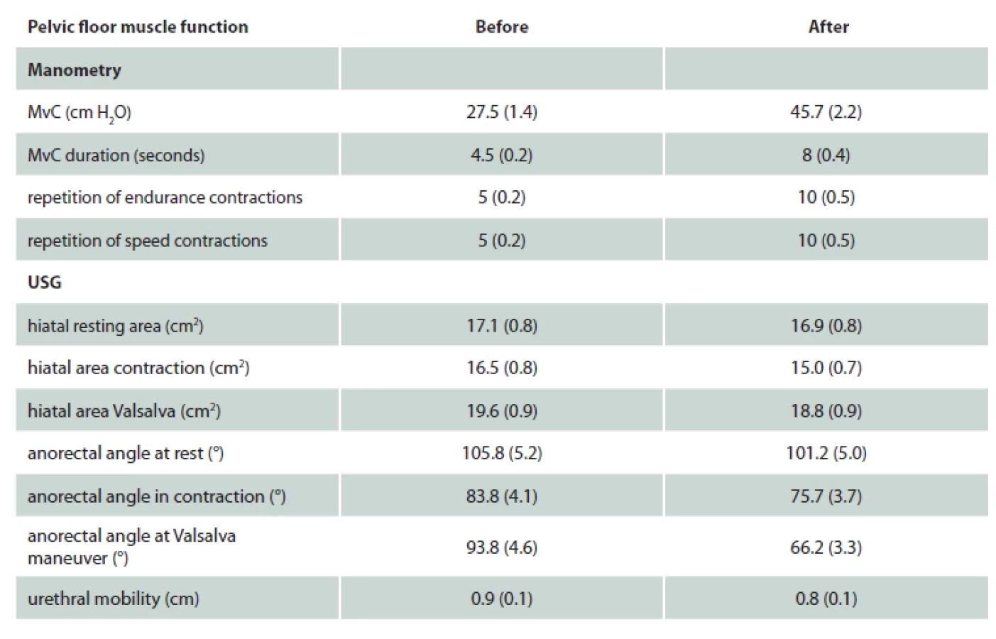
At the end of the 12-week horseback riding programme, the participant’s maximal voluntary contraction (MvC) value increased from 27.5 to 45.7 cm H2O. The duration of MvC also improved, rising from 4.5 to 8 s, as did the number of repetitions of all types of contractions, which increased from 5 to 10.
2D and 3D/4D ultrasound examinations
At the end of the 12-week horseback riding programme, ultrasound examination showed the following reductions in hiatal area compared with the initial assessment: from 17.1 to 16.9 cm2 at rest, from 16.5 to 15.0 cm2 during contraction, and from 19.6 to 18.8 cm2 during the Valsalva maneuver. Similarly, the participant’s anorectal angle showed the following decreases: from 105.8° to 101.2° at rest, from 83.8° to 75.7° during contraction, and from 93.8° to 66.2° during the Valsalva maneuver. Additionally, urethral mobility decreased from 0.9 to 0.8 cm over the 12 weeks (Tab. 1).
Statistical analysis of data from eight FemFit ® sensors
IVPs were recorded from the first to sixth sensors, while abdominal pressure was recorded from the seventh and eighth sensors, both measured in mmHg. FemFit® recorded 40 measurements per second, and an average for each second was calculated based on the difference between the minimum and maximum pressure values in mmHg. Data are presented as a mean with standard deviation. Statistical significance was assessed using the t-test and analysis of variance (ANOVA). The significance level was set at P < 0.05.

Results
IVP profile using FemFit®
We assessed the participant’s IVP profile using the FemFit® device during horseback riding, with measurements taken before and after the 12-week intervention in the following positions: walking riding, balanced seat, rising trot, and gallop. The lowest pressure values were recorded during walking riding, higher values during the balanced seat, even higher values during the rising trot, and the highest values during the gallop.
Significant differences in IVP profile scores were observed between the gallop and all other riding positions, with the highest values recorded during the gallop.
In comparisons before and after the 12-week programme, significantly higher IVP profile values were recorded during the balanced seat from sensors 1–5 and during the rising trot from sensors 3–4, and during the gallop from sensors 1–6. However, no significant changes were observed during walking riding after 12 weeks of riding. Additionally, there were no changes in intra-abdominal pressure (IAP) from sensors 7–8 during the study (Tab. 2, Fig. 1).
Discussion
The primary aim of this case study was to measure changes in the vaginal pressure profile using FemFit® during horseback riding. The secondary aim was to quantify changes in pelvic floor function following the horseback riding programme in a woman with mild symptoms of SUI. We examined changes in vaginal pressure profiles using the FemFit® device during horseback riding with pelvic floor muscle activation. Measurements were taken before and after a 12-week program in the walking riding position, balanced seat position, rising trot position, and gallop. IPV values were lowest during walking riding, higher during the balanced seat, much higher during the rising trot, and highest during the gallop.
In comparisons before and after the 12-week programme, significantly higher IVP profile values were recorded during the balanced seat from sensors 1–5, during the rising trot from sensors 3–4, and during the gallop from sensors 1–6.
We examined changes in pelvic floor function after the horseback riding programme:
Manometry examination revealed increases in the MvC value (indicating improved pelvic floor muscle strength), vC duration (indicating improved muscle endurance), and number of repetitions performed across all types of contractions (indicating enhanced ability to perform rapid contractions).
Ultrasound examination demonstrated reductions in hiatal space at rest, during contraction, and during the Valsalva maneuver. Additionally, decreases were observed in anorectal angle values and urethral mobility over the 12-week period. These changes suggest an improved ability to manage load in the pelvic floor muscle area and better coordination of the pelvic floor muscles.
Before the intervention, the participant experienced urinary leakage on average twice a week during walking. The amount of leakage was minimal. The impact on quality of life was rated at 4.
After 12 weeks of regular horseback riding, no urinary leakage during walking was recorded. The impact on quality of life remained at 2, although she continued to express concern about a potential recurrence of symptoms.
Säfer et al. [9] measured pelvic floor muscle activity using EMG biofeedback during horseback riding in 14 healthy women. Horseback riding was performed and measured during walking, trot, and gallop maneuvers, with the pelvis positioned in both sitting and thigh sitting postures. EMG biofeedback results in the tested women were comparable, and different riding paces showed significantly different EMG activity levels. The highest activity was recorded during the gallop in the sitting position (26–42 µV), while the lowest was observed during horse walking in the sitting position (5–12 µV). During the trot in the sitting and thigh seat positions, activity ranged from 16–30 µV. In our study, before the intervention, IVP during walking riding was 9.7 mmHg and IAP was 8.3 mmHg. In the balanced seat, IVP was 11.3 mmHg and IAP was 13.7 mmHg. At the rising trot, IVP reached 52.2 mmHg and IAP was 36.4 mmHg. During the gallop, the highest pressures were observed, with IVP at 105.7 mmHg and IAP at 43.4 mmHg.
Carboni et al. [10] examined the effects of horseback riding and pelvic floor muscle exercises in 12 women with an average age of 24 years. Horseback riding was performed for 30 min per week over 12 weeks, alongside pelvic floor muscle training, which included exercises for MvC, endurance, and relaxation of the pelvic floor muscles. The mean modified Oxford score (measured by vaginal palpation) increased from 2.74 before the intervention to 4.04 after the intervention, and the MvC manometry reading improved from 63.54 cm H2O before the intervention to 98.17 cm H2O after the intervention. Both differences were statistically significant. The combination of therapeutic horseback riding and pelvic floor muscle training appears effective in treating and preventing pelvic floor dysfunction. Similarly, in our study, we observed a positive effect of horseback riding on pelvic floor muscle function in a woman with SUI. Manometry results before and after the intervention showed an increase in the participant’s readings from 27.5 to 45.7 cm H2O.
Limitations and strengths
It would be beneficial to perform similar measurements on larger groups of female horse riders. The strengths of this study include the objective examination of the rider’s pelvic floor functional status before and after 12 weeks of riding, as well as the objective measurement of the IVP profile along the entire length of the vagina during horseback riding.
Conclusion
During horseback riding, involuntary activation of the pelvic floor muscles was lowest during walking, higher during the balanced seat, higher still during the rising trot, and highest during the gallop. In comparisons before and after the 12-week programme, significantly higher involuntary activation was recorded during the balanced seat, rising trot, and gallop.
Following the 12-week horseback riding programme, pelvic floor muscle strength and endurance had improved, and the ability to perform rapid contractions had increased. There was also a greater capacity to manage load in the pelvic floor muscle area and improved coordination of the pelvic floor muscles.
After 12 weeks of horseback riding, no urinary leakage was recorded.
These results suggest that recreational horseback riding involving pelvic floor muscle activation may positively influence pelvic floor muscle function and alleviate symptoms of SUI.
Acknowledgements
The authors wish to thank to Mr. Horse riding trainer for cooperation on the study.
Konflikt zájmů: Autoři deklarují, že text článku odpovídá etickým standardům, byla dodržena anonymita pacientů a prohlašují, že v souvislosti s předmětem článku nemají finanční, poradenské ani jiné komerční zájmy.
Publikační etika: Příspěvek nebyl dosud publikován ani není v současnosti zaslán do jiného časopisu pro posouzení. Autoři souhlasí s uveřejněním svého jména a e-mailového kontaktu v publikovaném textu.
Dedikace: Kultúrna a edukačná grantová agentúra Ministerstva školstva, výskumu, vývoja a mládeže Slovenskej republiky (KEGA)
Kultúrna a edukačná grantová agentúra MŠVVaM SR (KEGA) KEGA č. 002UPJŠ-4/2023-Nový edukačný model pre univerzitné študentky o vplyve nadváhy na dysfunkciu urogenitálneho systému.
Redakční rada potvrzuje, že rukopis práce splnil ICMJE kritéria pro publikace zasílané do biomedicínských časopisů.
Conflict of Interest: The authors declare that the article/manuscript complies with ethical standards, patient anonymity has been respected, and they state that they have no financial, advisory or other commercial interests in relation to the subject matter.
Publication Ethics: This article/manuscript has not been published or is currently being submitted for another review. The authors agree to publish their names and e-mails in the published article/manuscript.
Dedication: Cultural and Educational Grant Agency of the Ministry of Education, Research, Development and Youth of the Slovak Republic (KEGA)
Cultural and Educational Grant Agency of the Ministry of Education, Research, Development and Youth of the Slovak Republic (KEGA) No. 002UPJŠ-4/2023-New educational model for university students on the impact of overweight on urogenital system dysfunction.
The Editorial Board declares that the manuscript met the ICMJE “uniform requirements” for biomedical papers.
Zdroje
- Bø K, Sundgot-Borgen J. Are former female elite athletes more likely to experience urinary incontinence later in life than non-athletes? Scand J Med Sci Sports 2010; 20(1): 100–104. doi: 10.1111/j.1600-0838.2008.00871.x.
- Beale L, Maxwell NS, Gibson OR et al. Oxygen cost of recreational horse-riding in females. J Phys Act Health 2015; 12(6): 808–813. doi: 10.1123/jpah.2012-0428.
- Rahbar M, Salekzamani Y, Jahanjou F et al. Effect of hippotherapy simulator on pain, disability and range of motion of the spinal column in subjects with mechanical low back pain: A randomized single-blind clinical trial. J Back Musculoskelet Rehabil 2018; 31(6): 1183–1192. doi: 10.3233/BMR-170832.
- Rigby BR, Grandjean PW. The Efficacy of Equine-Assisted Activities and Therapies on Improving Physical Function. J Altern Complement Med 2016; 22(1): 9–24. doi: 10.1089/ acm.2015.0171.
- Cerulli C, Minganti C, De Santis C et al. Therapeutic horseback riding in breast cancer survivors: a pilot study. J Altern Complement Med 2014; 20(8): 623–629. doi: 10.1089/ acm.2014.0061.
- Hilliere C, Collado-Mateo D, Villafaina S et al. Benefits of Hippotherapy and Horse Riding Simulation Exercise on Healthy Older Adults: A Systematic Review. PM R 2018; 10(10): 1062–1072. doi: 10.1016/j.pmrj.2018.03.019.
- Clayton HM, Hobbs SJ. The role of biomechanical analysis of horse and rider in equitation science. Appl Anim Behav Sci 2017; 190 : 123–132. doi: 10.1016/j.applanim.2017.02.011.
- Wanless M. Rider Biomechanics. Stroud: Kenilworth Press 2017.
- Schäfer D, Pannek J. Measurement of pelvic floor function during physical activity: a feasibility study. Scand J Urol Nephrol 2009; 43(4): 315–318. doi: 10.1080/00365590902925022.
- Alanee S, Heiner J, Liu N et al. Horseback riding: impact on sexual dysfunction and lower urinary tract symptoms in men and women. Urology 2009; 73(1): 109–114. doi: 10.1016/j.urology.2008.07.058.
- Kruger J, Budgett D, Goodman J et al. Measuring the vaginal pressure profile during exercise. Australian & New Zealand Continence Journal 2018; 24(3): 85.
- Hagovská M, Bukova A, Svihra J. Measurement of the Vaginal Pressure Profile with the Femfit® and Leakage Events Using a Newly Developed Pad Test during Selected Sports Activities: A Pilot Study. Int Urogynecol J 2025; 36(4): 789–797. doi: 10.1007/s00192-025-06051-y.
- McConnell J, Murtagh L, Lim M et al. A pilot study to evaluate changes in pelvic floor muscle tone following pelvic organ prolapse surgery using a novel intra-vaginal pressure sensor device. Int Urogynecol J 2023; 34(5): 1043–1047. doi: 10.1007/s00192-022-05312-4.
- Cacciari LP, Kruger J, Goodman J et al. Reliability and validity of intravaginal pressure measurements with a new intravaginal pressure device: The FemFit®. Neurourol Urodyn 2020; 39(1): 253–260. doi: 10.1002/nau.24179.
- Rahmani N, Mohseni-Bandpei MA. Application of perineometer in the assessment of pelvic floor muscle strength and endurance: a reliability study. J Bodyw Mov Ther 2011; 15(2): 209 – 214. doi: 10.1016/j.jbmt.2009.07.007.
- Dietz HP, Wong V, Shek KL. A simplified method for determining hiatal biometry. Aust N Z J Obstet Gynaecol 2011; 51(6): 540–543. doi: 10.1111/j.1479-828X.2011.01352.x.
- Avery K, Donovan J, Peters TJ et al. ICIQ: A brief and robust measure for evaluating the symptoms and impact of urinary incontinence. Neurourol Urodyn 2004; 23(4): 322–330. doi: 10.1002/nau.20041.
- Carboni C, Blanquet M, Jaramillo KB. Effectiveness of Horseback Riding in the Management of Pelvic Floor Dysfunctions. Int J Health Rehabil Sci 2014; 3(1): 1–6. doi: 10.5455/ ijhrs.000000045.
Štítky
Fyzioterapie Rehabilitační a fyzikální medicína Tělovýchovné lékařstvíČlánek vyšel v časopise
Rehabilitation & Physical Medicine

2025 Číslo 2
- Naděje budí časná diagnostika Parkinsonovy choroby založená na pachu kůže
- Bezpečná alternativa ke konvenční terapii osteoartrózy během pandemie COVID-19
- Flexofytol® – přírodní revoluce v boji proti osteoartróze kloubů
- Recentní pokroky ve fyzioterapii hemofiliků
- Turoktokog alfa pegol s prodlouženým poločasem v léčbě těžké hemofilie A
Nejčtenější v tomto čísle
- Dostupnost ergoterapeutické ambulantní péče v České republice
- Akupunktura jako součást moderní rehabilitace
- Sonografická diagnostika fasciitis plantaris
- Meranie vaginálneho tlakového profilu počas jazdy na koni a jeho vplyv na funkciu panvového dna u ženy s únikom moču: prípadová štúdia
Zvyšte si kvalifikaci online z pohodlí domova
Mazová zátka a její řešení
nový kurzVšechny kurzy