
Application of sibgle wireless holter to simultaneous EMG, MMG and eim measurement of human muscles activity
Authors:
Erik Vavrinsky 1,2; Helena Svobodova 2; Martin Donoval 1,3; Martin Daricek 1,3; Martin Kopani 2; Peter Miklovic 4; Frantisek Horinek 1; Peter Telek 1
Authors place of work:
Institute of Electronics and Photonics, Slovak University of Technology, Bratislava, Slovakia
1; Institute of Medical Physics, Biophysics, Informatics and Telemedicine, Comenius University, Bratislava, Slovakia
2; NanoDesign ltd., Bratislava, Slovakia
3; Technological Institute of Sports, Slovak University of Technology, Bratislava, Slovakia
4
Published in the journal:
Lékař a technika - Clinician and Technology No. 2, 2018, 48, 52-58
Category:
Original research
Summary
This paper describes presentation, application and design of wireless holter with innovative functionality, used it in field of human muscular monitoring. In our experiments we monitored EMG (electromyography), MMG (mechanomyography) and EIM (electrical impedance myography), all by single device. New design of our holter allows measure with high quality and ultra-low power consumption. In this study we compared fatigue, load, total power, mean frequency and dependency of amplitude of human muscles. It is the first time when these all parameters were monitored simultaneously taking advantage of the holter device data output in order to find the signals interconnection. Data were compared with normally used medical devices and signal quality was verified. Our results confirmed that our device can precisely monitor muscle activity. The holter has a scientific potential and it can be applied in kinesiology or for control of electrical devices such as robotic and prosthetic body-parts.
Keywords:
EMG, MMG, EIM, single device, holter monitoring
Introduction
Combination of microelectronics and medicine is promising and interesting field of study nowadays. Due to increase of computing power of new microprocessors, improvement of chips and latest findings in human health measured parameters, is expansion of new integrated devices very fast and smart oriented. These devices for monitoring human health offer efficient healthcare tools for consumers and for biomedical research groups. This article presents design of device, which is intended for measure of muscles activity. It is suitable for recording of electromyography (EMG), mechanomyography (MMG) and electrical impedance myography (EIM).
This type of experiment, where EMG, MMG and EIM are measured at the same time, we have not already found in any literature. Moreover, our experiments are done with only one holter device where all channels are controlled by a common internal trigger. By this way, we can maximize time synchronization between individual channels and there are no errors that could occur using three individual devices with external trigger. In this paper we demonstrate new idea how to use our holter or another modern devices — what is able to do using new technologies. Our aim is therefore not to provide a comprehensive analysis of results and expanded statistics. This step we very like leave for other research groups that have the capacity to do so. Our research group is more focused on the development of medical facilities.
EMG provides excellent information about health of muscles and function of motor neurons, which transmit electrical signals to muscle cells and allow their contraction. During contraction of muscle, the cells
shrink and electrodes on surface measure electrical potential of muscle contraction presents on the skin. This method makes possible to observe activity of a single muscle, muscle force, nerve dysfunction, muscle dysfunction or problem with signal transmission between nerve and muscle [1–5].
MMG method is used to measure of mechanical signal from the surface of the muscle. Muscle contraction puts signal, which is recorded and we can observe vibrations of single motor unit [6]. This method was used to measure of loading and fatigue of muscles [1, 7] or to measure of mechanical delay of muscle contraction [8].
EIM is another method used to obtain an information about muscles. It measures electrical impedance of electrical potential generated by muscle and neuron cells. We can observe muscle condition and health. For example, this method was reported for using in diagnostic of neuromuscular disorders [9, 10], or for detection of changes in muscle composition [10].
EMG is frequently applied in standard clinical research, applied research, physiotherapy, rehabili-tation, motion analysis, sport training, work conditions, human body interactions and it can be used to control of industrial products. EMG record is reported in ratio of percent rather than quantitative values in Newton units [7, 11]. The typical case is a positive linear relationship rising with higher load [7]. Investigation of relationship between EMG signal and loading is important for calculation used to bio-mechanical models or can be used to determine the neuromuscular (training) status of a muscle [2, 12–15]. Measured EMG signals are often used to control the signals for prosthetic devices such as prosthetic hands, arms, and lower limbs [16]. The detected signals can be accustomed well for other practical purposes, as to control electronic devices such as assisting robotic hands, mobile phones or PDA [3, 6, 9, 10, 17].
For specification and improvement of control devices or prostheses can be used more measurements than single EMG such as the MMG and the EIM measurements. Combination of EMG and MMG methods was already reviewed for robotic rehabilitation and control of prostheses [8, 11, 18, 19]. MMG used as a second detector reduces total error of devices and prostheses with EMG monitoring up to 50% [11].
Total of 99% of all myography measurements consist of “old and well known” EMG, but there are new perspective methods that are not discovered well yet, as the MMG and EIM. In literature we found few experiments that combined EMG with MMG or EMG with EIM, but none with all three together [9–11].
In this study we demonstrate physiological parameters of muscles, dependent on load and fatigue, measured by the proposed multifunctional device with data compared with normally used EMG device.
Methods
Used sensor system is based on our previously introduced ECG Holter [20]. This holter (Fig. 1) is founded on analog front-end TI ADS1292R and microcontroller ATxmega 128A3. The AD converter ADS1292R is two-channel, 24-bit, delta-sigma analog-to-digital converter with a built-in programmable gain amplifier, internal reference, and an on-board oscillator. The ADS1292R incorporates all features commonly required in portable, low-power medical electrocardio-gram, sports, and fitness applications. Power consump-tion of one channel is only 335 mW.

Used bio-amplifier ADS 1292R is designed for wide bio-frequency and biopotential range and it should work not only for primarily designed ECG monitoring, but also for wider biopotential monitoring area, like EMG, that we already used to this experiment, or even EEG.
Used version of analog-to-digital converter ADS1292R also includes a fully integrated impedance measuring function (Fig. 1b), the second biopotential channel of the holter can be used to measure the EIM according to Texas Instruments report for impedance measurement [21]. In our work we used internal 30 mA modulation current source, where 64 kHz modulating square wave signal with phase delay of 135° is driving through measured muscles and resistor Zk = 40 kW. After demodulation and 4 Hz low pass filtering we obtain the EIM impedance Zb [22]. This circuit has been previously modified and tested to measure the EDA (electrodermal activity) [23].
The designed holter system has been extended by the gyroscope L3GD20, accelerometer with magnetometer LSM303D and barometer with temperature sensor BMP180.
The LSM303D accelerometer is a precise 3D 16-bit sensor which can also detects relative low vibrations. Previously we used it to seismocardiography or body movement monitoring. In this experiment, where the holter is fixed to the human body using bandage, we can use this accelerometer for the monitoring of MMG.
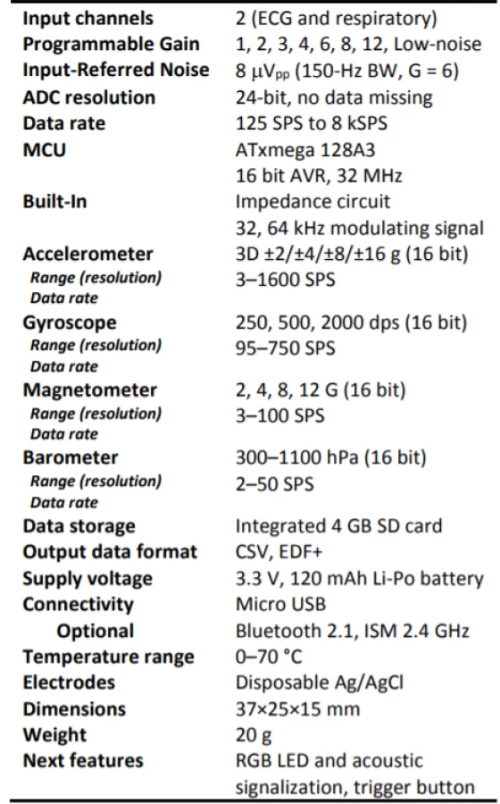
Therefore in this experiment we used two-channel analog front-end TI ADS1292R to monitor EMG and EIM, and built-in accelerometer LSM303D to MMG measurement. Detailed technical parameters of used holter are described in Table 1.
In the experiment we connect together EMG, MMG and EIM in single wireless holter monitoring system that offers perfect signal synchronization. We used standard sampling rate 2000 Hz for EMG and EIM and 400 Hz for MMG, the synchronization error was maximal 0.5 ms. MMG was measured in ± 4g range. For better signal quality, the received holter EMG signal was filtered in Labchart by using digital band-pass filter
1–500 Hz and EIM with MMG signal using 0.1 high-pass digital filter. The holter was bounded on muscles using Ag/AgCl electrodes, 2 cm distant, and for higher MMG precision the electrodes were also fixed using a bandage.
Whole test consisted of isometric and isotonic exercises of 6 probands (average age = 28, average BMI = 23, 4 male, 2 female, all healthy in good condition) done on biceps brachia and gastrocnemius muscle. In these results we provide only short information of one proband (male, age = 38, BMI = 22.4) measured on biceps brachia muscle, which demonstrates all technical opportunities of our sensor system.
Results and Discussion
On Fig. 2 is total time response of EMG, EIM and MMG. The exercise consisted of 6 series (load from 20% to 70% MVC) of 5 lifting’s. MVC (maximum voluntary concentration) was executed shortly before start of primary experiment. The isotonic exercises were followed after short relaxation by a 30 second 50% MVC isometric exercise.

Detailed time domain signals of 3 lifting’s, is visible on Fig. 3. We could see “EMG onset” (about 200 ms) between muscle activation (EMG start) and real body movement (total MMG shift), phase between the transition of isotonic movement to isometric. This time shift can be interesting for measuring of reflex and reactions in sport. It is interesting that EIM is just sensitive for isotonic signal and insensitive to isometric signal (Fig. 2, 3) what can be beneficial in a control of prosthesis [9–11].

We added measurements from professional device Neuro-MEP-4 (Fig. 4, 6, 7) to obtain well-balanced results. Our holter was connected with the muscles using Ag/AgCl electrodes in standard 2 cm EMG distance with parallel connected gold plated EMG electrodes with conductive paste, also in 2 cm distance. The active diameter of holter Ag/AgCl electrodes was 1.7 cm and active diameter of reference gold plated electrodes was 0.9 cm.

Fig. 5 shows frequency domain of isometric load. Left is PSD (power signal density) in 1st half of exercise, right in 2nd half. The comparison shows amplitude and mean frequency MNF shift of PSD due to fatigue factor.

Detailed analysis follows in Fig. 6. Here we showed time changes of myographic parameters (RMS - root mean square, Total power TP and MNF - mean frequency of PSD) at constant isometric load due to fatigue increasing. The total power spectrum was calculated using following parameters: FFT size = 500, which corresponds to 0.25 second actuator sampling speed, Data window = Hann (cosine-bell), Window overlap = 95%. The dependencies of RMS and TP of EMG are common known and perfectly correspond to theoretical models [3, 6, 8, 23–28].

The dependencies of MMG and fatigue factor are innovative and not enough examined in existing literature [2, 18, 19, 24–26, 29, 30] and it does not exist valid and proper theoretical models. We discovered that for fatigue examining seems useful the calculation of signal SD (standard deviation) and again TP. In complete analysis we used 2nd order polynomial fitting function.
Differences in the results between holter and reference device are mainly due to the different settings of band-pass filters (frequency shift: 500 Hz vs. 4000 Hz high cut-off frequency), the different sensor surfaces of the electrodes (different amplitudes: 17 mm vs. 9 mm diameter) and the possible slight differences in the location of the sensors on the respective muscle.
Fig. 7 presents typical signal isotonic changes at different MVC loads. For isotonic EMG to force ratio - EMG=f(F) we should ideally obtain linear relationships [24–26] but due to fatigue factor is the output slightly bent (about -0.5·MVC2). RMS and TP of EMG dependencies correlate to theoretical models [6, 18, 19]. The SD and TP of MMG signals and the RMS of EIM signals are also analysed. In complete analysis we again used only simple 2nd order polynomial fitting function.

Conclusion
In this paper we presented measurement of EMG, MMG and EIM using single wireless holter. Obtained results confirmed that our device is able to reliably and precisely monitor the muscle activity. Compared to the conventional devices our own designed system offers also MMG and EIM monitoring. Due to these innovative measuring methods we can offer improved results for robotics, sports and medicine research. Interesting could be found that EIM is sensitive for isotonic signal and insensitive to isometric signal which can be useful in robotics or prosthesis control.
High quality and scientific potential of the designed smart holter was experimentally proven. With ultra-low power consumption the novel design provides suitable and promising long-term testing and medical surveillance device.
The demonstration of the functionality of our system by measurement of the relationship between electrical activity and isometric muscle force, which can be applied in kinesiology or to control electrical devices such as robotic or prosthetic body-parts proves the application potential.
In the present study we have compared fatigue, load, total power, mean frequency and dependency of amplitude obtained with our device and referential medical device and we demonstrated efficiency of our system by measure of physiological parameters of human muscles.
Considering the capability of our device to simultaneously measure EMG, MMG and EIM signals of human muscles we will further focus on gender-related differences, which can be used to improvement of control algorithms for specialized actuators and progression in new healthcare diagnostic methods.
Acknowledgement
This work was supported in part by the Ministry of Education, Science, Research and Sport of the Slovak Republic under grant VEGA 1/0739/16 and VEGA 1/0886/17 and by the Slovak Research and Development Agency under the contracts APVV-15-0763 and APVV-16-0626.
Ing. Erik Vavrinsky, PhD.
Institute of Electronics and Photonics
Slovak University of Technology,
Ilkovicova 3, 81219 Bratislava, Slovakia
E-mail: erik.vavrinsky@stuba.sk
Phone: +421 (2) 60 291 65
Zdroje
[1] Bilgin, G., Hindistan, E. , Ozkaya, Y. G., Koklukaya, E., Polat, O., Colak, O. H.: Determination of Fatigue Following Maximal Loaded Treadmill Exercise by Using Wavelet Packet Transform Analysis and MLPNN from MMG-EMG Data Combinations. Journal of Medical Systems, 2015, vol. 39, no. 10.
[2] Konrad, P.: The ABC of EMG, no. March. 2006.
[3] Merlo, A., Campanini, I.: Technical Aspects of Surface Electromyography for Clinicians. The Open Rehabilitation Journal, 2010, vol. 3, no. 1, pp. 98–109.
[4] Kamen, G., Gabriel, D. A.: Essentials of electromyography: Champaign. Human Kinetics, 2010.
[5] Quasthoff, S.: Grundlagen der EMG Untersuchung. Das Neurophysiology, 2010, vol. 32, no. 1, pp. 1–27.
[6] Uchiyama, T., Hashimoto, E.: System identification of the mechanomyogram from single motor units during voluntary isometric contraction. Medical & Biological Engineering & Computing, 2011, vol. 49, no. 9, pp. 1035–1043.
[7] Esposito, F., Limonta, E., Cè, E.: Time course of stretching-induced changes in mechanomyogram and force characteristics. Journal of Electromyography and Kinesiology, 2011, vol. 21, no. 5, pp. 795–802.
[8] Longo, S., Cè, E., Rampichini, S., Devoto, M., Venturelli, M., Limonta, E., Esposito, F.: Correlation between stiffness and electromechanical delay components during muscle contraction and relaxation before and after static stretching. Journal of Electromyography and Kinesiology, 2017, vol. 33, pp. 83–93.
[9] Sanchez, B., Rutkove, S. B.: Present Uses, Future Applications, and Technical Underpinnings of Electrical Impedance Myography. Current Neurology &. Neuroscience Reports, 2017, vol. 17, no. 11, pp. 86.
[10] Li, X., Shin, H., Li, L., Magat, E., Li, S. and Zhou, P.: Assessing the immediate impact of botulinum toxin injection on impedance of spastic muscle. Med. Eng. Phys., vol. 43, pp. 97–102, 2017.
[11] Zhang, X., Li, X., Samuel, O. W., Huang, Z., Fang, P., Li, G.: Improving the robustness of electromyogram-pattern recogni-tion for prosthetic control by a postprocessing strategy, Front. Neurorobot., vol. 11, no. SEP, pp. 1–15, 2017.
[12] Abad, S. L. M., Maghooli, K.: Low Supply Voltage Electro-cardiogram Signal Amplifier.1st International Conference on Bioinformatics and Biomedical Engineering, 2007, pp. 798–801.
[13] De Luca, C. J.: Electromyography. Encyclopedia of Medical devices and Instrumentation, 2006, pp. 98–109.
[14] Stegeman, D. F., Blok, J. H., Hermens, H. J., Roeleveld, K.: Surface EMG models: Properties and applications. Journal of Electromyography and Kinesiology, 2000, vol. 10, no. 5, pp. 313–332.
[15] Merlo, A., Farina, D., Merletti, R.: A fast and reliable technique for muscle activity detection from surface EMG signals. IEEE Transactions on Biomedical Engineering, 2003, vol. 50, no. 3, pp. 316–323.
[16] Kimoto, A., Yamada, Y.: A new layered sensor for simultaneous measurement of EMG, MMG and oxygen consumption at the same position. Medical & Biological Engineering & Computing, 2015, vol. 53, no. 1, pp. 15–22.
[17] Lee, K.-S.: EMG-based speech recognition using hidden markov models with global control variables. IEEE Transactions on Biomedical Engineering, 2008, vol. 55, no. 3, pp. 930–940.
[18] Cifrek, M., Medved, V., Tonković, S., Ostojić, S.: Surface EMG based muscle fatigue evaluation in biomechanics. Clinical Biomechanics, 2009, vol. 24, no. 4, pp. 327–340.
[19] Hill, E. C., Housh, T. J., Smith, C. M., Cochrane, K. C., Jenkins, N. D. M., Cramer, J. T., Schmidt, R. J., Johnson, G. O.: Effect of sex on torque, recovery, EMG, and MMG responses to fatigue. Journal of Musculoskeletal and Neuronal Interactions, 2016, vol. 16, no. 4, pp. 310–317.
[20] Vavrinsky, E., Daricek, M., Horinek, F., Donoval, M.: Design of Very Precise and Miniature Low Power ECG Holter. In Electrocardiology 2014 : Proceedings of the 41st International Congress on Electrocardiology, 2014, pp. 257–260.
[21] Gupta, A. K.: Respiration Rate Measurement Based on Impedance Pneumography. Application Report, Texas Instruments, 2011, pp. 1–11.
[22] Kugelstadt, T.: Getting the most out of your instrumentation amplifier design. Analog Applications Journal, 2005, vol. 1, no. 5, pp. 25–30.
[23] Vavrinsky, E., Stopjakova, V., Donoval, M., Daricek, M., Svobodova, H., Mihalov, J., Hanic, M., Tvarozek, V.: Design of sensor systems for long time electrodermal activity monitoring. Advances in Electrical and Electronic Engineering, 2017, vol. 15, no. 2, pp. 184–191.
[24] Tosovic, D., Than, C., Brown, J. M. M.: The effects of accumulated muscle fatigue on the mechanomyographic waveform: implications for injury prediction. European Journal of Applied Physiology, 2016, vol. 116, no. 8, pp. 1485–1494.
[25] Ray, G. C., Guha, S. K.: Relationship between the surface e.m.g. and muscular force. Medical & Biological Engineering & Computing, 1983, vol. 21, no. 5, pp. 579–586.
[26] Enoka, R. M.: Muscle fatigue - from motor units to clinical symptoms.Journal of Biomechanic, 2012, vol. 45, no. 3, pp.
427–433.
[27] Calvert, T. W., Chapman, A. E.: The relationship between the surface EMG and force transients in muscle: Simulation and experimental studies. Proceedings of the. IEEE, 1977, vol. 65, no. 5, pp. 682–689.
[28] Han, H., Jo, S., Kim, J.: Comparative study of a muscle stiffness sensor and electromyography and mechanomyography under fatigue conditions. Medical &. Biological Engineering & Computing, 2015, vol. 53, no. 7, pp. 577–588.
[29] González-Izal, M., Malanda, A., Navarro-Amézqueta, I., Gorostiaga, E. M., Mallor, F., Ibañez, J., Izquierdo, M.: EMG spectral indices and muscle power fatigue during dynamic contractions. Journal of Electromyography and Kinesiology, 2010, vol. 20, no. 2, pp. 233–240.
[30] Al-Mulla, M. R., Sepulveda, F., Colley, M.: A review of non-invasive techniques to detect and predict localised muscle fatigue. Sensors, 2011, vol. 11, no. 4, pp. 3545–3594.
Štítky
BiomedicínaČlánek vyšel v časopise
Lékař a technika

2018 Číslo 2
Nejčtenější v tomto čísle
- Application of sibgle wireless holter to simultaneous EMG, MMG and eim measurement of human muscles activity
- Response by an automated inspired oxygen control system to hypoxemic episodes: assessment of damping
- Workflow for bioprinting of cell-ladem bioink
- Machine learning using speech utterances for parkinson disease detection
Zvyšte si kvalifikaci online z pohodlí domova
Mazová zátka a její řešení
nový kurzVšechny kurzy