
TRAUMA IN OCULOPLASTIC SURGERY A REVIEW
Authors:
A. Kopecký 1,2
![]() ; J. Němčanský 1,2
; J. Němčanský 1,2
Authors place of work:
Oční klinika Fakultní nemocnice Ostrava, Ostrava
1; Lékařská fakulta Ostravské univerzity, Ostrava, katedra Kraniofaciálních oborů
2
Published in the journal:
Čes. a slov. Oftal., 76, 2020, No. 3, p. 103-110
Category:
Přehledová práce
doi:
https://doi.org/10.31348/2020/18
Summary
The aim of this article is to present the basics of traumatology in oculoplastic surgery and to review the literature about this topic. This review sums up the problematic of injuries of the eyelid, lacrimal system and orbit. The most important types of trauma, their treatment options, and the most common complications are described.
In majority of oculoplastic traumas, surgical reconstruction is the treatment of choice. The surgery is often performer immediately, but sometimes the reconstruction of eyelid and lacrimal injuries can be postponed up to 48 hours, if the immediate surgery is not possible. Although the recommendations from the literature on this topic are variable, most of the patients require at least local antibiotics, more complex traumas systemic antibiotics.
Careful diagnostics and correctly performed surgical treatment, either only by ophthalmologist, or oculoplastic surgeon, or a multi-disciplinary team for more complex injuries, are the key to good functional and aesthetic results of the reconstruction.
Keywords:
trauma – oculoplastic surgery – eyelid trauma – orbital trauma – lacrimal trauma
INTRODUCTION
Oculoplastic surgery deals with the area of the eyelids, the periocular region, lachrymal pathways and eye socket. It is a traditional sub-specialisation of ophthalmology, the roots of which date back to antiquity, although its first mention in the literature is not found until 1925, in a textbook by E. Spaeth [1]. In 1969 this sub-specialisation was finally formally defined with the establishment of the American Society of Ophthalmic Plastic and Reconstructive Surgery [2]. Care for ocular traumas is an integral and important component of this specialisation.
EPIDEMIOLOGY
Although few studies have dealt with the epidemiology of ophthalmic traumas, we can state that the incidence of ocular traumas in adult patients is 38/100 000 per year. Injury to the eyelids constitutes approximately 8 % of traumas, injury to the eye socket approx. 5 %. Injury to the lachrymal pathways is less common [3].
Approximately 44 % of eyelid traumas are linked with injury to the eyeball (including contusions) [4]. In general, the upper eyelid is injured more frequently than the lower [5]. The margin of the eyelid may be affected in approximately 24 % of traumas, and in as many as 16 % of cases the lachrymal drainage pathways may be damaged.
In the category of occupational injuries, eyelid trauma is the most common type of ocular trauma [6]. The group most frequently affected by eyelid trauma is adult men.
A special category is traumas resulting from dog bites. These are more common in children than in adults, and are more often complicated by associated injury to the lachrymal canaliculus [7].
Around 70 % of injuries to the lachrymal pathways represent injury to the canaliculus. We encounter injury to the lachrymal sac only rarely (20 %), and injury to the nasolachrymal duct is the least common (10 %) [5]. We sporadically encounter injury to both canaliculi simultaneously, in approximately 12 % of cases [8].
We observe injury to the eye socket most frequently in individuals involved in traffic accidents (over 50 %), followed by falls and violent injuries. In children the most common causes of injuries to the eye socket are sports and games [9]. Orbital fractures are more common in men than in women, and occur most often in men aged between 21 and 30 years [10]. We most frequently encounter damage to the base and medial wall of the eye socket [11].
Burns and chemical injuries of the ocular region form approximately 7.5 to 27 % of all traumas treated at burns centres [12].
Clinical finding
Eyelid trauma
We divide eyelid traumas into:
- Blunt trauma (closed)
- Sharp trauma (open)
- Trauma from projectile
- Chemical and thermal trauma
We find closed traumas of the eyelids generally in the case of blunt traumas – usually as a result of contusions. A typical manifestation is the development of haematoma. Due to the anatomy of the eyelids, it is most often well bordered and may grow to large dimensions.
Abrasion is removal of the skin epithelium (or of deeper layers of the skin) upon sufficiently strong energy of blunt trauma.
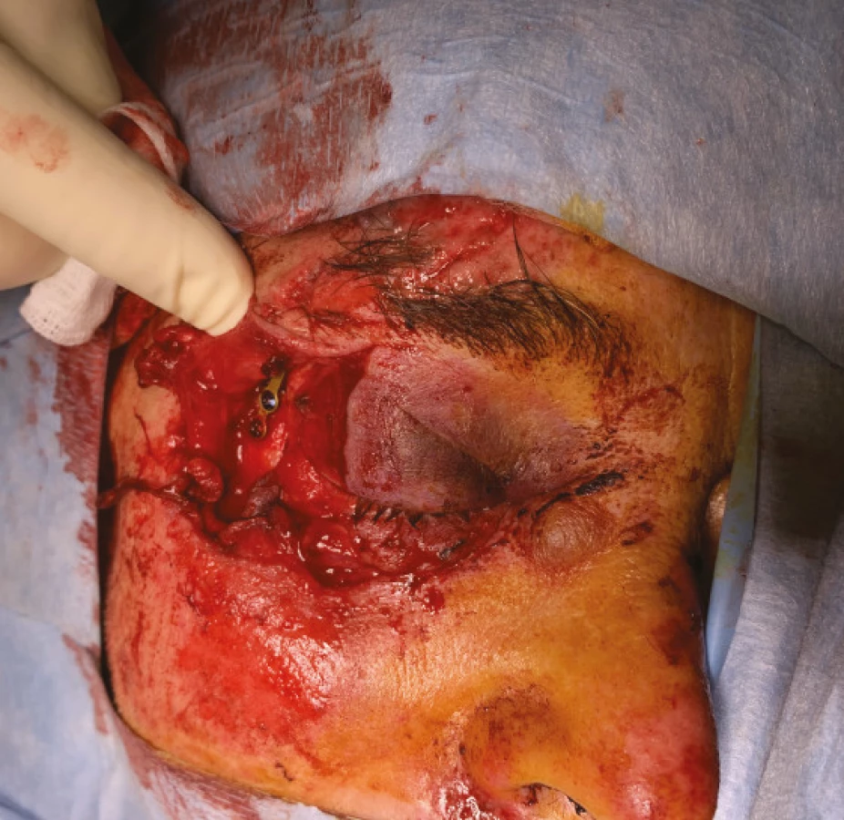
Open traumas of the eyelid include laceration (tearing injuries), cutting and slicing injuries, avulsion (detachment) and piercing injuries. Tearing injuries are irregular, of various depth, accompanied by swelling. Cutting and slicing injuries are generally well defined and are not accompanied by such pronounced swellings. Avulsions are injuries involving loss of tissue, caused by large force, and may lead to the exposure of deep anatomical structures. Piercing injuries may upon first appearance seem inconspicuous, but it is necessary to remove any serious hidden damage to the deeper structures (including the orbit), to remove the foreign body from the eye and any associated contamination.
Burns and chemical injuries are manifested by reddening of the eyelids, their reactive swelling or direct damage. It is always necessary first of all to eliminate damage to the conjunctiva and cornea, which directly threatens the patient’s sight.


Injury to lachrymal pathways
This may be direct or indirect, as mentioned above, in which injury to the canaliculus is unequivocally the most frequent. Injury to the medial part of the eyelid must always raise suspicion of this type of injury, and within the framework of diagnostics this should always be ruled out. Sometimes the clinical finding may be very inconspicuous, at other times the degree of injury to the medial part of the eyelid is so pronounced that injury to the canaliculus is clear already upon the basic examination.
In addition to the aforementioned dog bite, cases are described in the literature of injuries through various items, hooks, or even injuries during childbirth [7,13,14].
Injury to orbit
Isolated injuries to the orbit are rare, and mostly concern fractures of the base of the orbit. Orbital traumas can be differentiated according to whether the injury occurred only to soft structures, or also to the skeleton. The basic imaging method is CT (computer tomography) examination. Injuries to the orbit always require an interdisciplinary approach of an ophthalmologist, maxillofacial surgeon, ear, nose and throat specialist, neurosurgeon and radiologist, often according to what affiliated structures are afflicted in addition to the orbit.
A typical injury is a “blow-out fracture”, in which the mechanism of origin is a trauma-induced increase of intraorbital pressure (e.g. impact from a fist or ball). The most common is injury to the base of the orbit, which is manifested by diplopia and enophthalmos, as well as subcutaneous emphysema and in exceptional cases anaesthesia in the region of the infraorbital nerve.
Orbital fractures are often associated and may accompany other types of fractures of the facial skeleton, e.g. Le Fort type II and III fractures, fractures of the zygomaticomaxillary complex and nasoorbitoethmoid fractures [15].
Penetrating injuries to the orbit are differentiated anatomically according to localisation of the entry wound. According to this it is also possible to predict which structures of the orbit may be damaged [16]. Penetrating injuries are either with or without the presence of a foreign body in the orbit.
In the case of contusions of the orbit it is necessary to remember that according to some studies as many as 29.4 % may be linked with a blow-out fracture of the orbit [17]. As a result, these patients should be thoroughly examined, including the use of imaging methods.
DIAGNOSIS
Many patients report to an ophthalmologist with injury to the ocular adnexas, lachrymal pathways or orbit via a traumatology department, frequently as a consultancy examination in the case of more complex traumas.
In all patients it is of key importance to determine the mechanism of injury. The basis is therefore a carefully recorded medical history. From a general perspective it is necessary to exclude the threat to vital functions, from an ophthalmological perspective it is a priority to determine whether the eyeball and optic nerve are damaged or threatened. Penetrating injury of the eyeball must be unequivocally excluded. If the patient’s condition so allows, we must examine visual acuity and the ocular fundus.


A component of the examination is assessment of motility of the eyeballs, and in the case of suspicion of incarceration of the extraocular muscle it is necessary to perform a passive duction test, which helps us differentiate the restrictions of motility upon a background of paresis.
In the case of a fat prolapse upon injury to the upper eyelid, it is always necessary to exclude traumatic damage to the musculus levator palpebrae superioris or its aponeurosis. According to some studies, traumatic ptosis is the second most common of all types of ptosis, and its surgical correction, especially at an interval from the original trauma, may be complex and thus should be performed primarily after determination of the diagnosis [18].
In the case of orbital injury, we always add an imaging method, primarily CT [19]. Furthermore, in the case of an unclear finding, CT examination can help us exclude injury to the eyeball – in this case its sensitivity is around 75 % [20]. In addition, it has an irreplaceable role in identifying the foreign body in the eye socket. Only if a metal foreign body in the orbit is definitively ruled out can we proceed to examination by magnetic resonance imaging (MRI). MRI is useful in the detection of certain organic foreign bodies or for a better overview of a formed haematoma [19]. The role of MR is important also in the diagnosis of traumatic neuropathy of the optic nerve.
THERAPY
In the case of all traumas we should verify whether the patient has effective vaccination against tetanus, and whether it is possible to perform reconstruction under local anaesthesia. However, in general it applies that for extensive injuries to the eyelids, injuries to the lachrymal pathways, injury to the orbit and children’s injuries it is more suitable to perform the procedure under general anaesthesia.
Indication for general antibiotic treatment is a question for discussion. Some studies exist which have not confirmed the benefit of general antibiotics even in the case of closed orbital traumas without a foreign body, and recommend them only in the case of orbital operations [21]. However, in general we consider antibiotic prophylaxis to be important, at least in the case of injury by animals, organic material or in the case of presence of a foreign body [5].


Eyelid traumas
Minor superficial injuries to the eyelids can be resolved conservatively, with the application of antibiotic cream.

We can reconstruct open wounds of the eyelids immediately or after an interval of 1-2 days from the moment of injury [5]. This is important to remember especially with reference to the organisation of healthcare and the (un)availability of not only an oculoplastic surgeon, but also an ophthalmologist whatsoever in remote regions. If the margin of the eyelid is not affected, and if this does not concern an injury involving loss, we proceed anatomically and reconstruct the individual layers.
Upon damage to the edges of the eyelids, quality primary reconstruction consists in the prevention of future faulty positioning of the eyelid. We insert the primary suture into the “grey line” on the margin, and leave the ends long. The aim is to ensure slight hypercorrection in the sense of eversion of the edges of the wound on the margin, in order to prevent the formation of a sunken scar [22].
If the eyelid trauma involves such loss that it is not possible to use a direct suture, we must make use of advancement or rotation flaps, and in exceptional cases loose grafts. In addition to local shifts, in the case of eyelid traumas involving loss of tissue on a large scale, it is possible also to use techniques which are dominant for reconstructive tumour surgery, for example Hughes or Cutler - -Beard flaps, or if applicable modifications or combinations thereof with other techniques (for example with a Tenzel flap) [23,24].
In general we first of all reconstruct the deep structures such as the levator aponeurosis, tarsus or canthal ligament. We attempt to attach divided anatomical units in such a manner as to ensure a clearly accessible wound which will be then simple to reconstruct. The aim of the operation should always be to reconstruct not only the anterior lamella (skin and muscles), but above all the posterior lamella of the eyelid (tarsal plate and connecting canthal ligament), which is of key importance for preserving the correct position and function of the damaged eyelid [25].
Burns and chemical injuries to the eyelids
This is a specific issue in oculoplastic surgery, which requires treatment at specialised centres – burns centres, which have broad interdisciplinary teams of specialists at their disposal. Relatively rapid onset of contractions or lagophthalmos may lead to the necessity for early surgical intervention. According to Malhotra et al. we distinguish several surgical procedures which can be used at various times after the trauma – in the early phase this is tarsorrhaphy for difficult lagophthalmos upon a background of severe damage to the cornea, conjunctival advancement flaps in the case of lagophthalmos upon a background of destruction of the eyelids, or similarly functioning tenonplasty [12]. In the medium phase, retraction of the eyelids, ectropium or entropium is performed. From a long-term perspective the challenge is surgical solution of formed scars, contractions and faulty positioning of the eyelids, or trichiasis. Grafts, Z-plasty and a range of other reconstruction techniques are used according to the experience of the surgeon and the degree of damage to the tissue of the patient [26].
Lachrymal pathway traumas
In regular clinical practice we most often perform reconstruction of the torn canaliculus. Injury to the lower canaliculus is more common [27]. A surgical microscope is suitable for the operation. We find both parts of the torn canaliculus and with the aid of a suture and inserted stent we attempt to reconstruct the canaliculus. Injury to the canaliculus should be reconstructed as primary surgery, if immediate reconstruction is not possible, it is not recommended to wait more than 48 hours [5]. Although the success rate of secondary reconstructions is stated at 89 % in the literature, it is recommended always to attempt to perform reconstruction as primary [28].
Upon injury to one canaliculus, we most often insert a monocanalicular stent, and in exceptional cases a “ring” intubation is used, in which the stent as a ring passes through both canaliculi [27,29]. In general, the advantage of a monocanalicular stent is easier surgical technique and the insertion only into the injured canaliculus, while the disadvantage is higher probability of loss of the stent. In the case of ring intubation, by contrast, there is a higher risk of irritation and excessive tightening of the ring, usually the stent holds better [30].
In the case of injury to the lachrymal sac and tear duct, intubation by stent is indicated at least, but in the case of larger injuries, dacrycystorhinostomy (DCR) is often indicated upon primary reconstruction. Later complications must often be resolved by conjunctivocystorhinostomy [30]. If primary intubation is unsuccessful, it is usually recommended to wait for up to 4-5 months from the primary trauma before performing DCR.
We remove the inserted stents after 3 months at the earliest.
Orbital traumas
Complete therapy of orbital traumas is beyond the scope of our summary article. It often concerns a multidisciplinary problem in which, depending on the anatomical localisation, the ophthalmologist meets together with other specialists such as a neurosurgeon, ear, nose and throat specialist or maxillofacial surgeon. In the following section, we shall therefore focus primarily on therapy of fractures of the base of the orbit, with which general ophthalmologists encounter most frequently in clinical practice.
It is not necessary to operate on all fractures of the base of the orbit. Various recommendations exist regarding indication for surgery on the base of the orbit. According to consensus, the following three indications for reconstruction predominate in the literature: [31,32,33]
- Restriction of motility of the eyeball, accompanied with positive passive duction test and positive finding on CT with incarceration of extraocular muscle, presence of diplopia
- Defect of base is larger than half its surface, evident on CT
- Enophthalmos more than 2 mm
The time planning of reconstruction often differs according to the customs of the centre. In non-urgent situations it is recommended to wait 14 days until the swelling has ceased and then, according to the clinical finding, decide on indication for any applicable surgery [34]. An indication for early reconstruction of the base of the orbit is a situation in which incarceration of the muscle generates an oculocardiac reflex. Such a case may constitute a direct threat to the patient’s life [33]. Similarly, early indication is chosen for children. In the planning of surgery, it is necessary at the same time to consider the fact that after 14 days any potential future interventions may be complicated by incipient fibrosis. On the other hand, studies exist which describe a successful operation even as long as 6-8 weeks after the trauma [35]. According to the available literature, the timing of the operation on a fracture is a controversial topic, also due to the relatively insufficient number of prospective studies [35]. It is appropriate to mention that it is also necessary to take into account the patient’s general condition and sometimes, e.g. in the case of multiple traumas, it is suitable to perform reconstruction of the base within the framework of a single anaesthesia in the primary treatment.
It is also possible to find relative contraindications for surgery on a fracture of the base of the orbit in the literature. The most frequently mentioned are: hyphaema, retinal cracks, perforation of the eyeball or generally unstable condition of patient [32].

COMPLICATIONS
With regard to the scope of the issue and the diversity of possible traumas, it is possible to describe a broad spectrum of complications in oculoplastic traumatology. In the following overview we shall focus on the most significant of these.
Retrobulbar haematoma
This is a relatively rare but serious sight-threatening complication. It represents an urgent condition in which there is an increase of pressure in the orbit and compression of the optic nerve upon a background of retrobulbar haemorrhage. In the case of suspicion of retrabulbar haematoma we immediately perform lateral canthotomy with cantholysis, we do not perform an imaging examination (CT) since any delay may lead to irreversible damage to the optic nerve [36].

The prevalence of retrobulbar haematoma is generally within the range of 0.4-1.7 % [36,37,38].
In the literature we may also encounter the term orbital compartment syndrome [39]. This more generally describes a condition in which there is a dangerous increase of intraocular pressure – not only upon a background of retrobulbar haematoma, but also as a complication of orbital cellulitis, pronounced emphysema, or an accumulation of fluid (for example in the case of a burn) [40].
Traumatic optic neuropathy
This may be caused by a direct mechanism, e.g. damage to the optic nerve by a fragment of bone of the orbit, or often by an indirect mechanism, in which the force of the injury is transmitted via the soft and hard tissue of the face and orbit, resulting in damage to the nerve, primarily in places where the fixed part of the nerve passes into the loose part. This usually occurs on the transition of the intraorbital and intracanalicular portion of the optic nerve [41,42]. The diagnosis is clinical, in the case of suspicion primarily CT should be performed. MR may be initially negative and is more appropriate for monitoring affected patients [43]. It is suitable to use visual evoked potentials for confirmation of the diagnosis.
In the case of indirect injury, improvement of visual acuity is described in 40-60 % of cases, which is comparable to patients treated with steroids or the surgical decompression of the optic nerve. In general it is difficult to determine whether treatment by steroids or surgical decompression genuinely improves the prognosis of this complication [44]. For example, some studies found no difference between patients treated with steroids and those in whom steroids were combined with surgery [45]. Nevertheless, the literature often contains the recommendation that patients who have no contraindication for treatment with steroids for other medical reasons should receive the maximum daily dose of methylpredisolone 1 g [44]. This approach may be important also from a forensic perspective.
Infraorbital hypoesthesia
This is a relatively common complication of fracture of the base of the orbit, above all in cases in which fracture of the edge of the orbit also occurs [35,46]. Numerous authors consider this complication to be a further indication for surgical reconstruction of the fracture of the base of the orbit, since at least a partial improvement had been described following performed surgery, above all in patients with pronounced and progressing hypoesthesia [47].
Infection
The most serious infectious complication is inflammation progressing into the orbit or central nervous system [48]. In exceptional cases, such complications are also described as late, and as a result patients should be observed even if in outpatient care, especially after larger traumas [49]. The degree of use of antibiotics differs within the framework of individual regions. It had been demonstrated that prophylactic use of antibiotics following oculoplastic procedures leads to a lower number of infectious complications [50]. As a result, the use of antibiotics in the care of oculoplastic traumas can be recommended. Smaller injuries can be treated with local antibiotics, deeper traumas and injuries of a larger scale, orbital traumas or injuries caused by organic material or animals require general antibiotics.
CONCLUSION
Oculoplastic traumatology covers a wide range of injuries to various different anatomical structures.
Careful diagnosis and correctly performed surgical solution, either purely directed by an ophthalmologist, an ophthalmologist focusing on oculoplastic issue, or by an entire multidisciplinary team in the case of more complex injuries, is the key to a good functional and aesthetic result. However, of primary importance is the prevention of future complications.
Every ophthalmologist should know the basic diagnostics and possibilities of therapy, and at the same time must decide sufficiently in time as to whether it is possible to resolve the trauma completely by himself/herself or to send the patient to a specialised centre.
Zdroje
1. Watts MT. The history of oculoplastic surgery. Facial Plast Surg. 1993 Apr;9(2):151–6.
2. Ruben S. The incidence of complications associated with retrobulbar injection of anaesthetic for ophthalmic surgery. Acta Ophthalmol (Copenh). 1992 Dec;70(6):836–8.
3. Sahraravand A, Haavisto AK, Holopainen JM, Leivo T. Ocular trauma in the Finnish elderly - Helsinki Ocular Trauma Study. Acta Ophthalmol. 2018 Sep;96(6):616–622.
4. Herzum H, Holle P, Hintschich C. Eyelid injuries: epidemiological aspects. Ophthalmologe. 2001 Nov;98(11):1079–82.
5. Koch KR, Kopecky A, Heindl LM. Management of Periocular Soft Tissue Injuries. Klin Monbl Augenheilkd. 2016 Sep;233(9):1071–87.
6. Chang CH, Chen CL, Ho CK et al. Hospitalized eye injury in a large industrial city of South-Eastern Asia. Graefes Arch Clin Exp Ophthalmol 2008; 246 : 223–228
7. Sadiq MA, Corkin F, Mantagos IS. Eyelid Lacerations Due to Dog Bite in Children. J Pediatr Ophthalmol Strabismus. 2015 Nov-Dec;52(6):360–3.
8. Heichel J, Struck HG, Viestenz A. [Primary Reconstruction of Lacrimal Ducts after Trauma - Bicanaliculonasal Intubation Using Autostable Tubes]. Klin Monbl Augenheilkd. 2019 May 2. doi: 10.1055/a-0839-5172.
9. Jatla KK, Enzenauer RW: Orbital fractures: a review of current literature. Curr Surg 2004, 61 : 25–29.
10. Shin JW, Lim JS, Yoo G, Byeon JH. An analysis of pure blowout fractures and associated ocular symptoms. J Craniofac Surg. 2013 May;24(3):703–7.
11. Joseph JM, Glavas IP. Orbital fractures: a review. Clin Ophthalmol. 2011 Jan 12;5 : 95–100.
12. Malhotra R, Sheikh I, Dheansa B. The management of eyelid burns. Surv Ophthalmol. 2009 May-Jun;54(3):356–71.
13. Fannin LA, Fitch CP, Raymond WR, Flanagan JC, Mazzoli RA. Eye injuries from merchandise display hooks. American Journal of Ophthalmology. 1995. 120(3)397–399.
14. Harris GJ. Canalicular Laceration at Birth. 1988. 105(3):322–323.
15. Cruz AA, Eichenberger GC. Epidemiology and management of orbital fractures. Curr Opin Ophthalmol. 2004 Oct;15(5):416–21.
16. Mashriqi F, Iwanaga J, Loukas M, D‘Antoni AV, Tubbs RS. Penetrating Orbital Injuries: A Review. Cureus. 2017 Sep 29;9(9):e1725
17. Kreidl KO, Dennis YK, Mansour SE: Prevalence of significant intraocular sequelae in blunt orbital trauma. Am J Emerg Med 2003, 21 : 525–528.
18. Lim JM, Hou JH, Singa RM, Aakalu VK, Setabutr P. Relative incidence of blepharoptosis subtypes in an oculoplastics practice at a tertiary care center. Orbit. 2013 Aug;32(4):231–4.
19. Lin KY, Ngai P, Echegoyen JC, Tao JP. Imaging in orbital trauma. Saudi J Ophthalmol. 2012 Oct;26(4):427–32.
20. Joseph DP, Pieramici DJ, Beauchamp NJ Jr. Computed tomography in the diagnosis and prognosis of open-globe injuries. Ophthalmology. 2000 Oct;107(10):1899–906.
21. Wang JJ, Koterwas JM, Bedrossian EH Jr, Foster WJ. Practice patterns in the use of prophylactic antibiotics following nonoperative orbital fractures. Clin Ophthalmol. 2016 Oct 27;10 : 2129–2133.
22. Perry JD, Aguilar CL, Kuchtey R. Modified vertical mattress technique for eyelid margin repair. Dermatol Surg. 2004 Dec. 30(12 Pt 2):1580–2
23. Hishmi AM, Koch KR, Matthaei M, Bölke E, Cursiefen C, Heindl LM. Modified Hughes procedure for reconstruction of large full-thickness lower eyelid defects following tumor resection. Eur J Med Res. 2016 Jun 30;21(1):27.
24. Kopecky A, Koch KR, Bucher F, Cursiefen C, Heindl LM. Results of Cutler-Beard procedure for reconstruction of extensive full thickness upper eyelid defects following tumor resection. Ophthalmologe. 2016 Apr;113(4):309–13.
25. Kopecky A, Rokohl AC, Heindl LM. Techniques for the Reconstruction of the Posterior Eyelid Lamella. Klin Monbl Augenheilkd. 2018 Dec;235(12):1415–1428.
26. McIndoe AH. Total reconstruction of the burned face. Br J Plastic Surg. 1983;36 : 410–20
27. Kohlhaas M, Wiegmann L, Gaszczyk M, Walter A, Schaudig U, Richard G. Lacrimal duct treatment with ring intubation in injuries of the upper and lower eyelids. Ophthalmologe. 2001 Aug;98(8):743–6.
28. Bai F, Tao H, Zhang Y et al. Old canalicular laceration repair: a retrospective study of the curative effects and prognostic factors. Int J Ophthalmol 2017; 10 : 902–907
29. Kim T, Yeo CH, Chung KJ, Lee JH, Kim YH. Repair of Lower Canalicular Laceration Using the Mini-Monoka Stent: Primary and Revisional Repairs. J Craniofac Surg. 2018 Jun;29(4):949–952.
30. Komínek, P.: Poranění slzných cest. In Komínek, P., Červenka, S., Müller, K.: Nemoci slzných cest. Maxdorf 2003, Praha, s 170–75
31. Gart MS, Gosain AK. Evidence-based medicine: Orbital floor fractures. Plast Reconstr Surg. 2014 Dec;134(6):1345–55.
32. Kim, H. S., Jeong, E. C. (2016). Orbital Floor Fracture. Archives of craniofacial surgery, 17(3), 111–118.
33. Pham CM, Couch SM. Oculocardiac reflex elicited by orbital floor fracture and inferior globe displacement. Am J Ophthalmol Case Rep. 2017 Jun;6 : 4–6
34. Burnstine MA. Clinical recommendations for repair of orbital facial fractures. Curr Opin Ophthalmol. 2003 Oct;14(5):236–40.
35. Beigi B, Khandwala M, Gupta D. Management of pure orbital floor fractures: a proposed protocol to prevent unnecessary or early surgery. Orbit. 2014 Oct;33(5):336–42.
36. Kopecky A, Rokohl AC, Nemcansky J, Koch KR, Matousek P, Heindl LM. Retrobulbar Haematoma - a Complication that May Impair Vision. Klin Monbl Augenheilkd. 2019 Aug 15.
37. Edge KR, Nicoll JM. Retrobulbar hemorrhage after 12,500 retrobulbar blocks. Anesth Analg. 1993 May;76(5):1019–22.
38. Ruben S. The incidence of complications associated with retrobulbar injection of anaesthetic for ophthalmic surgery. Acta Ophthalmol (Copenh). 1992 Dec;70(6):836–8.
39. Ujam A, Perry M. Emergency management for orbital compartment syndrome-is decompression mandatory? Int J Oral Maxillofac Surg. 2016 Nov;45(11):1435–1437.
40. Hurst J, Johnson D, Campbell R, Baxter S, Kratky V. Orbital compartment syndrome in a burn patient without aggressive fluid resuscitation. Orbit. 2014 Oct;33(5):375–7.
41. Cirovic S, Bhola R M, Hose D R. et al.Computer modelling study of the mechanism of optic nerve injury in blunt trauma. Br J Ophthalmol. 2006;90(6):778–783.
42. Walsh F B. Pathological-clinical correlations. I. Indirect trauma to the optic nerves and chiasm. II. Certain cerebral involvements associated with defective blood supply. Invest Ophthalmol. 1966;5(5):433–449
43. Li J, Shi W, Li M, Wang Z, He H, Xian J, et al. Time-dependent diffusion tensor changes of optic nerve in patients with indirect traumatic optic neuropathy. Acta Radiol. 2014;55 : 855–63.
44. Kumaran, A. M., Sundar, G., & Chye, L. T. (2015). Traumatic optic neuropathy: a review. Craniomaxillofacial trauma & reconstruction, 8(1), 31–41.
45. Yang WG, Chen CT, Tsay PK, de Villa GH, Tsai YJ, Chen YR. Outcome for traumatic optic neuropathy--surgical versus nonsurgical treatment. Ann Plast Surg. 2004 Jan;52(1):36–42.
46. Boush GA, Lemke BN. Progressive infraorbital nerve hypesthesia as a primary indication for blow-out fracture repair. Ophthalmic Plast Reconstr Surg. 1994 Dec;10(4):271–5.
47. Homer N, Glass LR, Lee NG, Lefebvre DR, Sutula FC, Freitag SK, Yoon MK. Assessment of Infraorbital Hypesthesia Following Orbital Floor and Zygomaticomaxillary Complex Fractures Using a Novel Sensory Grading System. Ophthalmic Plast Reconstr Surg. 2019 Jan/Feb;35(1):53–55.
48. Howe L, Jones NS. Guidelines for the management of periorbital cellulitis/abscess. Clin Otolaryngol Allied Sci. 2004 Dec;29(6):725–728.
49. Starska K, Lukomski M, Starska-Dawidowskda D. Preseptal orbital abscess as a post-traumatic late complication-review of the literature and case report. Otolaryngol Pol. 2007;61(1):331–334.
50. Olds C, Spataro E, Li K, Kandathil C, Most SP. Postoperative Antibiotic Use Among Patients Undergoing Functional Facial Plastic and Reconstructive Surgery. JAMA Facial Plast Surg. 2019 Oct 24.
Štítky
OftalmologieČlánek vyšel v časopise
Česká a slovenská oftalmologie
2020 Číslo 3
- Selektivní laserová trabekuloplastika nesnižuje nitroční tlak více než argonová laserová trabekuloplastika
- Progresi glaukomu je třeba hodnotit strukturálními i funkčními parametry
- Ztráta centrálního vidění po filtrujících operacích glaukomu
- Od PGF-2 alfa-isopropyl esteru k latanoprostu: přehled vývoje Xalatanu
- Compliance u pacientů s glaukomem
Nejčtenější v tomto čísle
- PRES SYNDRÓM
- ZEVNÍ OFTALMOMYIÁZA ZPŮSOBENÁ LARVOU STŘEČKA OESTRUS OVIS
- TRAUMATOLOGIE V OKULOPLASTICKÉ CHIRURGII PŘEHLEDOVÝ ČLÁNEK
- ZMĚNY FOVEÁLNÍ AVASKULÁRNÍ ZÓNY A MAKULÁRNÍ MIKROVASKULATURY V RÁMCI VYŠETŘENÍ OCT ANGIOGRAFIE U MLADÝCH DIABETIKŮ 1. TYPU (PILOTNÍ STUDIE)
Zvyšte si kvalifikaci online z pohodlí domova
Mazová zátka a její řešení
nový kurzVšechny kurzy