-
Články
- Vzdělávání
- Časopisy
Top články
Nové číslo
- Témata
- Kongresy
- Videa
- Podcasty
Nové podcasty
Reklama- Kariéra
Doporučené pozice
Reklama- Praxe
Idiopathic Chodoidal Neovascular Membrane in a 12-year-old Girl
Authors: I. Krejčířová 1; R. Autrata 1; D. Vysloužilová 2; K. Šenková 1
Authors place of work: Dětská oční klinika, PDM, FN a LF MU v Brně, Černopolní 9, 613 00 Brno, Přednosta prof. MUDr. Rudolf Autrata, CSc., MBA 1; Oční klinika, PMDV, FN Brno, Jihlavská 20, 625 00 Brno, Přednosta prof. MUDr. Eva Vlková, CSc. 2
Published in the journal: Čes. a slov. Oftal., 74, 2018, No. 6, p. 249-252
Category: Kazuistika
doi: https://doi.org/10.31348/2018/6/6Summary
Choroidal neovascularization (CNV) is a rare but serious cause of visual impairment in children.
The case report of a girl with a unilateral classical choroidal neovascular membrane has been presented. The differential diagnosis of possible etiology and the clinical course of this sight threatening condition during treatment have been documented. The similar cases in pediatric patients in foreign literature have been discussed.
Current anti VEGF therapy is also available for pediatric patients and play a key role in improvement and stabilization of visual acuity in children with this disease.
Keywords:
Choroidal neovascularisation – choroidal neovascular membrane – CNV – idiopatic CNV – CNV in children
INTRODUCTION
Choroidal neovascularisation (CNV) in children constitutes a rare, sight-threatening ocular pathology [15]. It may lead to a serious reduction of central visual acuity and severe impairment of sight. A neovascular membrane forms, composed of newly formed capillaries, which spread from the choroid and penetrate through the Bruch's membrane subretinally or into the region beneath the retinal pigment epithelium [1]. Almost every ocular abnormality which afflicts the RPE and breaches the integrity of the Bruch's membrane may lead to the occurrence of CNV [6].
Whereas the main causes of the development of CNV in older adult patients include age-related macular degeneration (VPMD) [2], in younger individuals there is more frequently a presence of another specific background ocular pathology, which may predispose the individual to the formation of CNV [4]. The main etiologically defined units in connection with the origin of CNV include: 1) inflammatory/infectious; 2) degenerative; 3) traumatic; 4) neoplastic; 5) idiopathic; and 6) hereditary dystrophic retinal pathologies [6].
There is no “evidence based“ protocol specifically established for the treatment of CNV in paediatric patients [1]. In the literature there is only a small amount of data on the causes of CNV in paediatric patients, and various therapeutic schemes have been published in a number of isolated cohorts of patients and case reports [1, 4, 13]. With regard to the rarity of this pathology in children, it is not possible to expect or envisage any prospective randomised clinical trial [1]. For this reason it is very important to publish and gather individual data and clinical parameters for children treated for CNV.
CASE REPORT
In January 2017 a girl with deterioration of vision in the right eye was sent from the district outpatient department of ophthalmology to the Children's Ophthalmology Clinic (COC) at the University Hospital Brno. The onset of the complaints is stated in the anamnesis from the beginning of December 2016, when the girl began to notice a “fleck” in front of her right eye. In her anamnesis the girl has mild to medium myopia. Overall she is healthy. Her other anamnesis was without remarkable features.
Initial best corrected visual acuity (BCVA) in the right eye (RE) was 1.5 m - fingers, BCVA in the left eye (LE) was 1.0. The anterior segment was bilaterally biomicroscopically intact, refraction (AR) in cycloplegia: RE -2.75 -0.25 ax 50; LE -3.25 -0.25 ax 86; intraocular pressure (IOP) RE 17 mm Hg LE 19 mm Hg. On the fundus of RE the papilla had a physiological appearance, in the juxtafoveolar temporally lower region of the macula there was a perceptible greyish rounded deposit of the size of 1/3 PD with pronounced surrounding serous haemorrhagic saturation (fig. 1). The finding on the fundus of the LE was physiological. Spectral HD OCT (Zeiss Cirrus) was performed, on the basis of which we diagnosed unilateral active choroidal neovascular membrane (CNVM) (fig. 2). Fluorescence angiography (FAG) was subsequently performed on the girl (fig. 3), which confirmed the diagnosis of classic active choroidal neovascular membrane with juxtafoveolar localisation. In differential diagnostics we considered myopic, post-inflammatory or idiopathic etiology.
Fig. 1. Initial fundus photography OD: choroidal neovascularisation in juxtafoveolar localisation with significant haemorrhagic serous edema 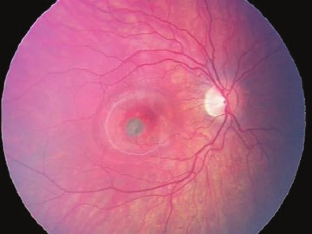
Fig. 2. Spectral HD OCT: Linear horizontal transfoveolar scan of right eye. Significant thickening and increase of volume of macular area, active CNV with medium reflectivity, flat serous ablation of sensory epithelium around. Thickness of juxtafoveolar area 680 μm at the time of diagnosis 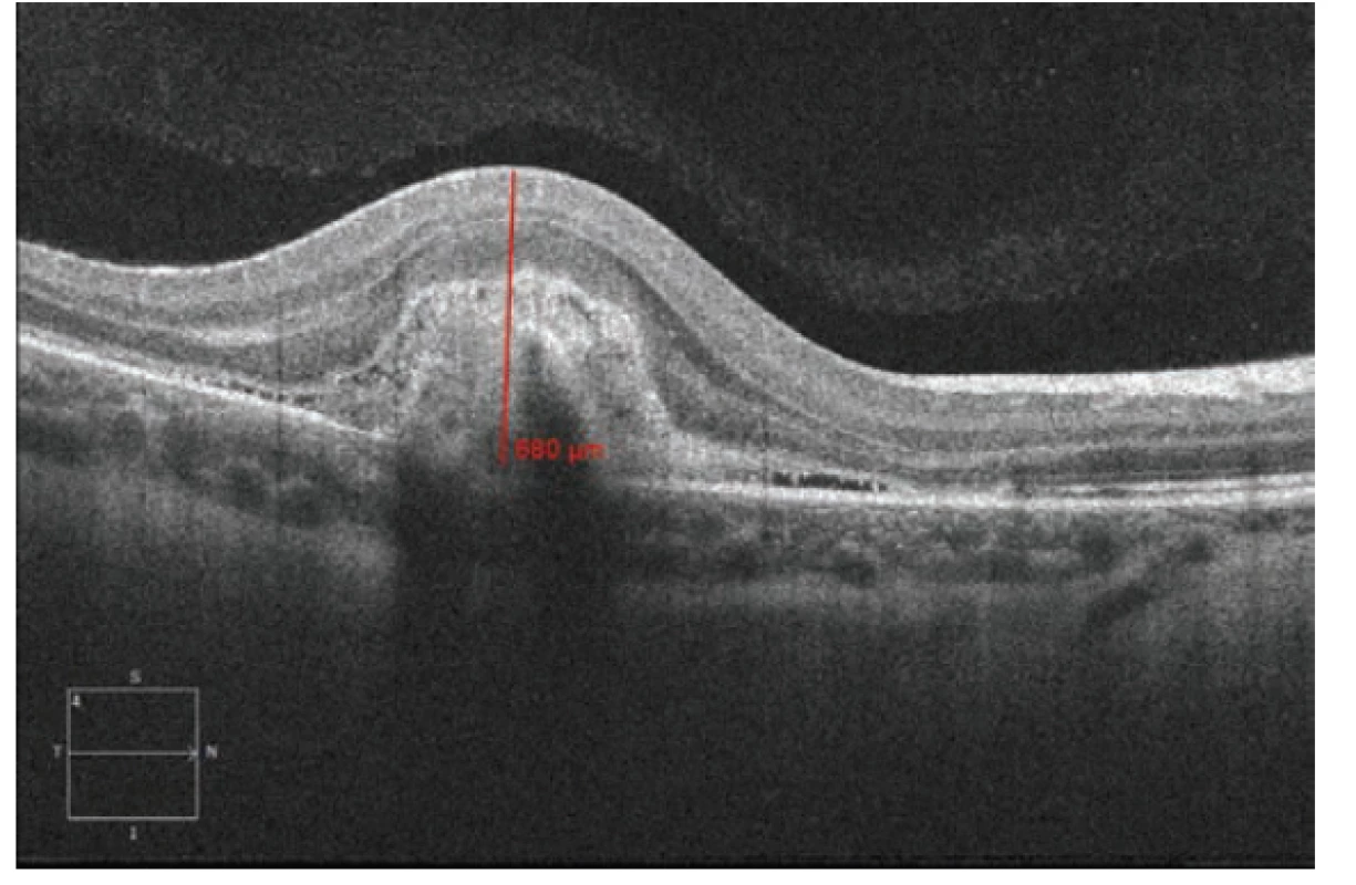
Fig. 3. Fluorescein angiography (FAG) OD: active classic choroidal neovascularisation in juxtafoveolar area 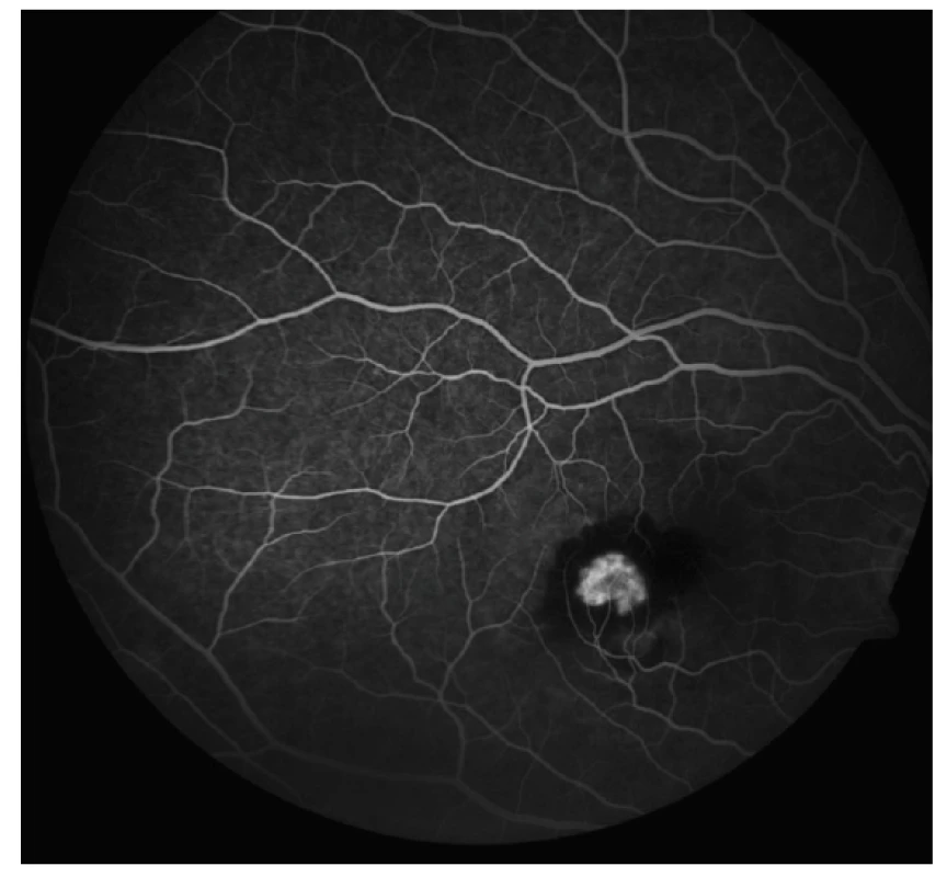
Basic samples were taken from the girl, including specific laboratory tests for uveitis and anthropozoonosis, all without a pathological finding. With regard to mild myopia and the absence of a finding of a primary ocular pathology, we concluded an idiopathic etiology of CNV. At COC, following consultation with the macular centre of the Clinic of Eye Treatments at Brno Bohunice University Hospital, we applied to the girl 3 injections of ranibizumab (Lucentis) intravitreally, 0.5 mg in 0.05 ml at monthly intervals. With regard to the girl's sensitivity and following consultation with her parents, application took place in standard aseptic conditions always under brief general anaesthesia. No complications were recorded after application.
The development of the pathology was regularly documented by means of spectral HD OCT, photo documentation of the ocular fundus using a fundus camera Oculus and RetCAM.
At a follow-up examination one week after the first intravitreal injection of ranibizumab, according to OCT there was a pronounced anatomical effect with a reduction of the thickness of the juxtafoveolar region from 680 um to 540 um. A functional effect was not yet perceptible, BCVA remained unchanged at 1.5 metres - fingers. At a follow-up examination after the following 2 weeks, a functional effect was now perceptible, i.e. BCVA RE 0.1, and the girl stated a marked subjective improvement. A further regression of the macular thickness of the retina in the juxtafoveolar region was evident to 460 um.
One week after the second IVT injection of Lucentis, a further pronounced functional effect was perceptible, in which BCVA RE was 0.5. Two weeks after the third intravitreal application of ranibizumab, there was a functional effect with improvement to BCVA RE 0.9 and a reduction of macular thickness in the juxtafoveolar region to 365 um (fig. 4). No complications were recorded during the course of observation. In long-term observation (˃18 months) a stabilisation took place, without progression of the pathology (fig. 5).
Fig. 4. Spectral HD OCT Linear horizontal macular scan after 3 intravitreal injections of ranibizumab (Lucentis) at monthly intervals with significant regression of macular thickness and volume. Regression of macular thickness of juxtafoveolar area from 680 μm to 365 μm 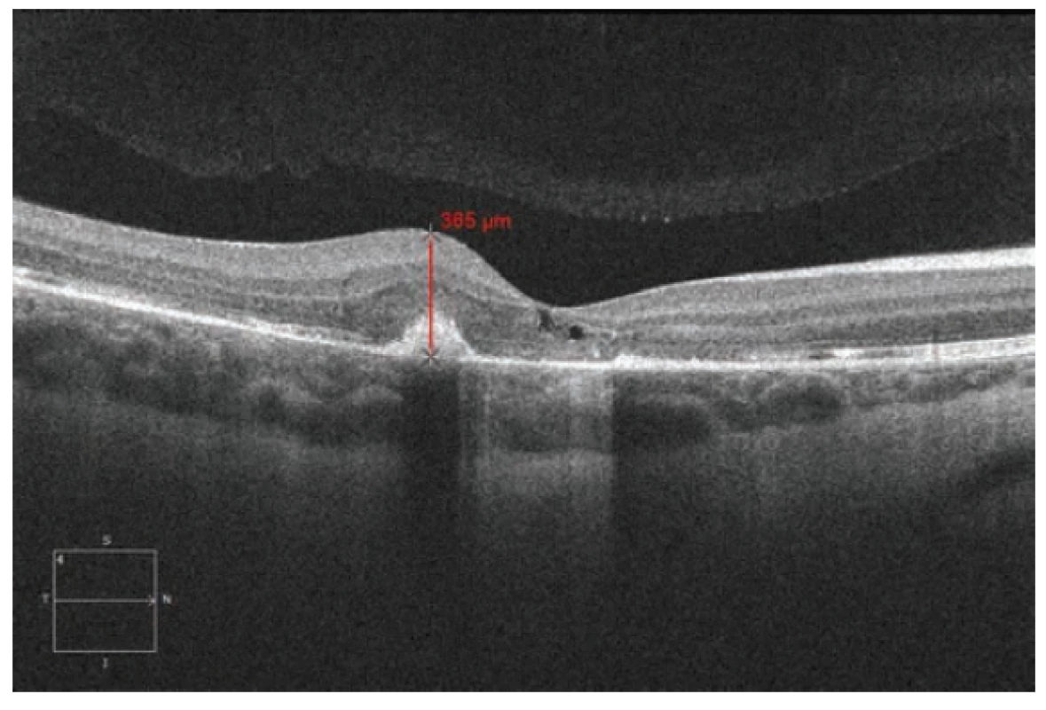
Fig. 5. Spectral HD OCT Linear horizontal macular scan after 3 intravitreal injections of ranibizumab (Lucentis) at long-term follow-up (˃ 12 months) with stable macular finding, without progression 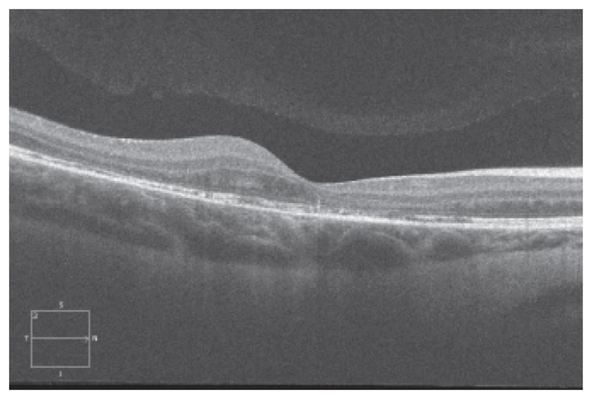
DISCUSSION
Barth et al. [1] in their monocentric retrospective study (2008-2016) document the etiology, clinical course and results of treatment in 10 child patients aged under 18 years (average age 13.9 ± 1.9 years; range 11-16 years) with CNV. The most common cause of CNV in this cohort of children was choroidal osteoma (30%), followed by hereditary macular dystrophies (20%). In other child patients there was a development of CNV as a consequence of degenerative myopia, punctate inner choroidopathy (n=1) or angioid streaks (n=1). In 20% of the children (n=2), CNV was indicated as idiopathic, without evidence of other ocular pathology. Other publications state the most frequent cause as post-infection and uveitic affection [11]. Rishi et al. [11] retrospectively document a cohort of 36 eyes of 27 children aged under 18 years with CVN over the period of 1978–2008. The most frequently stated cause is post-inflammatory (41% eyes), followed by Best's macular dystrophy (25% eyes), 4 patients (11% of eyes) are described with idiopathic CNV. Idiopathic CNV as a clinical unit in child patients is described also in other studies [2, 7, 14].
In the study by Barth et al. [1], in all children this primarily concerned unilateral CNV. In 20% of patients, CNV developed also in the other eye during the course of observation. This concerned patients with choroidal osteoma and hereditary macular dystrophy, and the pathology in the other eye developed within the course of 4 and 2 years respectively since the first visit. Rishi et al. [11] state bilateral CNV in 33% of children. In the studies the classic type of CNV predominates (60% and 100% respectively) [1, 10]. This may be connected with the good condition of the Bruch's membrane and preserved RPE in child patients in contrast with changes in patients with ARMD [1]. In the studies there is a predominance of subfoveolar localisation of CNV over extrafoveal localisation [1, 11].
No precise therapeutic procedure is stipulated for CNV in children, and this depends on the activity of the process, localisation of the lesion, as well as on the type of background ocular pathology. In previous studies, photodynamic therapy (PDT) was published as the fundamental primary treatment [5, 9-11]. With the possibility of application of anti-VEGF preparations, some paediatric centres have published favourable results of this treatment in children with CNV [1, 2, 8, 11, 12]. Barth, Rishi and Spaide [1, 11, 14] state that the course and development of CNV in child patients is far more favourable than in adult patients with ARMD. Barth et al. [1] did not record any complications of treatment with the aid of intravitreally applied injections of anti-VEGF preparations during the course of observation (average observation period 35 months, range 6-120 months). The preparations used are Bevacizumab (Avastin) 1.25 mg in 0.05 ml, and ranibizumab (Lucentis) 0.5 mg in 0.05 ml [1]. For stabilisation of 90% of the treated patients, 3 injections per eye were applied at monthly intervals [1]. Barth et al. [1] published average initial BCVA at 0.3 (0.1-0.8). At the end of the observation period, average final BCVA was only 0.4 (0.1-0.8). In 5 patients there was an improvement by 1-5 rows. Kim et al. [8] state an improvement of BCVA from 5/30 to 5/15. In our case report we recorded a highly beneficial effect of treatment of CNV with the aid of anti-VEGF, and an improvement of BCVA from 1.5 metres – fingers - to 5/5 sl.
CONCLUSION
Idiopathic active choroidal neovascular membrane in paediatric patients is a very rare ocular pathology. It most frequently afflicts the patient unilaterally, and without treatment represents serious damage to sight. No precisely recommended procedure is stipulated in the treatment of CNV in children. The available therapeutic modality is nevertheless provided by anti-VEGF preparations, which represent an effective stabilisation of this clinical unit and enable a marked improvement of central visual acuity.
Received by the Editorial Department on: 14 October 2018
Accepted for printing on: 5 December 2018
MUDr. Inka Krejčířová, Ph.D.
Children's Ophthalmology Clinic, PDM, University Hospital and Faculty of Medicine of the Masaryk University in Brno
Černopolní 9
613 00 Brno
Zdroje
1. Barth, T., Zeman, F., Helbig, H. et al.: Etiology and treatment of choroidal neovascularization in pediatric patients. Eur J Ophthalmol, 26(5); 2016 : 388-93.
2. Carneiro, AM., Silva, RM., Veludo, MJ. et al.: Ranibizumab treatment for choroidal neovascularization from causes other than age-related macular degeneration and pathological myopia. Ophthalmologica, 225(2); 2011 : 81-88.
3. Chhablani, J., Jalali, S.: Intravitreal bevacizumab for choroidal neovascularization secondary to Best vitelliform macular dystrophy in a 6-year-old child. Eur J Ophthalmol, 22(4); 2012 : 677-679.
4. Cohen, SY., Laroche, A., Leguen, Y. et al.: Etiology of choroidal neovascularization in young patients. Ophthalmology, 103(8); 1996 : 1241-1244.
5. Giansanti, F., Virgili, G., Varano, M., et al.: Photodynamic therapy for choroidal neovascularization in pediatric patients. Retina, 25(5); 2015 : 590-596.
6. Goshorn, EB., Hoover, DL., Eller, AW. et al.: Subretinal neovascularization in children and adolescents. J Pediatr Ophthalmol Strabismus, 32(3); 1995 : 178-182.
7. Jain, K., Shafiq, AE., Devenyi, RG.: Surgical outcome for removal of subfoveal choroidal neovascular membranes in children. Retina, 22; 2002 : 412–417.
8. Kim, R., Kim, YC.: Intravitreal ranibizumab injection for idiopathic choroidal neovascularization in children. Semin Ophthalmol, 29(3); 2014 : 178-181.
9. Lipski, A., Bornfeld, N., Jurklies, B.: Photodynamic therapy with verteporfin in paediatric and young adult patients: long-term treatment results of choroidal neovascularisations. Br J Ophthalmol, 92(5); 2008 : 655-660.
10. Mimouni, KF., Bressler, SB., Bressler, NM.: Photodynamic therapy with verteporfin for subfoveal choroidal neovascularization in children. Am J Ophthalmol, 135(6); 2003 : 900-902.
11. Rishi, P., Gupta, A, Rishi, E. et al.: Choroidal neovascularization in 36 eyes of children and adolescents. Eye, 27(10); 2013 : 1158-68
12. Sisk, RA., Berrocal, AM., Albini, TA. et al.: Bevacizumab for the treatment of pediatric retinal and choroidal diseases. Ophthalmic Surg Lasers Imaging, 41(6); 2010 : 582-592.
13. Sivaprasad, S., Moore, AT.: Choroidal neovascularisation in children. Br J Ophthalmol, 92(4); 2008 : 451-454.
14. Spaide, RF.: Choroidal neovascularization in younger patients. Curr Opin Ophthalmol, 10(3); 1999 : 177-181.
15. Wilson, ME., Mazur, DO.: Choroidal neovascularization in children: report of five cases and literature review. J Pediatr Ophthalmol Strabismus, 25(1); 1088 : 23-29.
Štítky
Oftalmologie
Článek SILENT SINUS SYNDROM
Článek vyšel v časopiseČeská a slovenská oftalmologie
Nejčtenější tento týden
2018 Číslo 6- Stillova choroba: vzácné a závažné systémové onemocnění
- Familiární středomořská horečka
- První schválený léčivý přípravek pro terapii Leberovy hereditární optické neuropatie dostupný rovněž v ČR
- Diagnostický algoritmus při podezření na syndrom periodické horečky
- Možnosti využití přípravku Desodrop v terapii a prevenci oftalmologických onemocnění
-
Všechny články tohoto čísla
- VIRTIOL – SIMULACE KVALITY VIDĚNÍ S MULTIFOKÁLNÍMI A EDOF INTRAOKULÁRNÍMI ČOČKAMI
- KORTIKOSTEROIDY INDUKOVANÁ ZADNÍ SUBKAPSULÁRNÍ KATARAKTA
- OČNÍ PROJEVY U PACIENTŮ S HIV INFEKCÍ
- VÝZNAM ZHODNOCENÍ VÝVOJE OCT OBRAZU PŘI KONZERVATIVNÍM ŘEŠENÍ VITREOMAKULÁRNÍ TRAKCE KOMPLIKOVANÉ MAKULÁRNÍ DÍROU
- SILENT SINUS SYNDROM
- IDIOPATICKÁ CHOROIDÁLNÍ NEOVASKULÁRNÍ MEMBRÁNA U 12LETÉ DÍVKY
- SCREENING, LÉČBA A DLOUHODOBÉ SLEDOVÁNÍ RETINOPATIE PŘEDČASNĚ NAROZENÝCH DĚTÍ V ČR
- OČNÍ KLINIKA 1. LÉKAŘSKÉ FAKULTY UNIVERZITY KARLOVY A VŠEOBECNÉ FAKULTNÍ NEMOCNICE V PRAZE SLAVÍ 200 LET OD SVÉHO ZALOŽENÍ
- Vážený a milý pán doc. MUDr. Tomáš Mazalán, CSc.
- Česká a slovenská oftalmologie
- Archiv čísel
- Aktuální číslo
- Informace o časopisu
Nejčtenější v tomto čísle- OČNÍ PROJEVY U PACIENTŮ S HIV INFEKCÍ
- SILENT SINUS SYNDROM
- VIRTIOL – SIMULACE KVALITY VIDĚNÍ S MULTIFOKÁLNÍMI A EDOF INTRAOKULÁRNÍMI ČOČKAMI
- KORTIKOSTEROIDY INDUKOVANÁ ZADNÍ SUBKAPSULÁRNÍ KATARAKTA
Kurzy
Zvyšte si kvalifikaci online z pohodlí domova
Autoři: prof. MUDr. Vladimír Palička, CSc., Dr.h.c., doc. MUDr. Václav Vyskočil, Ph.D., MUDr. Petr Kasalický, CSc., MUDr. Jan Rosa, Ing. Pavel Havlík, Ing. Jan Adam, Hana Hejnová, DiS., Jana Křenková
Autoři: MUDr. Irena Krčmová, CSc.
Autoři: MDDr. Eleonóra Ivančová, PhD., MHA
Autoři: prof. MUDr. Eva Kubala Havrdová, DrSc.
Všechny kurzyPřihlášení#ADS_BOTTOM_SCRIPTS#Zapomenuté hesloZadejte e-mailovou adresu, se kterou jste vytvářel(a) účet, budou Vám na ni zaslány informace k nastavení nového hesla.
- Vzdělávání


