
Transverse spinal cord lesion after embolization of a vascular lesion at the Th11 level: a case report of a severe complication of interventional treatment
Autoři:
A. Krasny; A. Sukop; M. Ouzky
Působiště autorů:
Department of Plastic Surgery, University Hospital Královské Vinohrady, Prague, Czech Republic
Vyšlo v časopise:
ACTA CHIRURGIAE PLASTICAE, 68, 1-2, 2026, pp. 42-45
doi:
https://doi.org/10.48095/ccachp202642
Krasny A, Sukop A, Ouzky M. Transverse spinal cord lesion after embolization of a vascular lesion at the level of Th11: a case report of a severe complication of interventional treatment. Acta Chir Plast 2026; 68 (1–2): 42–45.
Introduction
Vascular lesions of soft tissues and the spine represent a heterogeneous group of pathologies, including both hemangiomas and vascular malformations. These two entities differ fundamentally in pathophysiology, growth behavior, and therapeutic approach.
Hemangiomas are benign proliferating endothelial tumors, typically occurring in childhood and often undergoing spontaneous regression. In adults, they are less common and usually stable. Histologically, they consist of proliferating capillaries or larger vessels with a characteristic appearance. Spinal hemangiomas, particularly vertebral, are often asymptomatic and diagnosed incidentally on imaging. However, with significant expansion or posterior vertebral involvement, they may cause pain, neurological deficit, or pathological fracture [1,2].
Vascular malformations are congenital structural anomalies of the vascular system. According to International Society for the Study of Vascular Anomalies (ISSVA) classification, they are categorized as arteriovenous, venous, capillary, or lymphatic malformations. From the perspective of spinal ischemia, arteriovenous malformations (AVMs) and dural arteriovenous fistulas (dAVFs) are the most high-risk, as they may have direct connections to the spinal vascular network. These lesions grow with the patient, do not undergo involution, and may remain asymptomatic until complications arise [3,4].
Diagnosis relies primarily on imaging. Contrast-enhanced MRI is the modality of choice for assessing the extent, localization, and relation to the spinal cord and soft tissues. Hemangiomas typically show high signal on both T1 - and T2-weighted images, sometimes with cortical changes or focal sclerotic zones. Malformations often present variably, frequently with flow-void phenomena [5].
Definitive differentiation between hemangiomas and vascular malformations often requires digital subtraction angiography (DSA), which allows detailed visualization of vascular supply, arteriovenous shunts, and potential connections to spinal vasculature. Selective spinal angiography with detailed phases is the only method capable of revealing hidden feeding arteries, e. g., connection to the artery of Adamkiewicz [6].
Treatment depends on lesion type, clinical condition, and localization. Symptomatic hemangiomas may require surgical resection, often combined with preoperative embolization to reduce intraoperative bleeding risk. Vascular malformations require a multidisciplinary approach, including embolization, radiosurgery, or open resection. For spinal AVMs and dAVFs, the therapeutic goal is occlusion of the pathological shunt while preserving physiological spinal artery perfusion [7,8].
Particularly for lesions near the spine and spinal cord, extreme caution is necessary, both in indication for intervention and interpretation of imaging findings. Ideally, multimodal imaging should be combined with repeated multidisciplinary review by neuroradiologists, neurosurgeons, and interventional radiologists.
Case report
Patient: 45-year-old female, without significant medical or neurological history (Fig. 1).
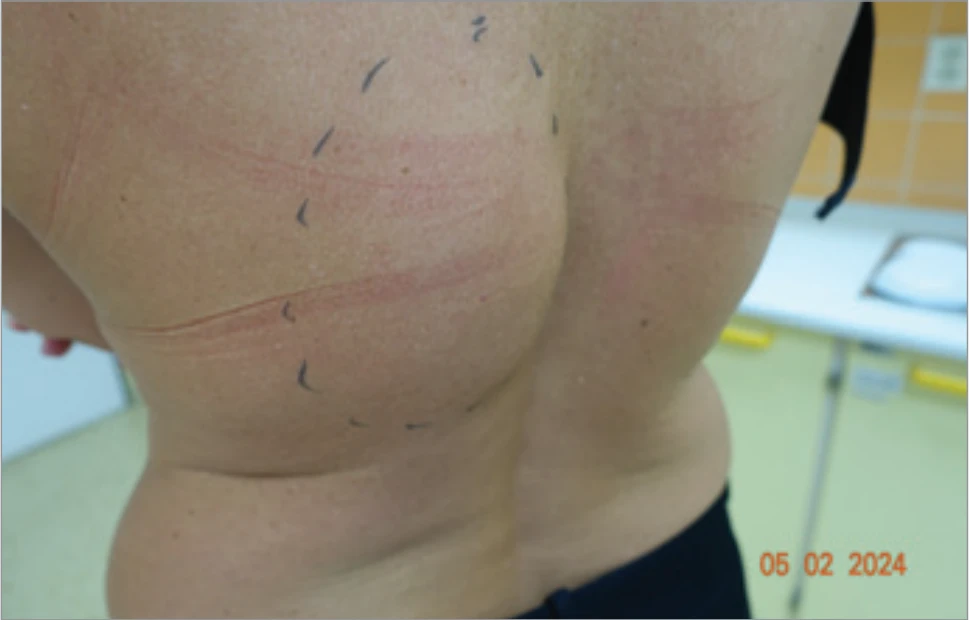
The patient presented with a palpable mass in the right paravertebral region (Th10–Th12), present for several years, without neurological deficit. MRI (Fig. 2) revealed a hypervascularized lesion consistent with hemangioma of the erector spinae muscle. The lesion was interpreted as a hemangioma without direct contact with the spinal canal. Images were subsequently reviewed at our institution, where a radiologist confirmed the original interpretation.
After multidisciplinary consultation (plastic surgery, neurosurgery, interventional radiology), preoperative embolization was planned to minimize intraoperative blood loss. In the case of complete embolization of the two main feeding arteries, surgical resection might not be required. The interventional radiologist assessed vascular supply on MRI and indicated embolization.
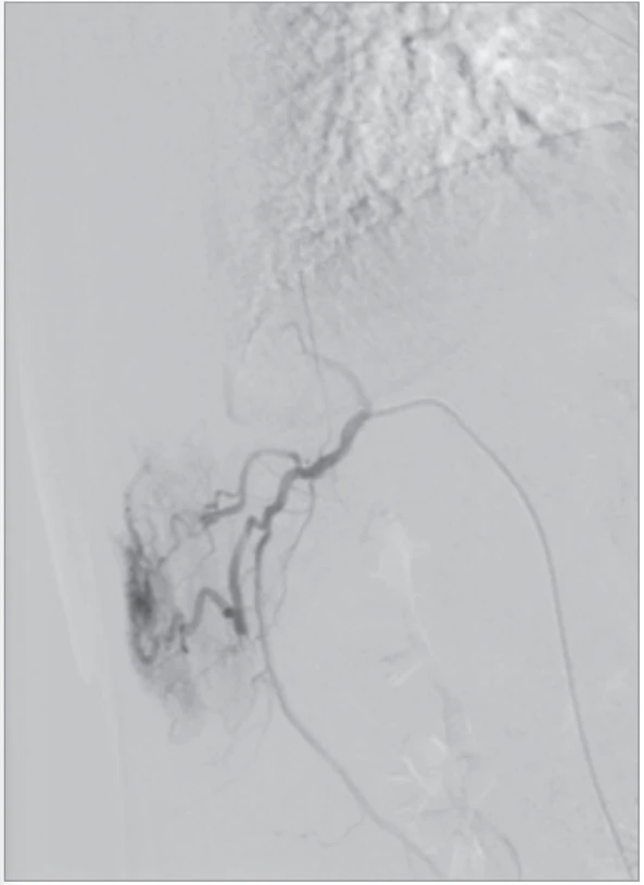
The patient was admitted to the ward the day before surgery, with embolization scheduled the same day. Angiography (Fig. 3) confirmed the MRI findings –the lesion was supplied by the right intercostal artery Th11. No spinal branches were apparent. Selective catheterization and embolization of the feeding vessel were performed.
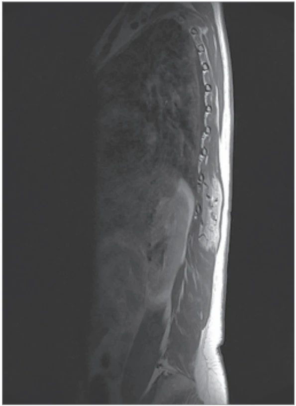
Immediately after embolic agent application, the patient experienced severe back pain with transient alteration of consciousness. Analgosedation was administered (tramadol, midazolam, metamizole). At the end of the procedure, she reported only residual discomfort. The procedure was considered technically successful. Within hours of returning to the ward, the patient developed hypesthesia of the lower limbs. Urgent neurological consultation and spinal MRI revealed signal changes in the gray matter from Th8 to Th11, with mild swelling of the spinal cord at Th11 and signs of ischemic edema. The lesion showed marked vascularization with expansile features.
Clinically, acute transverse spinal cord lesion with paraplegia was diagnosed. Sensory level was at Th12/L1; upper limbs were unaffected. The patient was urgently transferred to the neurological intensive care unit for acute spinal cord injury management. After stabilization, she was transferred to the spinal unit at University Hospital Motol, where she continued neurorehabilitation under specialized care.
The patient’s neurological deficit proved persistent. At the latest follow-up (18 months after the event), she remains paraplegic with sensory level at Th12/L1 and is dependent on a wheelchair for mobility. There has been no meaningful recovery of voluntary motor function in the lower limbs despite an intensive neurorehabilitation program addressing spasticity, bladder management and activities of daily living. She requires assistance for transfers and personal care. The extent and pattern of injury on MRI (diffuse ischemic/edematous changes of the gray matter from Th8–Th11) correlate with poor prognosis for motor recovery; long-term management focuses on secondary prevention, complication surveillance and functional adaptation.
Discussion
At our institution, management of symptomatic spinal/paraspinal vascular lesions is individualized and relies on a multidisciplinary team including neuroradiology, interventional radiology, neurosurgery, plastic surgery, anesthesiology and rehabilitation medicine. For symptomatic hemangiomas we favor surgical resection when feasible; preoperative embolization is considered only when (DSA clearly demonstrates a dominant feeding vessel or vessels that can be safely occluded, and careful angiographic analysis excludes feeders to the spinal arterial circulation, including the anterior spinal artery or radiculomedullary branches. When embolization is indicated, it is usually scheduled immediately prior to resection (commonly within 24 h) to reduce the chance of bleeding. Interdisciplinary case review and repeat imaging (including selective spinal angiography with a dedicated spinal phase when available) are recommended before any embolization near the vertebral column.
This case highlights the risk of severe complications, even when following standard recommendations. The presumed hemangioma was, in retrospect, more consistent with a vascular malformation with spinal arterial connections that were not visualized on overview or selective angiography.
Similar cases [9,10], though rare, have been described in the literature, particularly with arteriovenous shunts and undetected feeder branches to the anterior spinal artery, where embolization may result in irreversible spinal cord ischemia.
The rationale for minimally invasive embolization to reduce surgical morbidity is understandable, yet for vascular lesions near the spine, extreme caution is essential. Advanced diagnostic modalities, including functional MRI or intraoperative DSA with spinal phase (if available), should be considered.
Conclusion
Preoperative embolization prior to surgical resection may cause severe, irreversible neurological complications if the lesion is misdiagnosed as a hemangioma when in fact it is a vascular malformation with spinal arterial communication. Despite modern imaging, such complications cannot always be avoided. Awareness of these risks is essential for thorough diagnostic evaluation and treatment planning.
Preoperative embolization of presumed hemangiomas can cause severe, irreversible neurological injury if the lesion is in fact a vascular malformation with occult spinal arterial communication. This report is notable because it describes an elective preoperative embolization of a presumed paraspinal hemangioma (based on MRI) that produced catastrophic transverse myelopathy despite apparently negative angiography for spinal feeders. The case therefore contributes a cautionary example to the literature and advocates for stricter pre-embolization protocols, consideration of dedicated spinal angiographic phases, and reinforced multidisciplinary decision-making for lesions adjacent to the spine.
This case is unique as it documents a rare but devastating neurological complication following standard preoperative embolization of a presumed paraspinal hemangioma. Despite modern imaging and adherence to accepted protocols, the patient developed acute transverse myelopathy resulting in permanent paraplegia. The case highlights the diagnostic challenges in distinguishing hemangiomas from vascular malformations, the limitations of current imaging techniques, and the critical importance of a thorough multidisciplinary approach. Reporting such rare adverse events provides valuable insights for clinicians and emphasizes the need for extreme caution when performing embolization near the spinal cord.
Roles of the authors
Adam Krásný – first author, data collection, manuscript drafting; Andrej Sukop – corresponding author, supervision, manuscript revision; Martin Ouzký – coauthor, data collection.
Disclosure
The authors have no conflicts of interest to disclose. The authors declare that this study has received no financial support. All procedures performed in this study involving human participants were in accordance with ethical standards of the institutional and/or national research committee and with the Helsinki declaration and its later amendments or comparable ethical standards.
Zdroje
1. Mulliken JB., Glowacki J. Hemangiomas and vascular malformations in infants and children: a classification based on endothelial characteristics. Plast Reconstr Surg. 1982, 69 (3): 412–422.
2. Fox MW., Onofrio BM. The natural history and management of symptomatic and management of symptomatic and asymptomatic vertebral hemangiomas. J Neurosurg. 1993, 78 (1): 36–45.
3. Lasjaunias P., ter Brugge KG., Berenstein A. Surgical Neuroangiography: Vol. 3. Clinical and Interventional Aspects in Children. 2nd ed. New York: Springer, 2006.
4. Krings T., Geibprasert S. Spinal dural arteriovenous fistulas. AJNR Am J Neuroradiol. 2009, 30 (4): 639–648.
5. Da Ros V., Picchi E., Ferrazzoli V., et al. Spinal vascular lesions: anatomy, imaging techniques and treatment. Eur J Radiol Open. 2021, 8 : 100369.
6. Hong MK., Hong MK., Pan WR., et al. The angiosome territories of the spinal cord: exploring the issue of preoperative spinal angiography. Laboratory investigation. J Neurosurg Spine. 2008, 8 (4): 352–364.
7. Maimon S., Luckman Y., Strauss I. Spinal dural arteriovenous fistula: a review. Adv Tech Stand Neurosurg. 2016, 43 : 111–137.
8. Da Costa L., Dehdashti AR., terBrugge KG. Spinal cord vascular shunts: spinal cord vascular malformations and dural arteriovenous fistulas. Neurosurg Focus. 2009, 26 (1): E6.
9. Srinivasan G., Moses V., Padmanabhan A., et al. Utility of spinal angiography and arterial embolization in patients undergoing CT guided alcohol injection of aggressive vertebral hemangiomas. Neuroradiology. 2021, 63 (11): 1935–1945.
10. Fiore G., Bertani GA., Tariciotti L., et al. Vertebral body infarction after transarterial preoperative embolization of a vertebral hemangioma. J Neurol Surg A Cent Eur Neurosurg. 2023, 84 (6): 588–592.
Štítky
Chirurgie plastická Ortopedie Popáleninová medicína TraumatologieČlánek vyšel v časopise
Acta chirurgiae plasticae
2026 Číslo 1-2
- Potenciál vinpocetinu v léčbě kardiovaskulárních onemocnění
- Aktuální evropská doporučení pro léčbu renální koliky v důsledku urolitiázy
- Hybridní kooperující komplexy v léčbě osteoartrózy
Nejčtenější v tomto čísle
- A systematic review of sensory outcomes in nipple reinnervation after implant-based breast reconstruction
- Principles of nasal framework reconstruction for complex nasal defects: review
- Self-esteem and quality of sexual life in women undergoing breast reconstruction due to mastectomy: a cross-sectional study in a non-European setting
- Cutting-edge biotechnological strategies to enhance autologous fat graft survival