
Experimental surgery as part of the development of degradable biomaterials for cardiovascular surgery
Experimentální chirurgie jako součást vývoje degradabilních biomateriálů v kardiovaskulární chirurgii
Úvod: Kardiovaskulární choroby jsou zodpovědné za významnou morbiditu i mortalitu ve společnosti. Užití umělých cévních materiálů je často nezbytnou součástí v rámci chirurgické léčby, ať již je tato radikální nebo paliativní. V současné době dochází k vývoji řady nových biodegradabilních materiálů určených pro tyto účely. Preklinické testování každého nového materiálu je naprosto nezbytné, je prováděno jak in vitro, tak in vivo. Z tohoto důvodu jsou zvířecí experimentální modely nadále nutnou součástí testování před klinickým užitím. Cílem této práce je prezentovat možnosti užití různých zvířecích modelů na poli kardiovaskulární chirurgie a jejich extrapolace do klinické medicíny.
Metody: Autoři prezentují jejich obecné zkušenosti s experimentální chirurgií, na jejich podkladě diskutují optimální výběr zvířecího modelu pro testování nových materiálů pro kardiovaskulární chirurgii a stejně tak optimální lokalitu implantace.
Výsledky: Jako optimální experimentální zvířecí modely pro testování hemokompatibility a degradability nových materiálů uvádějí autoři modely potkana, králíka a prasete. Intraperitoneální implantace u potkana je snadná a lehce proveditelná procedura, stejně tak jako arteriální bandáž na aortě králíka či prasete. Rovněž karotické tepny jsou dobře využitelné. Bandáž na prasečí pulmonální tepně je již složitější zákrok s četnějšími komplikacemi. Explantované bandážované cévy po předem definované době jsou vhodné pro další mechanické testování ve smyslu biomechanických analýz, např. inflačně-extenzního testu.
Závěr: V posledních fázích preklinického testování nových materiálů se nelze nadále obejít bez in-vivo experimentů. Naší snahou je však striktně dodržovat koncept 3R – Replacement, Reduction a Refinement. V tomto smyslu je třeba využít co nejvíce potenciál každého zvířete tak, abychom mohli redukovat počty zvířat.
Klíčová slova:
experimentální chirurgie – kardiovaskulární chirurgie – model potkana – model králíka – model prasete
Authors:
J. Moláček 1,5; L. Vištejnová 1; P. Klein 1; T. Suchý 2; L. Horný 3; E. Kuželová Košťáková 4; M. Kindermann 1; H. Chlup 3; V. Jenčová 4; D. Lukáš 4; M. Šupová 2; I. Říha 5; V. Soukupová 5; V. Třeška 5
Authors place of work:
Biomedicínské centrum, Lékařská fakulta Karlovy University v Plzni
1; Oddělení kompozitních a uhlíkových materiálů, Ústav struktury a mechaniky hornin, Česká akademie věd, Praha
2; Strojní fakulta, České vysoké učení technické v Praze
3; Katedra chemie, Technická univerzita Liberec
4; Chirurgická klinika, Fakultní nemocnice Plzeň
5
Published in the journal:
Rozhl. Chir., 2022, roč. 101, č. 12, s. 599-606.
Category:
Původní práce
doi:
https://doi.org/10.33699/PIS.2022.101.12.599–606
Summary
Introduction: Cardiovascular diseases are responsible for significant morbidity and mortality in the population. Artificial vascular grafts are often essential for surgical procedures in radical or palliative treatment. Many new biodegradable materials are currently under development. Preclinical testing of each new material is imperative, both in vitro and in vivo, and therefore animal experiments are still a necessary part of the testing process before any clinical use. The aim of this paper is to present the options of using various experimental animal models in the field of cardiovascular surgery including their extrapolation to clinical medicine.
Methods: The authors present their general experience in the field of experimental surgery. They discuss the selection process of an optimal experimental animal model to test foreign materials for cardiovascular surgery and of an optimal region for implantation.
Results: The authors present rat, rabbit and porcine models as optimal experimental animals for material hemocompatibility and degradability testing. Intraperitoneal implantation in the rat is a simple and feasible procedure, as well as aortic banding in the rabbit or pig. The carotid arteries can also be used, as well. Porcine pulmonary artery banding is slightly more difficult with potential complications. The banded vessels, explanted after a defined time period, are suitable for further mechanical testing using biomechanical analyses, for example, the inflation-extension test.
Conclusion: An in vivo experiment cannot be avoided in the last phases of preclinical research of new materials. However, we try to strictly observe the 3R concept – Replacement, Reduction and Refinement; in line with this concept, the potential of each animal should be used as much as possible to reduce the number of animals.
Keywords:
experimental surgery – cardiovascular surgery – rat model – rabbit model – porcine model
INTRODUCTION
Cardiovascular diseases (CVD) still remain one of major causes of death worldwide. They are responsible for significant morbidity and therefore both non-invasive and especially invasive treatments continue to be developed [1−3]. In the past, a significant change in surgical treatment of vascular disorders was achieved upon the introduction of artificial vascular grafts in vascular surgery in the fifties and later in cardiac surgery [4−6]. Since that time, foreign materials became indispensable in cardiovascular surgery. Related to that is the need for preclinical testing of each new material. Hundreds of experiments both in vitro and in vivo have to be performed before introducing a new material or device to clinical practice.
Animal experiments are crucial in testing new devices not only in cardiovascular surgery. Whatever is to be implanted in the human body must go through extensive testing processes. For many people, animal experiments are still controversial, but the truth is that current medical research of vascular pathology is unimaginable without these experiments [7−11]. Of course, many ethical aspects apply to animal experiments. Even in the 21st century, the rules postulated by zoologist William Russell and microbiologist Rex Burch in the late fifties are valid [12]. They summarized them as the 3Rs: Replacement, Reduction and Refinement. In recent years, great emphasis has been placed on refinement. Any procedures affecting the quality of life of experimental animals should be refined as much as possible. The goal is to avoid any painful and stressful stimuli during the whole experiment. Only employees with appropriate education and experience should work with the animals. These are then usually supervised by people with university education [13]. Very strict rules apply to the use of experimental animals in the Czech Republic as well as in the whole European Union.
We aim to develop a resorbable fabric based on a composite material consisting of a synthetic poly(lactide - co-caprolactone) copolymer (PLCL) nanofibrous reinforcement combined with a collagen matrix for use in different indications in cardiovascular surgery. The point is to perform a comprehensive in vitro, in vivo and ex vivo evaluation of the composite materials based on their biodegradability and changes in their structural and mechanical properties.
The aim of this paper is not to present our research data; we would like to present the options of using various experimental animal models in the field of cardiovascular surgery including their extrapolation to clinical medicine; additionally, to highlight their advantages and disadvantages and to discuss optimal animal models for various purposes, various vessel types and sizes. Finally, potential options for the replacement of animal models with an in vitro test are discussed.
Status quo
Cardiovascular surgery of the 21st century cannot exist without artificial vascular grafts. They are used for vascular bypasses, vascular patches, covered stent grafts or external vessel support. There are several procedures in the field of cardiovascular surgery which are based on the mechanical interaction of the arterial wall and the external support. Pulmonary artery banding (PAB) in infant patients can be mentioned as a typical example. PAB is a palliative procedure which reduces pulmonary over-circulation in neonates suffering from certain congenital heart diseases and makes up the first stage of intervention prior to complete repair of the cardiac defects [14]. Wrapping, which is usually accompanied by clipping (clip-wrapping), is another example. The clip-wrapping procedure is used to treat, for example, blood blister-like aneurysms of the supraclinoid internal carotid artery and fusiform aneurysms of the anterior cerebral artery. The wrap tightly surrounds the artery and prevents the aneurysm from expanding even further. The wrapping also reinforces the arterial wall and minimizes the risk of aneurysm rupture [15]. Other examples of the vascular banding indication include the banding of an arteriovenous shunt for hemodialysis to avoid the peripheral steal syndrome [16]. The banding of an abdominal aortic aneurysm after endovascular aneurysm repair (EVAR) where the endoleak or endotension is a risky complication can also be a potential indication for the use of an external vascular support. Ramadan et al. reported successful use of the aortic wrap in the treatment of retrograde aortic dissection in the ascending aorta [17]. All these clinical scenarios benefit from external banding of arterial walls. In addition to above mentioned clinical cases, it is worth noting that studies investigating the application of an external support in arterialized vein grafts have suggested that the reduction of the initial graft over-distension by the external support leads to reduced intimal hyperplasia in the graft. We believe that there will be other indications for the use of the resorbable material in cardiovascular surgery in the future.
Tested materials
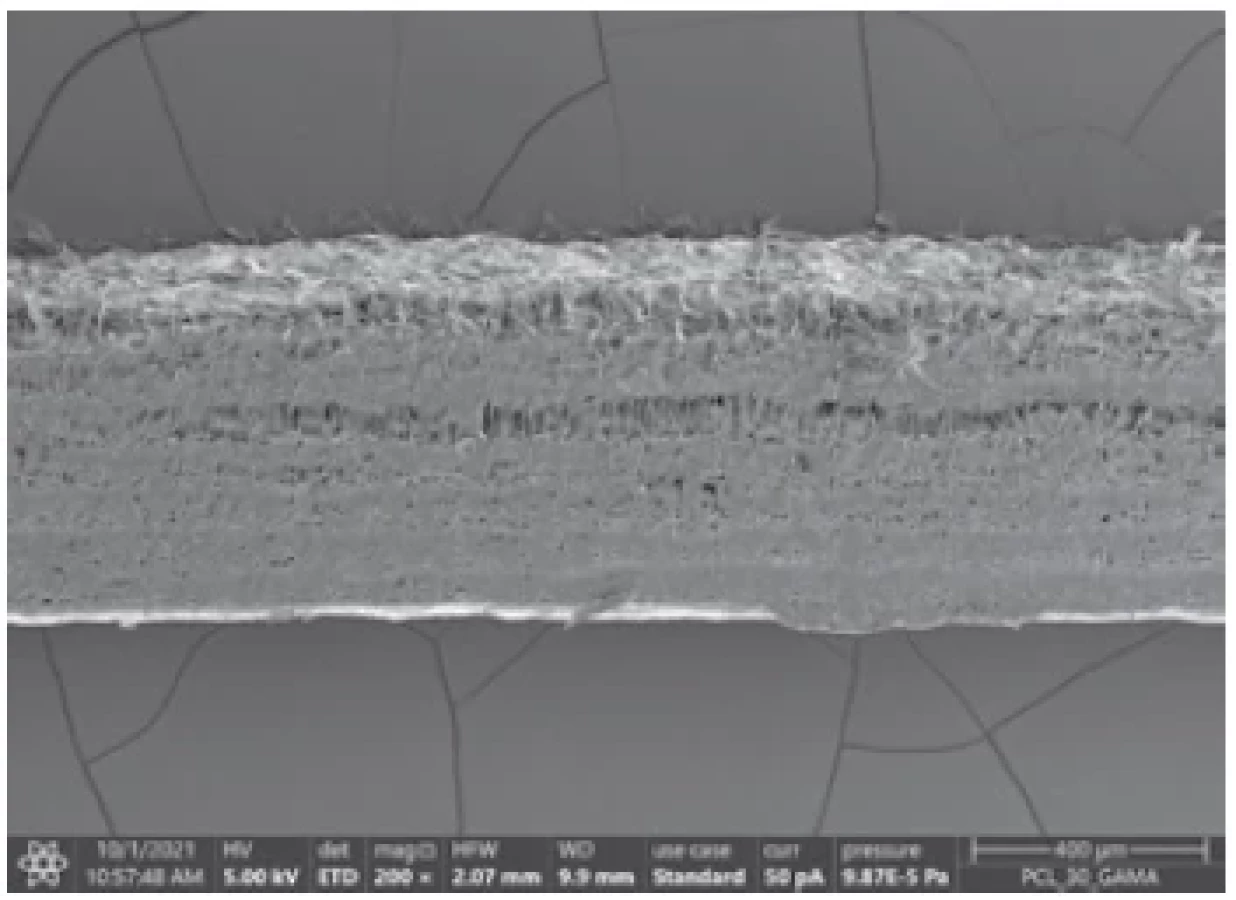
Composite bandings were prepared from nanofibrous layers based on L-lactide/caprolactone εcopolymer (PLCL; 70/30 mol/mol; Purasorb PLC 7015, Corbion, The Netherlands) or polycaprolactone (PCL; Merck, USA). The nanofibrous layers were prepared by needleless electrospinning (Production line NS 1S500U with precisely controlled air conditioning unit NSAC150, Elmarco, Czech Republic) under direct current from 10 wt% (PLCL) or from a 16 wt% (PCL) solution of chloroform and ethanol (Penta, Czech Republic) in the 8 : 2 weight ratio. Both kinds of the layers were prepared in two different surface densities, i.e. 15 or 30 g/m2, both with an average fiber diameter of 50–1,000 nm (Fig. 1). The matrix of the composite was composed of collagen type I (calf skin, VUP Medical, Czech Republic). Five separate layers of nanofibrous reinforcement were impregnated by 5 wt% of aqueous dispersion, placed in a form and left for 36 hours at room temperature. The volume fraction of all fibrous reinforcement in the composite was 70±10 vol%. The stability of the collagen matrix was further enhanced by cross-linking with a 95 wt% ethanol solution containing EDC (N-(3-dimethylaminopropyl) - N-ethylcarbodiimide hydrochloride) and NHS (N-hydroxysuccinimide) (4/1 w/w, Sigma Aldrich, USA). The final stage involved sterilization by gamma irradiation (25 kGy, BIOSTER, a.s., Czech Republic).
Degradability in vitro
Verification of degradability of a material using animal models is very limited for various ethical or financial reasons and is often one of the last stages of material development. For these reasons, and also in agreement with the 3R principles, in vitro laboratory simulations are used to replace animal models. Most often, this involves exposure to a certain type of medium under certain conditions (temperature, atmosphere, loading rate, etc.). A variety of solutions, culture media, body fluids, cell cultures or enzymes are used for this purpose. The results of these in vitro simulations should be able to be extrapolated to the in vivo situation at some confidence level. The rate of this extrapolation is the most problematic part. First, there is virtually no comparison of the effects of the different media with each other, and above all, there is almost no possibility of comparison with animal models. It is therefore not clear whether, for example, exposure in physiological solution can answer the question of what will happen to the material during implantation into an animal model or even into a human organism. Nevertheless, different solutions are widely used and the results from these simulations are considered relevant. Verification of the degradation properties of the bandages is an important stage of our project. In our degradation study we focused on the comparison of several simulated environments, namely simple salt solutions (phosphate buffer, physiological solution and Kokub’s SBF mimicking the ionic concentration of the inorganic part of blood plasma), enzymatic media (collagenase, proteinase K), protein-containing media (blood plasma, cell culture medium and culture medium with dermal fibroblasts) with in vivo environment (rat model, peritoneal implantation). Our aim is to select conditions that faithfully mimic the body environment, and in which extensive verification of the degradation of all studied materials in terms of changes in their mechanical and structural properties will be further performed.
Hemocompatibility
In addition to in vitro biodegradation analysis, it is necessary to apply hemocompatibility tests to analyze materials coming into contact with blood (Fig. 2). In our project, the materials are tested using the test of hemolysis – interaction with human erythrocytes, the thrombogenicity test – interaction with native platelets, and the coagulation test – interaction of the material with human plasma. Some materials can damage the erythrocyte membrane, causing hemoglobin to be released into the environment. Analysis of erythrocyte hemolysis after the interaction of materials with diluted whole human blood is based on spectrophotometric detection of hemoglobin at 570 nm. Testing is governed by the standard ČSN EN ISO 10993-4 (855220). The standard states that the percentage of hemolysis after contact of the material with blood must be up to 5%. Due to the contact of platelets with the materials, their activation becomes more or less accelerated. The degree of activation can be monitored by means of a cell viability measurement test (viability then corresponds to the rate of platelet activation). The interaction of materials with native platelets was assessed by measuring platelet viability with the CCK8 assay. Contact of the material with blood plasma may also affect the rate of coagulation. In the case of materials with an anticoagulant effect, the coagulation time is prolonged (the time when thrombin activation is detected). As part of the coagulation analysis, APTT (activated partial thromboplastin time, internal coagulation pathway monitoring) and PT (prothrombin time, also referred to as QUICK test, external coagulation pathway monitoring) tests were performed. It is necessary to test not only the composite bandages as a whole, but also their individual components, thus electrospun nanofibrous materials (Fig. 3) and the material forming the collagen matrix.


Experimental animal models
- Rat model (Rattus norvegicus) The rat is a favored laboratory animal model because of its suitable size, availability, handling and price. In this project, the rat strain Wistar (male, >6 months) is used as the model of a real body environment to analyze the degradation rates of developed composite materials. Two bands (42×6 mm) are implanted in the intraperitoneal wall of each animal and secured by nonabsorbable sutures. The bands are explanted 1, 3 and 6 months after implantation and submitted for histological, mechanical and structural analysis to monitor the interaction of the bands with real tissue and to evaluate the effect of body environment on degradation of the composite materials as reflected by their mechanical and structural changes (Fig. 4). Moreover, the changes in mechanical and structural properties of the composite materials caused by the real body environment are compared with those obtained in simulated body environments as part of in vitro tests. This comparison shed light on the application of simulated body environments with respect to their degree of similarity with the real body conditions.
Fig. 4. The composite material band before implantation (A), bands of composite material implanted intraperitoneally in the rat abdomen (B), the band explanted after 1 month and submitted for subsequent analysis (C) 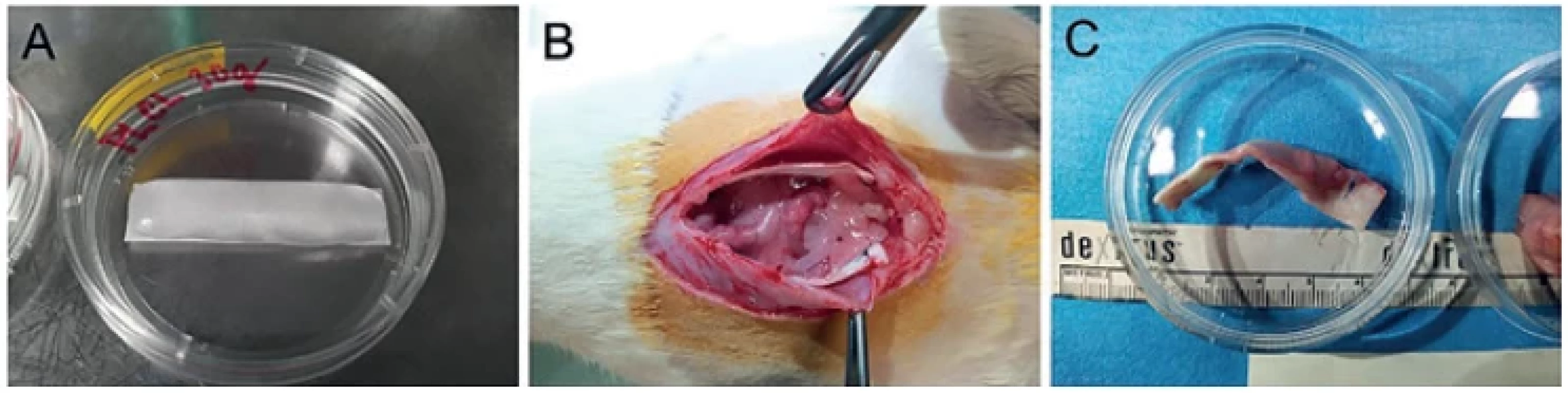
- Rabbit model (Oryctolagus cuniculus f. domesticus) The strain New Zealand White is used as the rabbit model (>4 months of age) to perform experimental vascular surgery on a rodent model. Implantation of the arterial band (cuff) made of a composite material in the abdominal aorta of rabbits and their subsequent 60-day follow-up is a suitable model for safety and functional study (Fig. 5). The rate of blood flow in the banded aorta is monitored using a Doppler machine (Fig. 6). After explantation the banded aorta (Fig. 7) is subjected to biomechanical analysis by means of inflation tests (see below) and the interactions of adventitia with the band are analyzed by histological techniques.
Fig. 5. Arterial bands on rabbit subrenal aorta 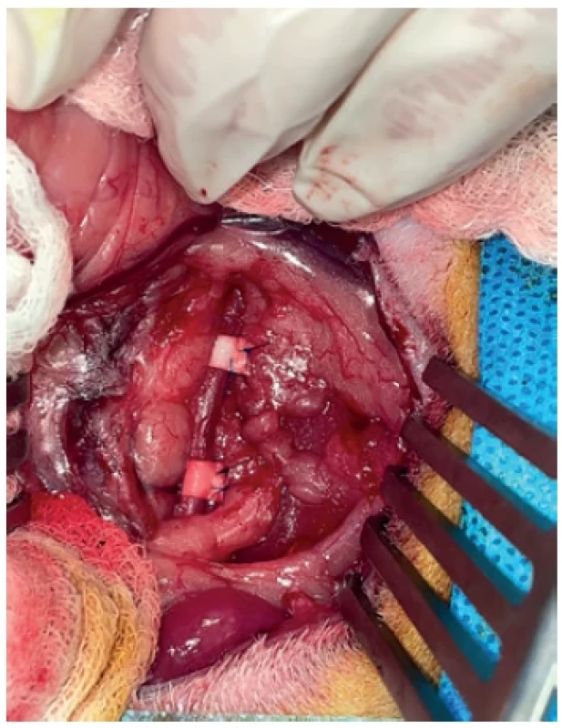
Fig. 6. Flow rate measurement on abdominal aorta of the rabbit 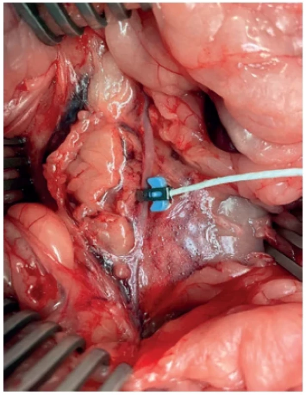
Fig. 7. Pulmonary artery band in the pig model 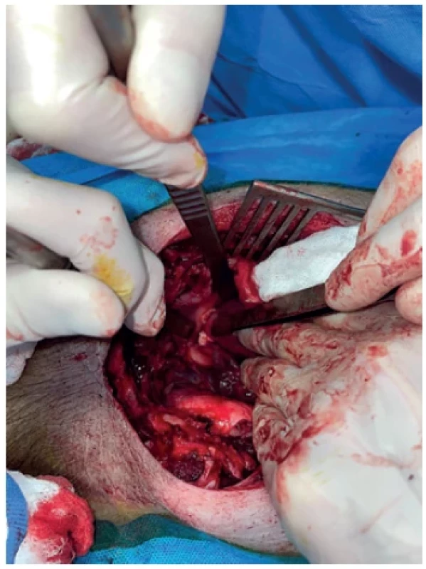
- Pig model (Sus scrofa f. domestica) The function of arterial bands made of the final composite material selected on the basis of previous tests in rat and rabbit models is evaluated in the domestic pig (approx. 25 kg). The size of the domestic pig and its vascular system are suitable for experimental surgery. The arterial bands are implanted in the pulmonary artery (Fig. 7), abdominal aorta (Fig. 8) and carotid artery of the piglets, and subsequent 60-day follow-up provides results that can be extrapolated to clinical practice. Similarly as in the rabbit model, the blood flow is monitored using a Doppler machine and the explanted banded arteries undergo both histological analysis as well as mechanical tensile and inflation tests.
Fig. 8. Arterial band on subrenal aorta of the pig 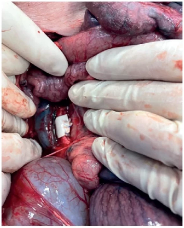
Fig. 9. Explanted rabbit abdominal aorta with the arterial band 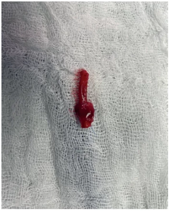


Mechanical testing
The principle of banding consists in the mechanical interaction between an arterial wall and a band. The band is tightly wrapped around an artery, resulting in narrowing of the arterial lumen. Blood flow and pressure change this way, which is followed by a mechanobiological reaction that adapts the arterial walls and the heart according to the new hemodynamical conditions. Mechanical induction of the remodeling process is the core of the treatment itself. From the biomechanical point of view, development of such bands must incorporate several steps. First, the properties of the band itself, associated with handling, need to be determined. The handling behavior of the band is mainly determined by its bending stiffness and extensibility. Second, the resorbability of the band must be documented. Another thing we have to deal with in the development of arterial bands is the remodeling of the banded artery. It should be verified that long-term mechanobiological interaction of the artery with the band does not lead to arterial sclerosis or other adverse reactions at the banding site.
The biomechanical evaluation conducted in our project is based on two types of experiments. Uniaxial tensile tests are carried out to study intact or partially, no matter if in vitro or in vivo, resorbed materials for band fabrication. The tensile test consists in uniaxial extension of a rectangular sample that is loaded in an experimental setup up to its failure. The measured loading force and deformation are subsequently used to determine the stress and strain at failure or to find out the constitutive model that generally describes the mechanical response of the material. In this case, the results are compared with those obtained with samples taken from sites not exposed to banding or with samples obtained from non-banded subjects.
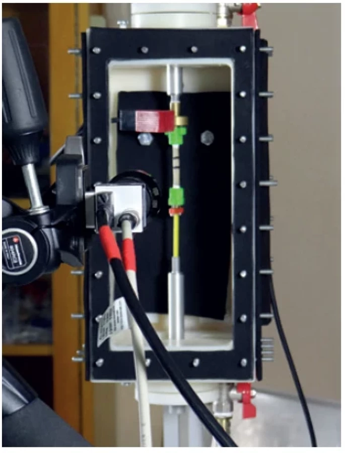
The second type of experiment is the inflation-extension test, which could be simplistically described as a pressurization test. In this experiment, the sample is a tubular segment of a blood vessel that is mounted into an adjustable frame where it is subsequently loaded with internal pressure (Fig. 10). During the experiment, values of applied pressure are recorded on the PC and the loaded geometry of the specimen is recorded by cameras. Image analysis of the acquired digital photographs is used to determine the deformed dimensions of the pressurized specimen. The results are pressure-deformation curves that can be used to compare banded and nonbanded vessels with each other. The resulting data can be used in regression analysis to determine material models suitable to be used in computational simulations of mechanical behavior of the banded arteries.
DISCUSSION:
Our research group has extensive experience with experimental surgery. We were engaged in abdominal aortic aneurysm modeling in the past. Porcine aorta was influenced by elastase infusion and increasing of turbulent flow. We influenced these aneurysms pharmacologically, as well [18,19]. In the field of transplant medicine, we study the problem of organ perfusion. We have focused on donors after cardiac death (DCD). DCD organs suffer from warm ischemia and require some type of reconditioning [20,21,22]. Both small and large animals (rabbit/pig) were used for DCD modeling and their kidneys were perfused using various approaches. Our team focuses also on a different type of liver parenchyma damage in the porcine model [23,24].
Based on our experience, we have decided to use definite animal models for testing new materials for cardiovascular surgery. These models are adequate for biocompatibility and degradability testing (all of them) as well as for evaluation of mechanical interaction between the arterial wall and the band (rabbit, pig). Especially large animals, such as porcine models, have a cardiovascular system that very well correlates with human anatomy and physiology and obtained results can be well extrapolated to human clinical practice. That is why porcine models are very favored in surgical experiments, not only for testing new materials and devices, but also for training of new surgical procedures [25−29]. We have no experience with an alternative option, the use of non-human primates, although this is also a well-established model [30,31]. Precise project planning and a search of recent literature data will reduce unnecessary errors and complications. We believe that we have chosen optimal experimental animal models and that relevant data will be obtained. We have not noticed any major technical problems or other difficulties so far. Sternotomy and pulmonary artery band placement in the porcine model is slightly technically demanding and we are considering replacing it with the carotid band.
Anyone who uses animal models must accept the key rules already mentioned above; researchers must understand how specific and challenging it is to carry out experimental work on animals. Ethical aspects must always come first and be considered at the beginning of the whole study design. The first “R” (replacement) must be considered. An in vivo experiment cannot be avoided in the last phase of our preclinical research in the field of new materials for cardiovascular surgery. However, we do try to use each animal´s potential as much as possible so that we can reduce the number of animals.
Tato práce vznikla za podpory grantu AZV NU20-02 - 00368.
Conflict of interests
The authors declare that they have not conflict of interest in connection with this paper and that the article has not been published in any other journal, except congress abstracts and clinical guidelines.
prof. MUDr. Jiří Moláček, Ph.D.
Department of Surgery
University Hospital in Pilsen
e-mail: molacek@fnplzen.cz
Rozhl Chir. 2022;101 : 599–606
Zdroje
1. Adair T, Lopez AD. The role of overweight and obesity in adverse cardiovascular disease mortality trends: an analysis of multiple cause of death data from Australia and the USA. BMC Med. 2020 Aug 4;18(1):199. doi: 10.1186/s12916-020 - 01666-y.
2. McAloon CJ, Boylan LM, Hamborg T, et al. The changing face of cardiovascular disease 2000-2012: An analysis of the World Health Organisation global health estimates data. Int J Cardiol. 2016 Dec 1;224 : 256−264. doi: 10.1016/j.ijcard. 2016.09.026. Epub 2016 Sep 15.
3. Jiang W, Rutherford D, Vuong T, et al. Nanomaterials for treating cardiovascular diseases: A review. Bioact Mater. 2017 Dec 6;2(4):185−198. doi: 10.1016/j.bioactmat. 2017.11.002.
4. Voorhess AB Jr, Jaretzky A, Blackemore AH. The use of tubes constructed from vinyon “N” cloth in bridging arterial defects. Ann Surg. 1952 Mar;135(3):332−336. doi: 10.1097/00000658-195203000-00006.
5. Blackemore AH, Voorhess AB Jr. Aneurysm of the aorta: a review of 365 cases. Angiology. 1954 Jun;5(3):209−231. doi: 10.1177/000331975400500305.
6. Collaghan JC. Replacement of the aortic and mitral valves using the Starr-Edwards ball-valve prosthesis: A report of 50 cases. Can Med Assoc J. 1964 Aug 29;91(9):411−421.
7. Valerianova A, Mlcek M, Grus T, et al. New porcine model of arteriovenous fistula documents increased coronary blood flow at the cost of brain perfusion. Front Physiol. 2022 Apr 27;13 : 881658. doi: 10.3389/fphys.2022.881658.
8. Nistor A, Jiga LP, Miclaus GD, et al. Experimental swine models for perforator flap dissection in reconstructive microsurgery. PLoS One 2022 Apr 11;17(4):e0266873. doi: 10.1371/journal. pone.0266873.
9. Grajciarová M, Turek D, Malečková A, et al. Are ovine and porcine carotid arteries equivalent animal models for experimental cardiac surgery: A quantitative histological comparison. Ann Anat. 2022 Jun;242 : 151910. doi: 10.1016/j. aanat.2022.151910.
10. Kim K, Anderson EM, Martin AJ, et al. Development of a murine iliac arteriovenous fistula model for examination of hemodialysis access-related limb pathophysiology. JVS Vasc Sci. 2021 Oct 6;2 : 247−259. doi: 10.1016/j.jvssci.2021.09.022.
11. Rasmussen J, Skov SN, Nielsen DB, et al. In-vitro and in-vivo evaluation of a novel bioprosthetic pulmonary valve for use in congenital heart surgery. J Cardiothorac Surg. 2019 Jan 9;14(1):6. doi: 10.1186/ s13019-019-0830-1.
12. Balls M, Goldberg AM, Fentem JH, et al. The three Rs: the way forward: the report and recommendations of ECVAM Workshop 11. Altern Lab Anim. 1995 Nov - Dec;23(6):838−866.
13. Liska V. (ed.), et al. Experimental surgery. Pilsen, Nava 2016. ISBN 978-80-7211-490 - 0.
14. Daley M, Brizard CP, Konstantinov IE, et al. Absorbable pulmonary artery banding: a strategy for reducing reoperations. Eur J Cardiothorac Surg. 2017 Apr 1;51(4):735−739. doi: 10.1093/ejcts/ ezw409.
15. Motoyama Y, Takamura Y, Yamada S, et al. Partial wrap-clipping of the entrance of the pseudolumen of a fusiform aneurysm in the posterior inferior cerebellar artery: a technical note. Acta Neurochir (Wien) 2017 May;159(5):861−864. doi: 10.1007/ s00701-017-3099-y. Epub 2017 Jan 31.
16. Braesco J, Le Paul Y, Mondine P, et al. Fistules artério-veineuses hypertrophiques d’hémodialyse. Réduction à l’aide d’un filet péri-veineux constrictif. Presse Med. 1991 May 11;20(18):866−867.
17. Ramadan R, Guihaire J, Verscheure D. Off-pump wrapping for acute aortic dissection in high-risk patients: A simplified procedure for a life-threatening condition. Ann Thorac Surg. 2020 Aug;110(2):750−751. doi: 10.1016/j.athoracsur. 2020.03.118. Epub 2020 May 21.
18. Molacek J, Treska V, Kobr J, et al. Optimization of the model of abdominal aortic aneurysm--experiment in an animal model. J Vasc Res. 2009;46(1):1−5. doi: 10.1159/000135659. Epub 2008 May 31.
19. Houdek K, Moláček J, Třeška V, et al. Focal histopathological progression of porcine experimental abdominal aortic aneurysm is mitigated by atorvastatin. Int Angiol. 2013 Jun;32(3):291−306.
20. Opatrný V, Třeška V, Zeithaml J, et al. Perfusion of a kidney graft from a donor after cardiac death based on immediately started machine perfusion: An experimental study on a big animal. Transplant Proc. 2021 Jul-Aug;53(6):2082−2090. doi: 10.1016/j.transproceed.2021.06.026. Epub 2021 Jul 15.
21. Moláček J, Opatrný V, Matějka R, et al. Retrograde oxygen persufflation of kidney - experiment on an animal. In Vivo 2016;30(6):801−805. doi: 10.21873/invivo. 10997.
22. Opatrný V, Moláček J, Třeška V, et al. Perfusion of a kidney graft from a donor after cardiac death based on immediately started pulsatile machine perfusion-an experimental study on a small animal. Transplant Proc. 2018 Jun;50(5):1544−1548. doi: 10.1016/j. transproceed.2018.02.067.
23. Vištejnová L, Liška V, Kumar A, et al. Mesenchymal stromal cell therapy in novel porcine model of diffuse liver damage induced by repeated biliary obstruction. Int J Mol Sci. 2021 Apr 21;22(9):4304. doi: 10.3390/ijms22094304.
24. Troup O, Skalicky A, Vistejnova L, et al. Bevacizumab does not inhibit the formation of liver vessels and liver regeneration following major hepatectomy: A large animal model study. In Vivo 2022 May-Jun;36(3):1083−1094. doi: 10.21873/ invivo.12806.
25. Donaldson RI, Buchanan OJ, Graham TL, et al. Development of a novel epidural hemorrhage model in swine. Mil Med. 2021 Oct 20:usab427. doi: 10.1093/ milmed/usab427. Epub ahead of print.
26. Basir A, Loncq de Jong M, Gründeman PF, et al. The early days of vascular and heart valve prostheses: a historical review. J Cardiovasc Surg. (Torino) 2020 Oct;61(5):528−537. doi: 10.23736/S0021 - 9509.19.11011-7. Epub 2019 Sep 3.
27. Munisso MC, Mahara A, Yamaoka T. Design of in situ porcine closed-circuit system for assessing blood-contacting biomaterials. J Artif Organs 2018 Sep;21(3):317−324. doi: 10.1007/s10047-018-1042-5. Epub 2018 Apr 11.
28. Williams PD, Malik N, Kingston PA. Coronary angiography and percutaneous coronary intervention in the porcine model: a practical guide to the procedure. Animal 2012 Feb;6(2):311−320. doi: 10.1017/S1751731111001650.
29. Cai J, Huang H, Zhou Y, et al. A new type of aortic valved stent with good stability and no influence on coronary artery. J Cardiothorac Surg. 2013 Nov 12;8 : 210. doi: 10.1186/1749-8090-8-210.
30. Zhou H, Liu Y, Long X, et al. Feasibility of ultrasound-induced blood-brain barrier disruption with a single-element transducer under three different frequencies in two non-human primates in vivo: Case report. J Neurosci Methods 2022 Jan 1;365 : 109383. doi: 10.1016/j.jneumeth. 2021.109383. Epub 2021 Oct 8.
31. Washida K, Hattori Y, Ihara M. Animal models of chronic cerebral hypoperfusion: From mouse to primate. Int J Mol Sci. 2019 Dec 7;20(24):6176. doi: 10.3390/ ijms20246176.
Štítky
Chirurgie všeobecná Ortopedie Urgentní medicínaČlánek vyšel v časopise
Rozhledy v chirurgii
2022 Číslo 12
- Využití HemaGelu při hojení ran v RVmedCentru privátní klinice s. r. o., zabývající se estetickou a laserovou chirurgií hlavy a krku, ušním, nosním, krčním a korektivní dermatologií
- Současnost a budoucnost péče o rány
- Umělý svěrač anu po deseti letech
- Aktuální poznatky ke spasmolytickým a analgetickým účinkům metamizolu na gastrointestinální trakt
- U pacientů s traumatem je častěji dosaženo adekvátních hladin anti-Xa při dávkování enoxaparinu podle hmotnosti
Nejčtenější v tomto čísle
- Domestic pig head and neck arteries from the viewpoint of imaging methods and experimental surgery
- Chylothorax treatment with thoracic duct embolization
- Introducing in vivo pancreatic cancer models for the study of new therapeutic regimens
- Permanent intravenous access in experimental surgery – our experience
Zvyšte si kvalifikaci online z pohodlí domova
Mazová zátka a její řešení
nový kurzVšechny kurzy