-
Články
- Vzdělávání
- Časopisy
Top články
Nové číslo
- Témata
- Kongresy
- Videa
- Podcasty
Nové podcasty
Reklama- Kariéra
Doporučené pozice
Reklama- Praxe
EXTERNAL OPHTHALMOMYIASIS CAUSED BY OESTRUS OVIS (A CASE REPORT)
Authors: L. Hartmannová 1; R. Mach 1; R. Záruba 2; M. Pavlovský 3
Authors place of work: Oční oddělení, Nemocnice Most o. z., Krajská zdravotní Ústí nad Labem, a. s. 1; Mikrobiologické oddělení, Nemocnice Most, o. z., Krajská zdravotní Ústí nad Labem, a. s. 2; Patologické oddělení, Nemocnice Most, o. z., Krajská zdravotní Ústí nad Labem, a. s. 3
Published in the journal: Čes. a slov. Oftal., 76, 2020, No. 3, p. 130-134
Category: Kazuistika
doi: https://doi.org/10.31348/2020/22Summary
The work deals with atypical conjunctival infection of Czech patient with Oestrus ovis larvae. Ophthalmomyiasis is infestation of mammalian eyes by the larvae or worms of some flies. The most common cause of human myiasis is the Sheep. Shepherds are infected in habitats, but human eye disease outside the areas of abundant hamsters is rare. We describe a case of eye disease in a middle-aged man from the Czech Republic who spent a summer holiday seven weeks before examination in the north of Greece. During the first examination he was completely treated and no further problems were reported. Ophthalmomyiasis externa should be considered as a possible infection of travelers to the southern endemic regions when returning with an acute causeless onset of a one-sided foreign body sensation in the eye.
Keywords:
Ophthalmomyiasis – myiasis – Oestrus Ovis – unilateral conjunctivitis – foreign body feeling
INTRODUCTION
We have practically never encountered sheep bot flies in ophthalmology departments in the Czech Republic. From school we most probably remember Hyperderma bovis, the northern cattle grub, which is linked with startled cattle. When laying eggs, fertilised female Hyperderma bovis flies emit a sound with a high frequency to which cattle are highly sensitive, with the result that they are startled and have fits of delirium.
However, Oestrus ovis belongs to a group of nasal and throat bot flies from the subfamily of Oestrinae. It is widespread in all regions of the world where sheep and goats are present. This covers the region of the Mediterranean Sea, the Middle East, North and Central America, Australia, Brazil and Southern Africa. The incidence of the fly has decreased in Northern Europe in recent years [1]. This year in Croatia, two new cases from an urban agglomeration from last year were described. A further 259 cases of human ophthalmomyiasis were reported in Mediterranean countries in the PubMed database. All the cases were caused by Oestrus ovis [2]. A total of 260 cases (99.62 %) were external, whereas 1 (0.38 %) had internal form of ophthalmomyiasis.
Adult Oestrus ovis are robust bot flies, slightly similar to slimmer bumblebees, with a size of 10–15 mm. The females do not lay eggs, but already living larvae. All the stages of larvae have mouth hooks, and also thorns on their rear segments, which serve for fixation onto the mucosa and prevent their expulsion from the nostrils of the host. Impregnated females target sheep or other animals (goats, mouflon, mountain goats, deer, roebuck) on hot sunny days from May to July. The females approach the nostrils of the host, and in flight spray clusters of larvae (30–40 larvae) via their ovipositor in a droplet into the nostrils or the surrounding area. Larvae of the first instar (an individual developmental phase between shedding the old exoskeleton for the purpose of enabling further growth of the insect), with a size of 1–2 mm (Fig. 1) immediately migrate into the nostrils of the host with the aid of hooks and thorns, where they settle and survive the winter. At the end of the winter and beginning of the spring, they progressively metamorphose into larvae of the second and third instar inside their host, and increase in size. The larvae may reach the paranasal sinuses, gullet and cranial cavities, and also damage the meninges. Larvae live off mucus, which is produced by the irritated mucosa. In the spring larvae of the third instar are expelled from the host and immediately form a cocoon. After 1 to 2 months adult flies emerge.
Fig. 1. Oestrus ovis larva in outer corner of left eye 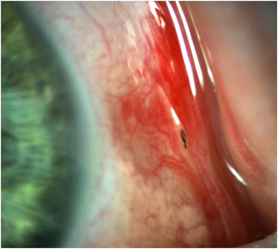
Ophthalmomyiasis is the infestation of mammalian eyes or the periorbital tissue by larvae or worms of certain flies, most often sheep bot flies. In localities of incidence shepherds are frequently infected, but human infection is rare outside areas of abundant incidence of bot flies. On the basis of the location of infestation, we classify three forms of ophthalmomyiasis. 1. external ophthalmomyiasis: colonisation of eyelids, lachrymal pathways, conjunctiva and cornea. This is most commonly manifested as acute catarrhal conjunctivitis with non-specific symptoms of a sensation of a foreign body, lachrymation, photophobia, erythema and potentially also periocular edema. Manifestations are usually short-term and self-limiting, because the larvae cannot develop further and die within 10 days. 2. internal ophthalmomyiasis, in which larvae penetrate into the eyeball, and are evident in the chamber fluid, in the area of the iris, vitreous body or subretinal space, in rare cases they may cause sight-threatening endophthalmitis and optic atrophy. 3. orbital ophthalmomyiasis: larvae penetrate into the orbit and affect the adnexa and optic nerve [3]. However, Oestrus ovis larvae are not capable of excreting proteolytic enzymes, and as a result are mostly limited to the outer surface of the eye [4]. Rare complicating cases are stated in the literature, such as: subconjunctival haemorrhage (Fig. 2), corneal ulcer, deterioration of sight, intraocular invasion with endophthalmitis, iridocyclitis and even blindness [5].
Fig. 2. Very rapidly moving living organisms are difficult to capture on photographic slit lamp 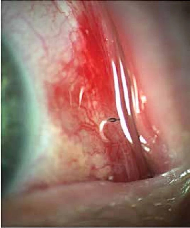
CASE REPORT
We describe the case of an ocular pathology in a middle-aged man from the Czech Republic who had spent a summer holiday in northern Greece seven weeks previously to being examined. Late in the evening in the last week of August 2019, the patient reported to an eye emergency clinic. Reportedly sawdust had flown into his left eye, which was lachrymating and painful. In addition to minor erosion of the corneal epithelium and mild hyperperfusion of the conjunctiva, the examination detected a rippling movement of more than twenty elongated 1.5–2 mm long large pale micro-organisms with a dark rostral end (eyes and organs) and a whitish-transparent pointed caudal end, especially in the inner corner of the eye, and upon a detailed examination throughout the entire surface of the bulbar conjunctiva and in the fornices. Progressively all the larvae were removed from the surface of the eye in installation anaesthesia on a slit lamp, after which they were fixed in formaldehyde (Fig. 3) and on the following day examined and microscopically documented (Fig. 4). We did not find any parasites beneath the conjunctiva, as stated by certain studies [6]. Afterwards the patient was without any further symptoms, and after healing of minor post - -traumatic erosions of the corneal epithelium required no further care. Nasal symptoms were negative. After a period of six months the patient is entirely without complaints.
Fig. 3. Caught individual Oestrus ovis larva in formaldehyde photographed on slit lamp 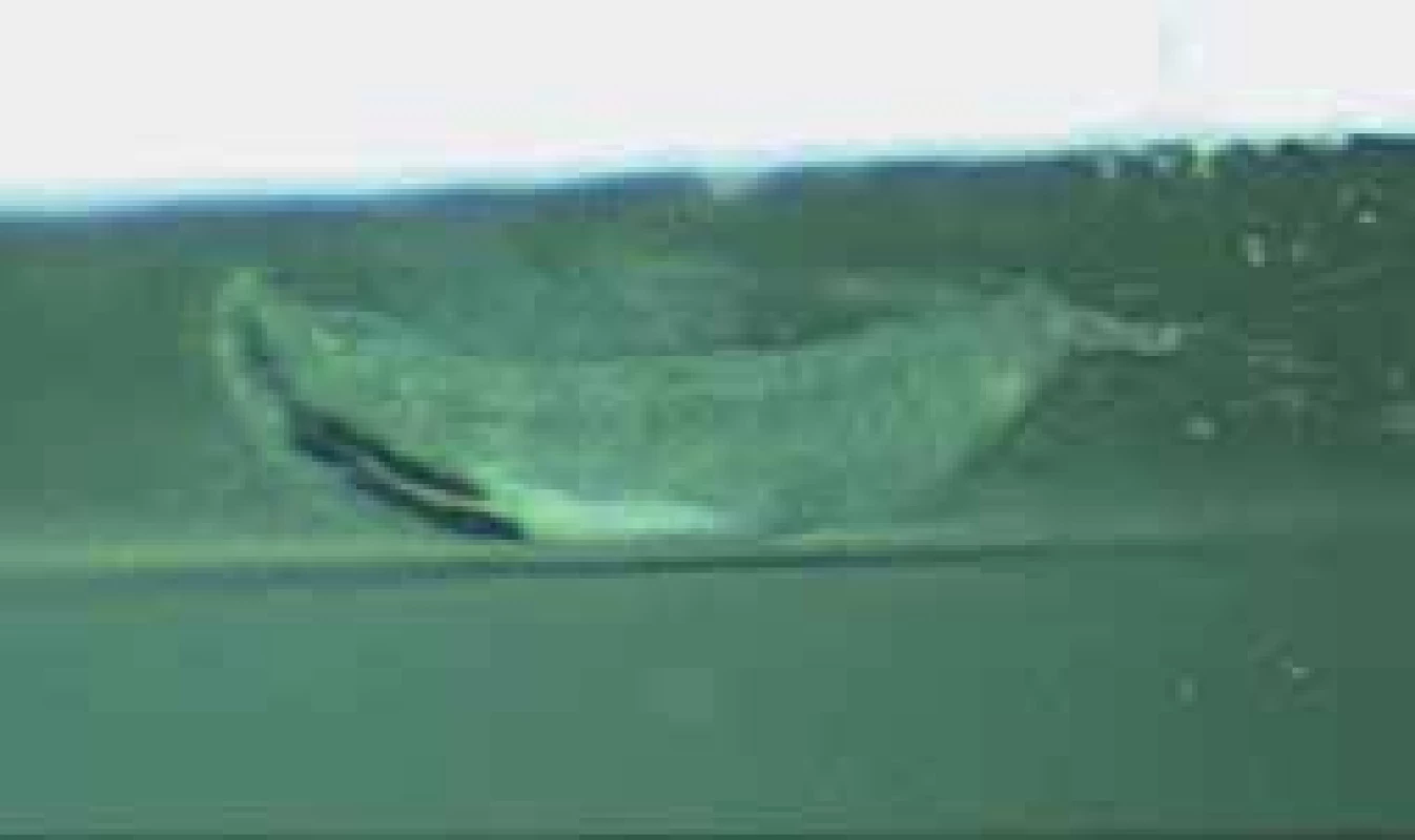
Fig. 4. Oestrus ovis larva (enlarged 100x, Nikon Eclipse) 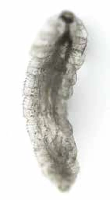
DISCUSSION
In our climatic conditions, we may probably encounter myiasis in travellers returning from southern countries where there are large numbers of sheep and therefore also bot flies. In non-endemic countries this may represent a diagnostic challenge for doctors who are unfamiliar with this infestation, especially if organs other than the skin are affected [7]. Human infestation by bot flies is stated in the case of impact of a fertile female into the patient’s eye and the surrounding area. This is commonly reported by shepherds and in rural areas in the Mediterranean region, in tropical and subtropical countries [8].
Myiasis ranks among the five most commonly reported dermatological pathologies in travel medicine, and ophthalmomiasis represents approximately 5 % of all cases of myiasis [7]. Oestrus ovis is the main causative agent of myiasis of cavities. It may cause complications such as allergic reactions, and especially very rare tissue-invasive internal ophthalmomyiasis. For this reason it is essential to ensure the complete removal of all larvae in order to prevent serious ocular consequences (loss of sight, endophthalmitis, meningitis) [9]. It is necessary to conduct a careful examination on a slit lamp in order to prevent erroneous diagnosis of bacterial inflammation of the conjunctivas instead of external ophthalmomyiasis [10]. It is also necessary to determine whether the larvae have penetrated beneath the conjunctiva. Subconjunctival parasites must subsequently be extracted under a surgical microscope. It is further recommended to search for larvae penetrating the sclera or infiltrating into the orbit [6]. The primary treatment of external ophthalmomyiasis is surgical extraction of larvae under local anaesthesia, with the use of a wet cotton swab or forceps. Merely rinsing the eye with physiological solution is not sufficient due to the firm grasp on the mucosal tissue by the larval hooks. Ocular paralytic agents have been proposed in the literature, which prevent deep embedding of the larvae into the ocular tissue and facilitate their later removal. Effective agents are 4-5 % cocaine, lidocaine or 1-4 % pilocarpine solution. However, as mentioned above, even despite paralysis larvae are able to cling onto tissues with their hooks (Fig. 5). The ophthalmic creams Neomycin, Bacitracin and Polymyxin B have been used successfully for the probable suffocation of larvae (Fig. 6 and 7). In addition to surgical extraction, three-day oral therapy with mebendazole or another available broad-spectrum antihelminthic agent from the group of benzimidazoles is recommended. Prophylactic corticosteroids and antibiotics may be applied locally [11].
Fig. 5. Detail of right rostral hook of Oestrus ovis larva in view from ventral side (enlarged 200x) 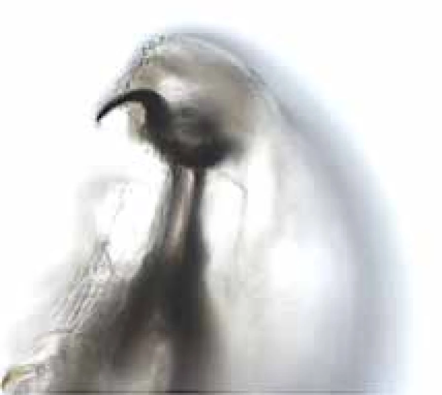
Fig. 6. Six individual Oestrus ovis larvae in outer corner of eye 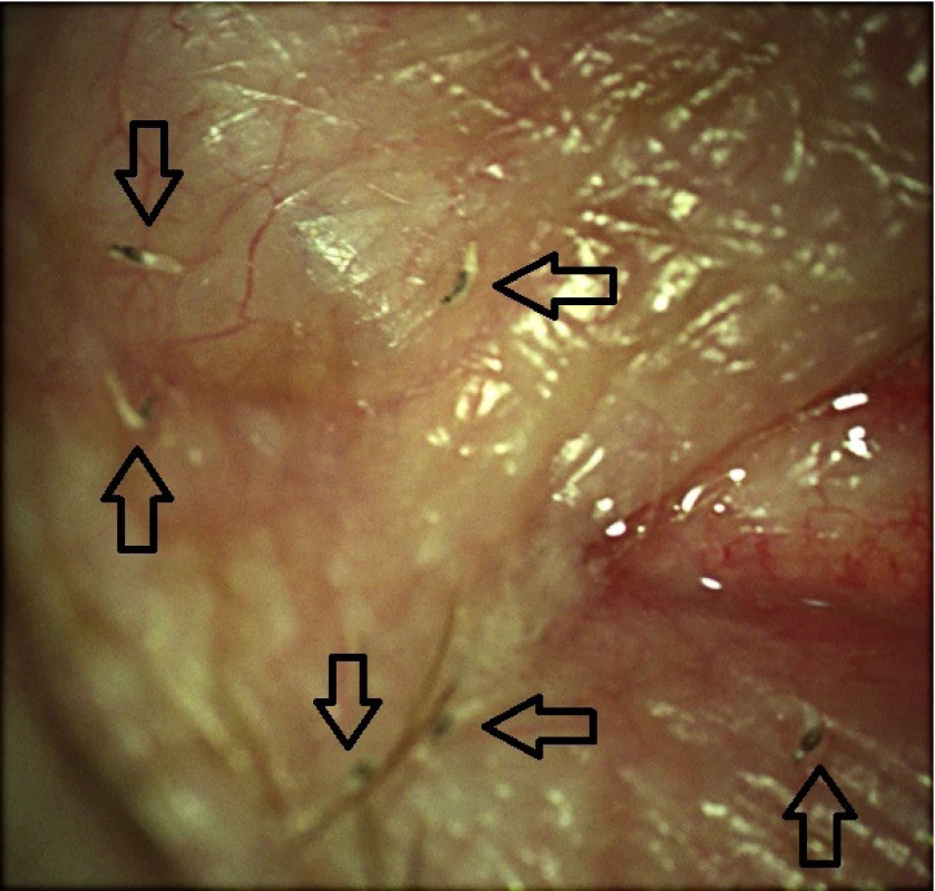
Fig. 7. Individual Oestrus ovis larva on edge of eyelid 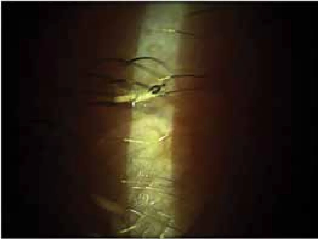
In a routine examination on a slit lamp, Oestrus ovis shows negative phototaxis, small and translucent larvae move fairly rapidly away from bright light, which may lead to incorrect diagnosis of the condition if they are overlooked. The morphological properties of Oestrus ovis larvae are relatively typical (Fig. 8 and 9), but may differ, and it is recommended to conduct molecular verification of testing [12]. However, this has been applied only rarely in human medicine [13]. With regard to public health, with regard to the quick diagnosis of human and veterinary cases of myiasis outside of the endemic regions, it is also important to prevent the spread of the bot fly into new regions where it did not previously appear [7].
Fig. 8. Oestrus ovis larva from side view (enlarged 100x) – well visible eyes and one hook 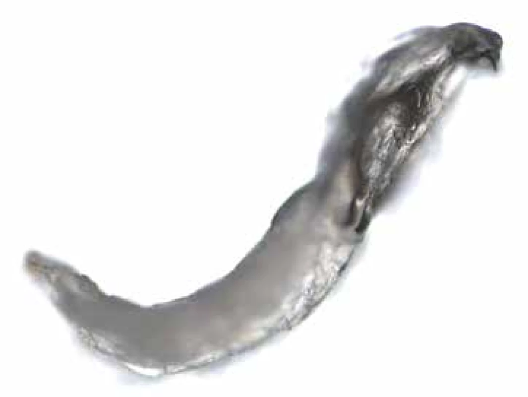
Fig. 9. Oestrus ovis larva from ventral view (enlarged 100x) with detail of internal organs 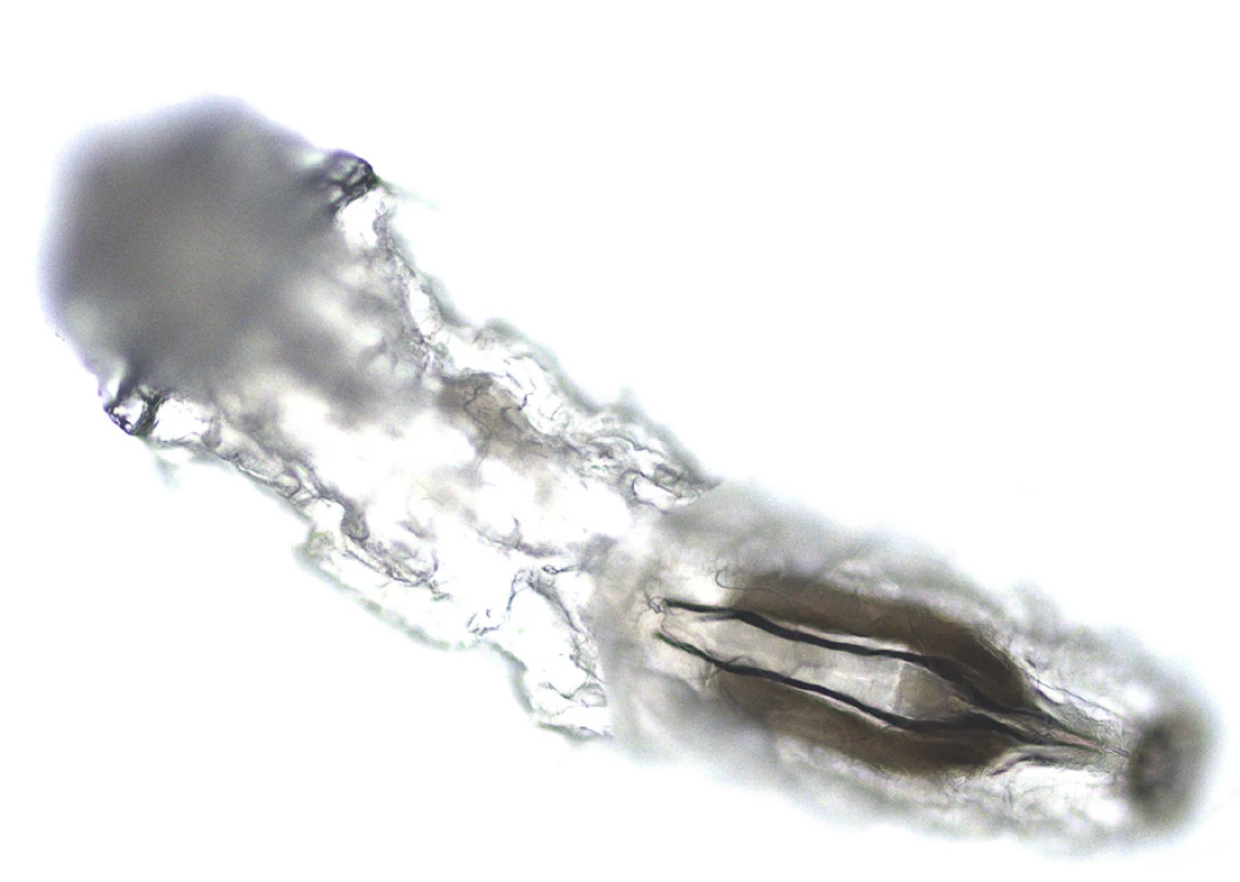
As concerns whirling disease of sheep and infestation of cattle and horses, it is possible to consider these manifestations of endoparasitosis (pathology caused by parasites attacking/affecting internal organs) caused by larvae of various types of bot flies to be genuinely very infrequent in our conditions. Images or preparations with bot fly larvae in the brain of an affected animal, beneath the skin or embedded in the stomach can be found rather only in textbooks and school cabinets. In this country hoofed forest animals (larger furred game), i.e. roebuck and deer, may also suffer from infestation. Unpredictable manifestations of an infested roebuck were described by the writer Jan Vrba; a roebuck was known in the hunting ground for “dancing” one step forward and two steps back. After the roebuck was shot, it was determined that it had bot fly larvae in its brain [14]. As a result, today hunters place de-worming preparations in feeding troughs for deer and roebuck. Humans are not at risk either directly or indirectly from bot flies. This means that they do not lay eggs beneath the skin or in the nose in humans, and there is no danger from any swallowing of bot fly larvae in meat.
We did not determine the manner of infiltration of larvae into the eye of our patient. With regard to the large number of larvae in one eye (almost one entire clutch), we would not assume a chance infection from soiled hands, as stated by a German patient who had recently returned from a mountaineering expedition on the Greek island of Kalymnos, where she had repeatedly touched sheep [15]. We presume a chance impact and release of larvae or poor choice of host by the female bot fly, in which the glistening eye was mistaken for the moist snout of a sheep, or other anatomical distances of the nose from the eye in sheep and humans. A surprising factor is the latency of seven weeks without subjective complaints since the patient’s stay in the endemic region following the chance infestation by parasites, followed by a minor injury to the same eye. We explain the long survival of a larger group of larvae by means of the more copious irritation of the mucosa caused by the “romping” of the larvae. The abundance of mucus in the conjunctival sac could be a rich source of nutrition for the surviving larvae.
CONCLUSION
External ophthalmomyiasis should be considered a potential infection following return from southern endemic regions (Turkey, Greece, Spain, Portugal) upon unexplained onset of a unilateral sensation of a foreign body in the eye or unilateral mucous conjunctivitis. Increased awareness of this condition among ophthalmologists and clinical microbiologists is the key to timely diagnosis and an effective clinical solution. Global warming may predetermine a future increase in the prevalence of Oestrus ovis in humans, which emphasises the need for mandatory reporting and observation of pathologies.
Zdroje
1. Wikipedia. Oestrus ovis. [internet]. Avaible from: https://en.wikipedia.org/wiki/Oestrus_ovis.
2. Pupić-Bakrač A, Pupić-Bakrač J, Škara Kolega M, Beck R. Human ophthalmomyiasis caused by Oestrus ovis-first report from Croatia and review on cases from Mediterranean countries. Parasitol Res. 2020 Mar;119(3):783–793.
3. Sharma K: Ophthalmomyiasis externa. A case report from Alkharj, Saudi Arabia. Saudi J Ophthalmol. 2018 Jul-Sep;32(3):250-252. Epub 2017 Nov 24.
4. Odat TA, Gandhi JS, Ziahosseini K. A case of ophthalmomyiasis externa from Jordan in the Middle East. Br J Ophthalmol. 2007;91. Video report. [Google Scholar].
5. Pal N, Majhi B. Unilateral conjunctivitis of unique etiology: A case report from Eastern India. J Nat Sci Biol Med. 2016 Jan-Jun;7(1):104–6.
6. Pather S, Botha LM, Hale MJ, Jena-Stuart S. Ophthalmomyiasis Externa: Case Report of the Clinicopathologic Features. Int J Ophthalmic Pathol. 2013 Feb 18;2(2). doi: 10.4172/2324-8599.1000106.
7. Francesconi F, Lupi O. Myiasis. Clin Microbiol Rev. 2012;25 : 79–105.
8. Panadero-Fontán R, Otranto D. Arthropods affecting the human eye. Vet Parasitol. 2015;208 : 84–93.
9. Dunbar J, Cooper B, Hodgetts T, et al. An outbreak of human external ophthalmomyiasis due to Oestrus ovis in southern Afghanistan. Clin Infect Dis. 2008;46:e124–6.
10. Akdemir MO, Ozen S. External ophthalmomyiasis caused by Oestrus ovis misdiagnosed as bacterial conjunctivitis. Trop Doct. 2013;43 : 120–3.
11. D‘Assumpcao C, Bugas A, Heidari A, Sofinski S, McPheeters RA. A Case and Review of Ophthalmomyiasis Caused by Oestrus ovis in the Central Valley of California, United States. J Investig Med High Impact Case Rep. 2019 Jan-Dec;7 : 2324709619835852. doi: 10.1177/2324709619835852.
12. Otranto D, Traversa D, Giangaspero A. Myiasis caused by Oestridae: serological and molecular diagnosis. Parassitologia. 2004;46 : 169–72.
13. Rukke BA, Cholidis S, Johnsen A, Ottesen P. Confirming Hypoderma tarandi (Diptera: Oestridae) human ophthalmomyiasis by larval DNA barcoding. Acta Parasitol. 2014;59 : 301–4.
14. Vrba J. Srnčí milování a jiné obrázky z chodské přírody. Praha (Czechoslovakia): Nakladatelství Odeon; 1968;346s.
15. Fries FN, Pattmöller M, Seitz B et al. Ophthalmomyiasis externa due to Oestrus ovis in a traveller returning from Greece. Travel Medicine and Infectious Disease 23. 2018;101–102.
Štítky
Oftalmologie
Článek PRES SYNDRÓM
Článek vyšel v časopiseČeská a slovenská oftalmologie
Nejčtenější tento týden
2020 Číslo 3- Stillova choroba: vzácné a závažné systémové onemocnění
- Familiární středomořská horečka
- Diagnostický algoritmus při podezření na syndrom periodické horečky
- Možnosti využití přípravku Desodrop v terapii a prevenci oftalmologických onemocnění
- Selektivní laserová trabekuloplastika nesnižuje nitroční tlak více než argonová laserová trabekuloplastika
-
Všechny články tohoto čísla
- TRAUMATOLOGIE V OKULOPLASTICKÉ CHIRURGII PŘEHLEDOVÝ ČLÁNEK
- ZMĚNY FOVEÁLNÍ AVASKULÁRNÍ ZÓNY A MAKULÁRNÍ MIKROVASKULATURY V RÁMCI VYŠETŘENÍ OCT ANGIOGRAFIE U MLADÝCH DIABETIKŮ 1. TYPU (PILOTNÍ STUDIE)
- OCT ANGIOGRAFIE A DOPPLEROVSKÁ SONOGRAFIE U NORMOTENZNÍHO GLAUKOMU
- ŠÍŘE CHIASMATU U NORMOTENZNÍCH A HYPERTENZNÍCH GLAUKOMŮ
- ZEVNÍ OFTALMOMYIÁZA ZPŮSOBENÁ LARVOU STŘEČKA OESTRUS OVIS
- PRES SYNDRÓM
- Česká a slovenská oftalmologie
- Archiv čísel
- Aktuální číslo
- Informace o časopisu
Nejčtenější v tomto čísle- PRES SYNDRÓM
- ZEVNÍ OFTALMOMYIÁZA ZPŮSOBENÁ LARVOU STŘEČKA OESTRUS OVIS
- TRAUMATOLOGIE V OKULOPLASTICKÉ CHIRURGII PŘEHLEDOVÝ ČLÁNEK
- ZMĚNY FOVEÁLNÍ AVASKULÁRNÍ ZÓNY A MAKULÁRNÍ MIKROVASKULATURY V RÁMCI VYŠETŘENÍ OCT ANGIOGRAFIE U MLADÝCH DIABETIKŮ 1. TYPU (PILOTNÍ STUDIE)
Kurzy
Zvyšte si kvalifikaci online z pohodlí domova
Autoři: prof. MUDr. Vladimír Palička, CSc., Dr.h.c., doc. MUDr. Václav Vyskočil, Ph.D., MUDr. Petr Kasalický, CSc., MUDr. Jan Rosa, Ing. Pavel Havlík, Ing. Jan Adam, Hana Hejnová, DiS., Jana Křenková
Autoři: MUDr. Irena Krčmová, CSc.
Autoři: MDDr. Eleonóra Ivančová, PhD., MHA
Autoři: prof. MUDr. Eva Kubala Havrdová, DrSc.
Všechny kurzyPřihlášení#ADS_BOTTOM_SCRIPTS#Zapomenuté hesloZadejte e-mailovou adresu, se kterou jste vytvářel(a) účet, budou Vám na ni zaslány informace k nastavení nového hesla.
- Vzdělávání


