-
Články
- Vzdělávání
- Časopisy
Top články
Nové číslo
- Témata
- Kongresy
- Videa
- Podcasty
Nové podcasty
Reklama- Kariéra
Doporučené pozice
Reklama- Praxe
A rare subtype of papillary kidney tumors (case report)
Authors: Justine Wacquet 1; Elisabeth Richard-Coulet 2; Justine Varinot 1; Ondrej Hes 3; Eva Compérat 1
Authors place of work: Department of Pathology, Tenon Hospital, HUEP, Sorbonne University, 75005 Paris, France 1; Sipath Unilabs, 63063 Clermont Ferrand, France. 2; Department of Pathology, Charles University in Prague–Faculty of Medicine in Plzen, Plzen, Czech Republic 3
Published in the journal: Čes.-slov. Patol., 56, 2020, No. 1, p. 35-37
Category: Původní práce
During this last decade the kidney cancer classification has undergone a real revolution. Increased understanding of underlying processes has taken place, many new types have been described and characterised and allow a better patient’s management. Some of these entities have been introduced into the latest WHO classification 2016, some have recently been recognized and are still under evaluation (1). Biphasic squamoid papillary renal cell carcinoma (RCC) is a rare type of renal tumor, recently described (2). This lesion is not yet introduced into the WHO classification 2016.
CASE PRESENTATION
A 57-years old woman with a prior history of right renal transplant, presented a kidney tumor on the left native kidney. The unifocal tumor measured 5 x 5 x 3 cm and had ivory encephaloid aspects. Macroscopically, this tumor pushed the renal capsule towards the perirenal fat, but didn’t grow through it, no infiltration of perirenal fat, the hilum or the vessels was seen. No necrosis or haemorrhage was observed.
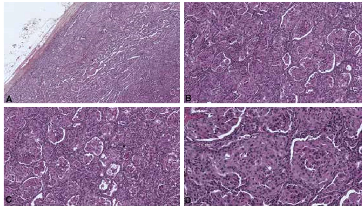
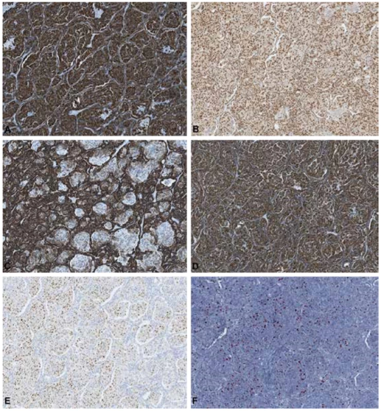
Microscopically, the lesion was well delimited by a fibrotic capsule (figure 1A). The tumor showed a dual cell population. A large cell contingent with eosinophilic cytoplasm and high-grade nuclei, often clustering in nests was observed. These large cells were surrounded by small cuboidal cell with scant cytoplasm giving a pseudo alveolar or glomeruloid-like aspect (figure 1B, C, D). The stroma was poorly abundant, sometimes containing some lymphocytes and calcifications. The second aspect of the tumor displayed a papillary architecture, which was close to papillary type 1 carcinomas. These papillary structures were surrounded by bigger eosinophilic cells. Immunohistochemistry showed a strong expression of CK7, PAX8, AMACR and vimentin for both cell types (figure 2 A, B, C, D). Cyclin D1 on the other hand was only expressed by the nuclei of the large cell (figure 2 E). The proliferation index Ki-67 was higher in large cells (Figure 2F).
Fig. 1. Histology of the lesion. A: the tumor is encapsulated (HES x5). B: HES x 10. C: HES x10. D: HES x20 
Fig. 2. Immunohistochemical study. A: CK7, membranous expression by the two cells population. B: PAX8, nuclear expression by the two cells types. C: AMACR. D: Vimentin. E: Cyclin D1, nuclear expression only by large cells. F: Ki67, nuclear expression 
These histological and immunohistochemical findings are suggestive of a biphasic squamoid papillary RCC of 5 cm with no fat or hilum vessels infiltration, classified as pT1b (pTNM 2017).
DISCUSSION
Biphasic squamoid papillary RCC or biphasic alveolo-squamoid RCC was originally reported as a separate entity by Peterson et al in 2012 (2). A recent review lists about 100 cases reported in the literature (1). A slight male predominance has been described with an age of onset ranging from 39 to 79 years, similar to conventional papillary type 1 carcinomas. These tumors occur mainly on native kidneys, but two unifocal cases of appearance on grafts have been described (3). They can occur in association with other types of RCC (papillary or clear cell carcinoma). Some rare multifocal cases have been reported, including one case from a mother and her son with a MET mutation context (4). C-MET is known to play a major role in the development of papillary renal cell carcinomas, mostly type 1, some authors have described an association between c-Met expression and clinical outcome and c-Met could be a prognostic marker (5). Moreover, a case of biphasic squamoid papillary RCC has been reported in a patient with Birt-Hogg-Dubé syndrome (6).
In one of the largest series of 21 cases described by Hes et al in 2016, the authors describe the aggressive potential of this entity with 3 patients displaying distant metastases and two patients, who died as a result of the spread of the disease (7). In another study in 2017, the 24-month median follow-up of 23 patients revealed one patient alive with disease and one dying of disease (6).
Macroscopically, these tumors are solid, whitish or brown. Mostly their size is small but bigger lesions up to 15 cm have been described (1). From a histological point of view, these generally solid growing tumors display as main characteristic two different cell populations. The first contingent consists of large cohesive cells with abundant eosinophilic cytoplasm and high nuclear atypia with nucleoli. They are mostly grouped in clusters consisting of small nests creating alveolar aspects. These cells are designated as “squamoid” because of their resemblance to squamous cells, but there is no evidence of squamous differentiation such as intercellular bridges or positivity of p63 or CK5/6 immunohistochemistry (1). The other population which form small amphophilic or clear cells, sometimes with cuboid scant cytoplasm, resemble to cells seen in type 1 papillary renal carcinoma. These cells are arranged around nests, which display glomeruloid-like aspect. Hes et al and Trpkov et al have observed images of emperipolesis within the contingent of large “squamoid” cells. Emperipolesis is defined by the presence of intact cell within the cytoplasm of another cell. Emperipolesis is unusual in RCC but has been described to be a morphologic hallmark of biphasic squamoid papillary RCC (6,7), but can also be found in high grade clear RCC with a syncytial-type multinucleated giant tumor cell component (8,9). In addition, necrosis, psammomas and areas with foam cells macrophages can be identified.
In immunohistochemistry, both cell populations are reactive for CK7, PAX8, AMACR, vimentin, CD10 and EMA. A useful marker to perform is cyclin D1 which is expressed only in the nuclei of large cells, highlighting the “squamoid” nests. Also, Ki-67 appears to be higher in the large cell contingent (6).
From a molecular perspective, FISH studies find gains in chromosomes 7 and / or 17 like in classical papillary renal cell carcinomas type 1 (1). On the basis of these histological, immunohistochemistral and molecular findings, some authors suggest that this entity is related to papillary RCC. Indeed, for Hes et al, 10 out of 21 tumors had areas of papillary RCC, mostly type 1, associated with biphasic squamoid papillary RCC (7). Alike, Chartier et al described a case with multifocal biphasic squamoid papillary RCC mixed with typical papillary RCC type 1 (4)
The main differential diagnosis are metanephric adenoma (MA) and mucinous tubular and spindle cell carcinoma (MTSC) (1). Nevertheless, these two entities lack the dual morphology, which is always seen in biphasic squamoid papillary carcinoma. MA show small acini with small lumina and even display areas of solid appearance but there are no « squamoid » cells. Moreover, the immunohistochemical profile is different with reactivity for WT1 and CD57. MTSC have mucin in their stroma, which is never present in biphasic squamoid papillary RCC. Immunohistochemistry present similarities with positive reactivity for CK7 and AMACR.
In summary, biphasic squamoid papillary RCC seems to be a rare distinctive neoplasm of RCC or a subtype of papillary RCC, which should be recognized because of his malignant potential. Further studies with larger series and longer follow-ups would be interesting to more accurately determine the prognosis of these lesions and potentially assert a continuous spectrum with papillary RCC supporting with molecular findings.
CONFLICT OF INTEREST
The authors declare that there is no conflict of interest regarding the publication of this paper.
Corresponding author:
Eva Compérat
Dpt. of Pathology
Hôpital Tenon, HUEP, Sorbonne University
Paris VI, France
tel: 003356017013
email: eva.comperat@aphp.fr
Zdroje
1. Trpkov K, Hes O. New and emerging renal entities: a perspective post-WHO 2016 classification. Histopathology 2019; 74(1): 31-59.
2. Petersson F, Bulimbasic S, Hes O, Slavik P, Martínek P, Michal M, et al. Biphasic alveolosquamoid renal carcinoma: a histomorphological, immunohistochemical, molecular genetic, and ultrastructural study of a distinctive morphologic variant of renal cell carcinoma. Ann Diagn Pathol 2012; 16(6):459-469.
3. Troxell ML, Higgins JP. Renal cell carcinoma in kidney allografts: histologic types, including biphasic papillary carcinoma. Hum Pathol 2016; 57 : 28-36.
4. Chartier S, Méjean A, Richard S, Thiounn N, Vasiliu V, Verkarre V. Biphasic Squamoid Alveolar Renal Cell Carcinoma: 2 Cases in a Family Supporting a Continuous Spectrum With Papillary Type I Renal Cell Carcinoma. Am J Surg Pathol 2017; 41(7): 1011-1012.
5. Erlmeier F, Weichert W, Autenrieth M, Ivanyi P, Hartmann A, Steffens S. c-MET Oncogene in Renal Cell Carcinomas. Aktuelle Urol 2016; 47(6): 475-479.
6. Trpkov K, Athanazio D, Magi-Galluzzi C, Yilmaz H, Clouston D, Agaimy A, et al. Biphasic papillary renal cell carcinoma is a rare morphological variant with frequent multifocality: a study of 28 cases. Histopathology 2018; 72(5): 777 - 785.
7. Hes O, Condom Mundo E, Peckova K, Lopez JI, Martinek P, Vanecek T, et al. Biphasic Squamoid Alveolar Renal Cell Carcinoma: A Distinctive Subtype of Papillary Renal Cell Carcinoma? Am J Surg Pathol 2016; 40(5): 664-475.
8. Williamson SR, Kum JB, Goheen MP, Cheng L, Grignon DJ, Idrees MT. Clear cell renal cell carcinoma with a syncytial-type multinucleated giant tumor cell component: implications for differential diagnosis. Hum Pathol 2014; 45(4): 735-744.
9. Rotterova P, Martinek P, Alaghehbandan R, Prochazkova K, Damjanov I, Rogala J, et al. High-grade renal cell carcinoma with emperipolesis: Clinicopathological, immunohistochemical and molecular-genetic analysis of 14 cases. Histol Histopathol 2018; 33(3): 277-287.
Štítky
Patologie Soudní lékařství Toxikologie
Článek PŘEDSTAVUJEME NOVÉ EDITORYČlánek Jessenius o srdci
Článek vyšel v časopiseČesko-slovenská patologie

2020 Číslo 1-
Všechny články tohoto čísla
- Prof. MUDr. Zdeňka Vernerová, CSc.
- State of the art in diagnostics of ischemic heart disease and current recommended therapeutic approach
- Differential diagnosis of heart tumours
- Current nomenclature and histopathological criteria for assessment of the noninflammatory degenerative diseases of the aorta
- Deset let redakční práce a změny do budoucna
- Echinococcus multilocularis: Diagnostic problem in a liver core biopsy
- A rare subtype of papillary kidney tumors (case report)
- The role of a pathologist in surgical staging for carcinoma of the cervix uteri
- PŘEDSTAVUJEME NOVÉ EDITORY
- Rosai and Ackerman’s Surgical Pathology
- Prof. Dr. Leo Taussig - zapomenutý průkopník komplexní cytologie mozkomíšního moku
- Jessenius o srdci
- Spomienka na prof. MUDr. Štefana Kopeckého, PhD.
- Monitor aneb nemělo by Vám uniknout, že...
- Česko-slovenská patologie
- Archiv čísel
- Aktuální číslo
- Informace o časopisu
Nejčtenější v tomto čísle- Differential diagnosis of heart tumours
- PŘEDSTAVUJEME NOVÉ EDITORY
- Echinococcus multilocularis: Diagnostic problem in a liver core biopsy
- State of the art in diagnostics of ischemic heart disease and current recommended therapeutic approach
Kurzy
Zvyšte si kvalifikaci online z pohodlí domova
Autoři: prof. MUDr. Vladimír Palička, CSc., Dr.h.c., doc. MUDr. Václav Vyskočil, Ph.D., MUDr. Petr Kasalický, CSc., MUDr. Jan Rosa, Ing. Pavel Havlík, Ing. Jan Adam, Hana Hejnová, DiS., Jana Křenková
Autoři: MUDr. Irena Krčmová, CSc.
Autoři: MDDr. Eleonóra Ivančová, PhD., MHA
Autoři: prof. MUDr. Eva Kubala Havrdová, DrSc.
Všechny kurzyPřihlášení#ADS_BOTTOM_SCRIPTS#Zapomenuté hesloZadejte e-mailovou adresu, se kterou jste vytvářel(a) účet, budou Vám na ni zaslány informace k nastavení nového hesla.
- Vzdělávání


