-
Články
- Vzdělávání
- Časopisy
Top články
Nové číslo
- Témata
- Kongresy
- Videa
- Podcasty
Nové podcasty
Reklama- Kariéra
Doporučené pozice
Reklama- Praxe
Death due to Arrhythmogenic Right Ventricular Dysplasia: A case report
Úmrtí v důsledku arytmogenní dysplasie pravé komory srdeční: Kazuistika
Arytmogenní dysplasie pravé komory srdeční je jednou z hlavních příčin náhlého úmrtí mladých sportovců. V práci je popisován případ mladého muže, který zmírá ve 28 letech bez jakýchkoliv anamnestických příznaků. Při příjezdu na pohotovostní příjem se cítí velice špatně. Prvotní vyšetření EKG vykazovalo ventrikulární extrasystoly a bylo proto doporučeno přijetí na kardiologickou kliniku. Následující den se jeho stav natolik zhoršil a na tuto kliniku byl přivezen již mrtev. Histologické vyšetření vzorků srdeční svaloviny odebrané z pravé i levé komory odhalilo masivní přeměnu svaloviny pravé komory ve fibrózní a vyzrálou tukovou tkáň. V tomto případě šlo o náhlé úmrtí bez přítomnosti jakýchkoliv symptomů či rodinné nebo osobní anamnézy.
Klíčová slova:
arytmogenní dysplázie pravé komory – náhlá srdeční smrt – pitva
Authors: Okan Akan 1; Selçuk Çetin 2; Bülent Eren 1; Dilek Durak 2; Nursel Türkmen 2; Ümit Naci Gündoğmuş 3
Authors place of work: Council of Forensic Medicine of Turkey, Bursa Morgue Department, Bursa, Turkey 1; Uludag University Medical Faculty, Forensic Medicine Department, Council of Forensic Medicine of Turkey Bursa Morgue Department, Bursa, Turkey 2; Istanbul University, Forensic Medicine Institute, Council of Forensic Medicine of Turkey, Istanbul, Turkey 3
Published in the journal: Soud Lék., 58, 2013, No. 3, p. 39-41
Category: Původní práce
Summary
Arrhythmogenic right ventricular dysplasia (ARVD) is both a myocardial disease that predominantly affects the right ventricle (RV) and one of the major causes of sudden death in the young and athletes. A 28-year-old man with no signicant medical history, applied to an emergency department with feeling very ill. After his initial examinations, electrocardiography (ECG) showed ventricular extra systoles and he was recommended for admission to a cardiology polyclinic. The next day, his condition worsened and he was dead on arrival at the hospital. A histological examination of heart samples, which were obtained from the RV and LV, revealed the massive replacement of myocardium by fibrous and mature adipose tissue in the RV. In this case, there were no symptoms, family and medical history and its clinical presentation was as an unexpected sudden death.
Keywords:
Arrhythmogenic right ventricular dysplasia, sudden cardiac death, autopsy
Arrythmogenic right ventricular dysplasia (ARVD) is one of a number of sudden death causes among the young and athletes (1,2). ARVD is a myocardial disease, affecting the right ventricle (RV), morphologically characterized by diffuse or segmental lack of myocardium in the RV free wall, which is replaced by fatty or fibro fatty tissue, and also histologically by fibro fatty degeneration of cardiomyocytes, which leads to electrical instability and contractility abnormalities (1,3–9). We described an autopsy case of a 28-year old man with sudden death due to ARVD.
CASE REPORT
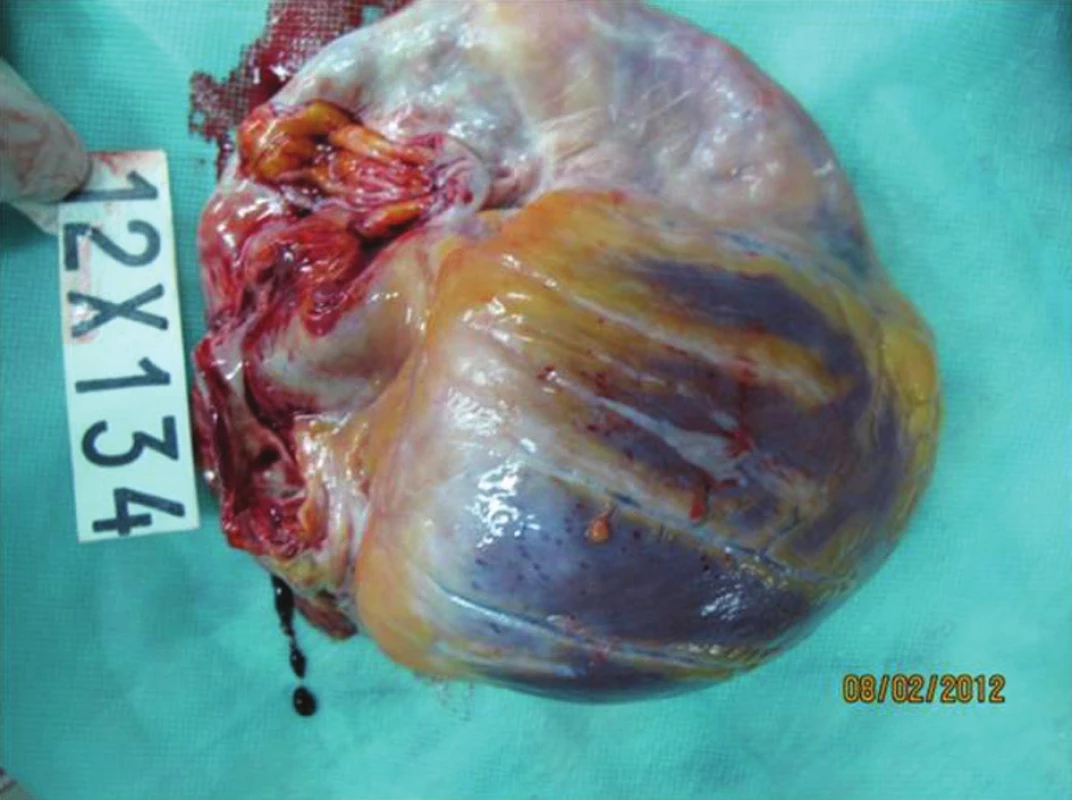
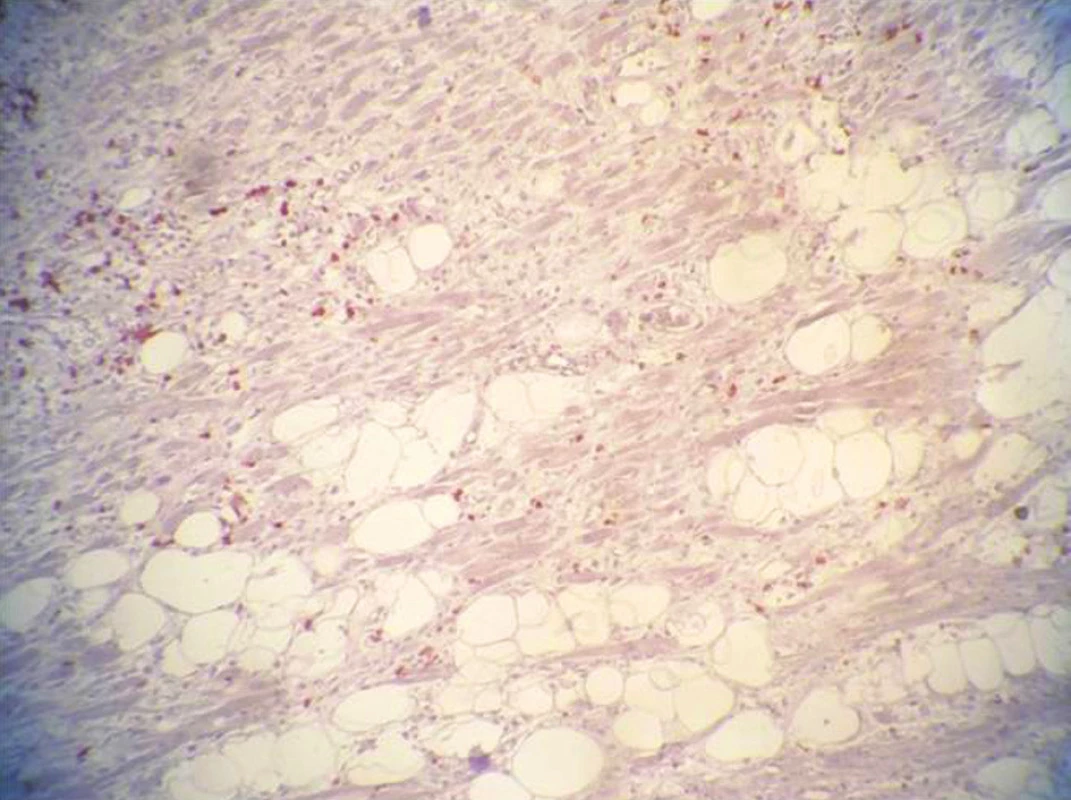
A 28-year-old man with no signicant medical history applied to the emergency department of provincial hospital with feeling very ill. An electrocardiography (ECG) performed in the emergency department the day before he died showed ventricular extra systoles as well as ventricular arrhythmia. After his initial physical examination, he was recommended for admission to a cardiology polyclinic for detailed investigation. The next day, his condition worsened and he was dead on arrival at the hospital. A medico-legal autopsy was performed to clarify the manner and cause of death as mandated by the local prosecutor. His clinical history was completely inconspicuous and his family history had no indication sudden cardiac death. An external examination showed that deceased was 182 cm in height and weighed 90 kg. White foam around the mouth and nostrils, injection marks on the inguinal and antecubital regions and the dorsal part of left hand as well as signs of defibrillation paddles on the anterior wall of the chest were detected during external examination. No significant injuries were observed on external examination. At gross macroscopic internal examination, the heart weighted 560 g, the heart chambers appeared dilated and pethechial hemorrhages were observed on the surface of the LV (Figure 1). Dissection of right ventricular wall revealed a yellowish discoloration of fat tissue. The sections of left ventricular wall and septum were normal in macroscopic investigation. There were only nonspecific signs, such as organ congestion, and pulmonary oedema as the pathological macroscopic findings of internal organs. Histological sections of formalin-xed tissue from the brain, cerebellum, lungs, liver, kidneys, and spleen revealed nonspecific postmortem findings. Pathologic investigation included hematoxylin-eosin, Mason-Trichrome staining and CD8, CD4 immunostaining. The slides were examined with a light microscope. Sections from the heart which were obtained from the RV and LV. Samples from RV revealed massive replacement of myocardium by fibrous and mature adipose tissue in the right ventricle (Figure 2). The predominance of CD4+ and CD8+ lymphocytes were observed microscopically. The gross and microscopic examinations were otherwise unremarkable.
A toxicological screening of tissue was performed and blood and urine samples were taken. The Headspace Gas Chromatography (GC/HS) technique was used for blood alcohol analysis which was at normal levels and Spot Test, Thin Layer Chromatography (TLC) and Cloned Enzyme Donor Immunoassay (CEDIA) techniques were used for drug screening in tissue, blood and urine samples which were negative for analyzed substances.
DISCUSSION
Arrhythmogenic right ventricular dysplasia (ARVD) is a myocardial disease, which predominantly affects the right ventricle (RV) and is one of the major causes of sudden death in the young and athletes (1,2). ARVD is morphologically characterized by diffuse or segmental lack of myocardium in the RV free wall which is replaced by fatty or fibro fatty tissue and also histologically by fibro fatty degeneration of cardiomyocytes which leads to electrical instability and contractility abnormalities (1,3–8). The replacement of the right ventricular myocardium by fibro fatty tissue is progressive process, starting from the epicardium or midmyocardium and then extending to become transmural pathology (2,9). Although several theories have been proposed and different genetic variants have been described, the accurate aetiopathogenesis of ARVD is still unknown (2–5,7–10). ARVD has been described as a disease of unknown cause, characterized by fibro fatty replacement of the RV myocardium (7). The diagnosis of ARVD is based on major and minor criteria including genetic, structural, histological, electrocardiographic, and familial factors which have been established by the Study Group on ARVC of the Working Group Myocardial and Pericardial Disease of the European Society of Cardiology and of the Scientific Council on Cardiomyopathies of the International Society and Federation of Cardiology (8,10). However, many of the cases, of which the first clinical presentation was sudden death, were diagnosed with an autopsy similar to our presented case. Sporadic and familial forms of the ARVD have been described (11). The presence of a family history of ARVD has been reported in 30 % to 50 % of cases (2,5,12,13,15). The autosomal dominant is the most common inheritance form of ARVD, although autosomal recessive forms like Naxos Disease and Carvajal Syndrome have also been reported in the literature (5,14,15). Twelve different genetic variants have been described associated with ARVD, resulting from mutations of genes encoding several components of cardiac desmosomes (15,16). ARVD is associated with highly variable clinical presentation such as ventricular tachycardia, syncope, RV dysfunction and sudden death (2,5,7,8,9). In the 7 % to 29 % of the cases, the first presentation of ARVD may be sudden cardiac death with no prior onset symptoms (15,17). In the presented case there were no symptoms, family and medical history and his first clinical presentation was unexpected sudden death. For that reason, an autopsy is essential to reveal the exact morphologic and histopathologic features of the disease and to provide a proper explanation of the cause of sudden death. Fatty and fibro fatty forms are the two morphologic variants of ARVD (8). While the residual islands of myocytes are surrounded by fibrosis, RV wall thinning with aneurismal dilatation, mononuclear inflammatory cell infiltrates and involvement of LV and septum in some cases are seen in the fibro fatty form, the fatty form is characterized by an almost complete replacement of myocardium by adipose tissue with sparing of the septum and left ventricle and without wall thinning (4,7). In this present study, a case of the morphologically fibro fatty form of ARVD without involvement of LV and septum was described. Microscopic evaluation revealed CD8+ lymphocytes on the RV myocardial samples. Thiene G et al. reported that myocardial inflammation may be seen in up to 75 % of hearts at autopsy (18). However, these findings to date have not been clearly understood and explained as to whether inflammation is cause of the cell death or a response to primary myocardial changes (is it a cause or consequence in reality?). We presented interesting autopsy case of ARVD whose first clinical presentation was sudden unexpected death.
Correspondence address:
Dr. Bülent Eren
Council of Forensic Medicine of Turkey
Bursa Morgue Department
Osmangazi, Heykel, 16010, Bursa, Turkey
tel.: +90 224 442 84 00 / 1632; +90 224 222 03 47
fax: +90 224 442 91 90; +090 224 225 51 70
e-mail:drbulenteren@gmail.com.
Zdroje
1. Marcus FI, Fontaine GH, Guiraudon G, Frank R, Laurenceau JL, Malergue C, Grosgogeat Y. Right ventricular dysplasia: a report of 24 adult cases. Circulation 1982; 65 : 384–398.
2. Thiene G, Corrado D, Basso C. Arrhythmogenic right ventricular cardiomyopathy/dysplasia. Orphanet J Rare Dis 2007; 2 : 45.
3. Hughes SE, McKenna WJ. New insights into the pathology of inherited cardiomyopathy. Heart 2005; 91 : 257–264.
4. Basso C, Thiene G, Corrado D, Angelini A, Nava A, Valente M. Arrhythmogenic right ventricular cardiomyopathy. Dysplasia, dystrophy or myocarditis? Circulation 1996; 94 : 983–991.
5. Corrado D, Fontaine G, Marcus FI, McKenna WJ, Nava A, Thiene G, Wichter T. Arrhythmogenic right ventricular dysplasia/cardiomyopathy: need for an international registry. Circulation 2000; 101 : 101–106.
6. Güdücü N, Kutay SS, Ozenc E, Ciftci C, Yigiter AB, Isci H. Management of a rare case of arrhythmogenic right ventricular dysplasia in pregnancy: a case report. J Med Case Reports 2011; 5 : 300.
7. Kayser HW, van der Wall EE, Sivananthan MU, Plein S, Bloomer TN, de Roos A. Diagnosis of arrhythmogenic right ventricular dysplasia: a review. Radiographics 2002; 22(3): 639–648; 649–650.
8. Corrado D, Basso C, Thiene G. Arrhythmogenic right ventricular cardiomyopathy: diagnosis, prognosis, and treatment. Heart 2000; 83 : 588–595.
9. Basso C, Corrado D, Marcus FI, Nava A, Thiene G. Arrhythmogenic right ventricular cardiomyopathy. Lancet 2009; 373 : 1289–1300.
10. McKenna WJ, Thiene G, Nava A, et al. Diagnosis of arrhythmogenic right ventricular dysplasia/cardiomyopathy. Task force of the working group myocardial and pericardial disease of the European Society of Cardiology and of the Scientific Council on Cardiomyopathies of the International Society and Federation of Cardiology. Br Heart J 1994; 71 : 215–218.
11. Goland S, Czer LS, Luthringer D, Siegel RJ. A case of arrhythmogenic right ventricular cardiomyopathy. Can J Cardiol 2008; 24 : 61–62.
12. Nava A, Thiene G, Canciani B, et al. Familial occurrence of right ventricular dysplasia: a study involving nine families. J Am Coll Cardiol 1988; 12 : 1222–1228.
13. Hermida JS, Minassian A, Jarry G, et al. Familial incidence of late ventricular potentials and electrocardiographic abnormalities in arrhythmogenic right ventricular dysplasia. Am J Cardiol 1997; 79 : 1375-1380.
14. Carvajal-Huerta L. Epidermolytic-palmoplantar keratoderma with woolly hair and dilated cardiomyopathy. J Am Acad Dermatol 1998; 39 : 418–421.
15. Khan A, Mittal S, Sherrid MV. Arrhythmogenic right ventricular dysplasia: from genetics to treatment. The Anatolian Journal of Cardiology 2009; 9 : 24–31.
16. Sato T, Nishio H, Suzuki K. Sudden death during exercise in a juvenile with arrhythmogenic right ventricular cardiomyopathy and desmoglein-2 gene substitution: a case report. Leg Med (Tokyo) 2011; 13 : 298–300.
17. Corrado D, Basso C, Rizzoli G, Schiavon M, Thiene G. Does sports activity enhance the risk of sudden death in adolescents and young adults? J Am Coll Cardiol 2003; 42 : 1959–1963.
18. Thiene G, Basso C. Arrhythmogenic right ventricular cardiomyopathy: An update. Cardiovasc Pathol 2001; 10 : 109–117.
Štítky
Patologie Soudní lékařství Toxikologie
Článek Expert Forensic ScienceČlánek Záchrana v zimě
Článek vyšel v časopiseSoudní lékařství

2013 Číslo 3-
Všechny články tohoto čísla
- Gender differences in alcohol affection on an individual
- Death due to Arrhythmogenic Right Ventricular Dysplasia: A case report
- Expert Forensic Science
- Traumatic changes of intrathoracic organs due to external mechanical cardiopulmonary resuscitation. Case reports
- Záchrana v zimě
- Sudden death due to high take-off right coronary artery
- Signs of self-inflicted wounds; how accurate they are
- Nejlepší monografie a nejlepší článek (Kritéria pro nominaci na cenu ČSSLST)
- Soudní lékařství
- Archiv čísel
- Aktuální číslo
- Informace o časopisu
Nejčtenější v tomto čísle- Traumatic changes of intrathoracic organs due to external mechanical cardiopulmonary resuscitation. Case reports
- Gender differences in alcohol affection on an individual
- Sudden death due to high take-off right coronary artery
- Death due to Arrhythmogenic Right Ventricular Dysplasia: A case report
Kurzy
Zvyšte si kvalifikaci online z pohodlí domova
Autoři: prof. MUDr. Vladimír Palička, CSc., Dr.h.c., doc. MUDr. Václav Vyskočil, Ph.D., MUDr. Petr Kasalický, CSc., MUDr. Jan Rosa, Ing. Pavel Havlík, Ing. Jan Adam, Hana Hejnová, DiS., Jana Křenková
Autoři: MUDr. Irena Krčmová, CSc.
Autoři: MDDr. Eleonóra Ivančová, PhD., MHA
Autoři: prof. MUDr. Eva Kubala Havrdová, DrSc.
Všechny kurzyPřihlášení#ADS_BOTTOM_SCRIPTS#Zapomenuté hesloZadejte e-mailovou adresu, se kterou jste vytvářel(a) účet, budou Vám na ni zaslány informace k nastavení nového hesla.
- Vzdělávání


