
Accuracy of a modified CMT+ for assessing pelvic floor muscle contraction in pregnancy
Authors:
S. H. Stensgaard 1; K. M. Bek 2; K. M. Ismail 3
Authors‘ workplace:
Department of Obstetrics and Gynecology, Perinatal Research unit, Aarhus University Hospital, Denmark
1; Department of Obstetrics and Gynecology, Aarhus University Hospital, Denmark
2; Department of Gynecology and Obstetrics, Charles University Medical Faculty and University Hospital in Pilsen, Czech Republic
3
Published in:
Ceska Gynekol 2025; 90(4): 293-298
Category:
Original Article
doi:
https://doi.org/10.48095/cccg2025293
Overview
Objective: The ability to locate pelvic floor muscles (PFM) is essential for the effectiveness of pelvic floor muscle training programs. The aim of this study was to investigate the accuracy of a modified coccygeal movement test that incorporates an objective assessment of abdominal muscle co-activation (CMT+) (Index test) compared to transabdominal ultrasound (TAU) scanning (Gold standard) in diagnosing accurate PFM contraction. Methods: Pregnant women attending the hospital for a routine in the middle 2nd trimester scan who are able to understand the study rationale and information were considered eligible for inclusion. TAUs were performed by one out of two trained operators. CMT+ was performed by an experienced physiotherapist. The CMT+ assessor and participants were blind to the TAU result. Results: A total of 117 participants were recruited into the study with a mean BMI of 30.86 kg/m² (4.5) and 23.16 kg/m² (3.7), resp. CMT+ (Index test) correctly identified 5 out of the 9 participants who were not able to contract and 107 out of the 108 who were able to contract their PFM resp. (sensitivity = 55.6%, specificity = 99.1%, positive predictive value = 83.3% and negative predictive value = 96.4%; LR+ = 60 and LR – = 0.45). Conclusion: CMT+ is an easy to perform test with high specificity and negative predictive value that has the additional benefit of assessing any concomitant abdominal muscle co-activation. Therefore, CMT+ is a potentially useful initial screening test to identify those who cannot perform a correct pelvic floor muscle contraction, and would benefit from specialized assessment and structured training.
Keywords:
pelvic floor – transabdominal ultrasound – pelvic floor muscles – co-activation – CMT+
Introduction
There is a recognized association between pregnancy and pelvic floor disorders. Studies show that 25–30% of women report urinary incontinence after childbirth. Moreover, most women who have symptoms of incontinence by 3 months postnatal will continue to have symptoms and the negative impact on their quality of life in the longer term [1]. Pelvic floor exercises are an important element in the prevention and treatment of pelvic floor dysfunction [2]. In their systematic review, Bø et al. concluded that there was no strong evidence that exercise regimes, apart from pelvic floor muscle training (PFMT), can reduce stress urinary incontinence in women [3]. There is also a positive link between the increase in pelvic floor muscle (PFM) strength and improvement in stress urinary incontinence [3].
Currently, it is recommended that pregnant women are advised to perform PFMT early in their antenatal care [4]. Furthermore, people hear about benefits of pelvic floor exercises from the media and social networks, yet this information is seldom backed up by guidance as to how to ensure that they can locate their PFMs correctly. This issue is of great importance in view of evidence from several studies confirming that 25–30% of women are unable to locate and contract their PFMs correctly, where they often contract other muscle groups (gluteal, thigh or abdominal muscles) instead [5–7].
Vaginal examination has been the traditional clinical test for assessing the ability to contract the PFM. However, ultrasound and magnetic resonance imaging (MRI) are considered the gold standard objective tests to assess and quantify this [8]. Using dynamic MRI, Bø et al. showed that during a PFM contraction, there was an inward lift of the coccyx in a ventral – cranial direction, whereas during straining, the coccyx was displaced in a caudal-dorsal direction [9]. This finding was later supported by Grassi et al. and Fujisaki et al. [10,11]. Based on these findings, we previously explored the feasibility of clinically assessing the movement of the coccyx as an indicator of an accurate PFM contraction (CMT – the Coccygeal Movement Test) [12]. In our first study we demonstrated that CMT was a reproducible, easy to perform test, that has the potential to be a useful screening test for the assessment of PFM contraction. However, we observed that although some women can locate and contract their PFMs, they seemed to co-contract their abdominal muscles at the same time. Hence, we proposed modifying the CMT to include the simultaneous assessment of abdominal wall muscles for any co-contraction (CMT+).
Transabdominal ultrasound (TAU) is a noninvasive reliable and valid method for assessing pelvic floor function by observing a cranial movement of the bladder base as an indicator of a correct PFM contraction [13] and was shown to have significant correlation to digital palpation when measured simultaneously [14]. Therefore, the aim of this study was to investigate the accuracy of CMT+ using TAU as the gold standard comparator.
Methods
The study plan was reviewed by the Regional Scientific Ethical Committee and an ethical approval waiver was issued due to the low risk and noninvasive nature of the study. A valid informed written consent was obtained from all participants prior to recruitment.
Participants
All pregnant women attending the hospital for their routine in the middle 2nd trimester scan appointment, who were able to understand the reason for the study, and in the presence of one of two specially trained TAU assessors, were considered eligible for inclusion into the study.
Women were approached and informed about the study whilst waiting for their routine ultrasound scan and were only included if they agreed and signed a consent to participate in the study. For each participant, the weight in kilograms and height in centimeters were recorded and the BMI was calculated. Once included, participants were given an information leaflet about pelvic floor exercises and a short (3–5 min) verbal instruction about what a correct pelvic floor contraction entail. They were specifically asked to relax the gluteal muscles, hip adductors and stomach muscles and were instructed to try to contract their pelvic floor as if they were trying to stop passing wind.
TAU – gold standard
A Pro Focus UltraView – 2202-7 ultrasound machine was used to acquire all TAU scans of bladder base elevation, as an indicator of PFM contraction. Two ultrasound assessors were specially trained before the study to undertake this assessment. Ultrasound assessments were performed after completion of the mid trimester scan while the woman was lying in a supine position with a full bladder. The assessor turned the screen away from the women and the transducer was placed over the supra-pubic region and slightly angled caudally until a satisfactory view of the inferior/posterior part of the bladder was obtained. The TAU assessor then asked the participant to “squeeze” with no further comments, instructions, or feedback. Movement of the bladder base was observed and documented as surrogate markers for PFM and abdominal wall muscle contractions (Fig. 1).
Based on the direction (s) of bladder base displacement, the TAU assessor categorized participants as:
1. Valsalva;
2. nothing;
3. PFM contraction with abdominal muscle co-activation;
4. correct isolated PFM contraction.
The TAU assessment was repeated twice and the higher score of the two assessments was used as the result for the transabdominal scan assessment (gold standard).
CMT+
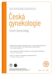
All CMT+ assessments were performed by the same trained assessor who was blind to the TAU assessor’s findings. CMT+ assessments were performed on top of the participants’ normal clothing while they were in a standing or sitting position. The assessor placed the thenar and hypothenar eminences against the upper part of the subject’s sacrum with the index and ring finger on the gluteal muscles and the middle finger on or close to the coccyx as previously described [12].

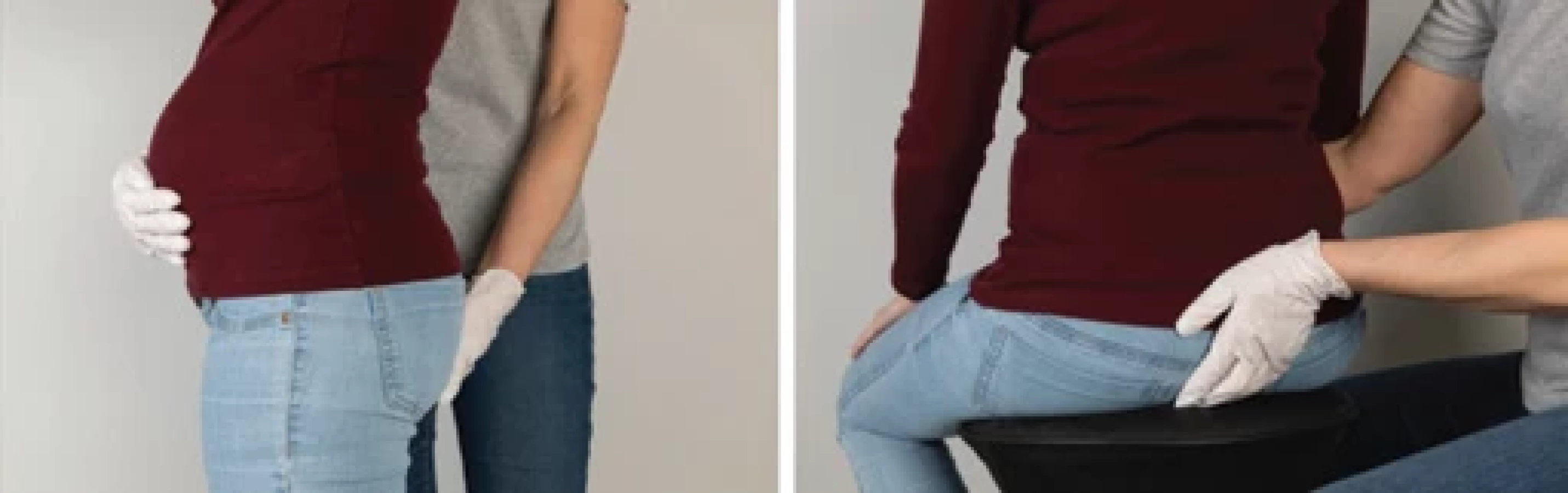
The assessor then placed the non-dominant hand on the participant’s lower abdomen to palpate any contraction of the lower abdominal muscles, as an indicator of abdominal muscle co-contraction (co-activation) (Fig. 2). The CMT+ assessor asked the patient to “squeeze” her PFM and then decided if the test indicated:
1. Valsalva;
2. nothing;
3. PFM contraction with abdominal muscle co-activation;
4. correct isolated PFM contraction.
An outward or no coccygeal displacement indicated “straining” or “nothing” respectively (positive test – no PFM contraction). An inward displacement of the coccyx, or the tissues around it, was considered indicative of a “correct PFM contraction” (negative test). We documented if there was an inward movement associated with abdominal wall contraction but still classified them as able to contract their PFM. The assessor was allowed to guide the patient if they were using their gluteal muscles excessively hindering their ability to palpate the tissues around the coccyx.
Statistical analysis
All measurements were entered using EpiData and analyses was performed using SPSS 11.0 statistical software. Results are given with their 95% confidence intervals. A significance level of 0.05 was chosen. Based on the number of true positive, true negative, false positive and false negative CMT+ observations compared to TAU, the sensitivity, specificity, positive and negative predictive values, and likelihood ratio of a positive (LR+) and negative (LR–) were calculated.
Results
A total of 122 subjects were informed about the study whilst waiting for their in the middle 2nd trimester scan. Of these, 120 consented to participate and were included into the study. Complete data were available for 117 participants who contributed to the final analysis. Participants had a mean BMI of 23.16 kg/m² (SD 3.7; range 18–35), mean age of 20.86 (SD 4.5; range 20–44) years and 70 of the participants were primigravidas. Because the main aim of the study was to evaluate if there was a pelvic floor contraction or not, the findings of both the TAU and CMT+ were dichotomized to either “not able to contract” (Valsalva or nothing = positive test) or “able to contract” (PFM contraction with abdominal muscle co-activation or correct PFM contraction = negative test) (Tab. 1).


On TAU, 105 (90%) had the same result on both attempts. However, 12 (10%) had two different observations on ultrasound scanning, and the test result category changed, from not being able to contract to being able to contract, in 4 of these particiants. Compared to TAU (gold standard), CMT+ (Index test) correctly identified 5 of the 9 participants who were not able to contract and 107 of the 108 who were able to contract their PFM respectively (sensitivity 55.6%, specificity 99.1%, positive predictive value 83.3% and negative predictive value 96.4%; LR+ = 60 and LR – = 0.45) (Tab. 2).
Discussion
Based on this study, CMT+ had a better specificity (99.1%) than sensitivity (55.6%). Furthermore, the test had a high negative predictive value, so it was fairly accurate in identifying women who could locate their PFM. Despite the lower positive predictive value (83.3%), there was only one participant out of 108 was mis-diagnosed as not able to contract their pelvic floor (False positive). We believe that this is an acceptable false positive rate, particularly, that the only clinical consequence is potentially an unnecessary second confirmatory test, such as a vaginal examination or pelvic ultrasound scan. Nevertheless, in this study we obtained a false negative (incorrect diagnosis that they are able to contract their PFM when the gold standard test suggested they were not able to) in 4 participants. It is important for screening tests to have very few false negatives. In the initial CMT study the assessor was only allowed to give the command “squeeze” twice [12]. In this study the CMT+ assessor could guide the patient if they were using their gluteal muscles excessively hindering the ability to palpate the tissues around the coccyx. Although only speculative, it is possible that such guidance might have enabled these participants to perform a correct contraction, having not been able to do so when assessed a few minutes prior to that on TAU. In a real clinical setting, the assessor will have the opportunity to do multiple testing while giving feedback, and this is expected to reduce false positive and false negative results.
When asked to do a “PFM contraction”, only 73% of participants did an isolated contraction of the pelvic floor according to TAU assessment. Hence, 27% did a Valsalva, nothing or abdominal muscle co-activation when attempting a PFM contraction. These findings are supported by other studies showing that approximately 25–30% of all women are unable to contract their PFMs correctly based on a short verbal instruction [5–7]. Although studies by Sapsford et al. [15] have indicated that specific abdominal exercises activate the PFMs (in subjects with no symptoms of pelvic floor muscle dysfunction) and some physiotherapists use activation of the transversus abdominis as an indicator for “pelvic floor muscle activity”, it is important to emphasize that the pubococcygeus is the primary muscle that must be trained. Indeed, contraction of the transversus abdominis will potentially increase the intrabdominal pressure and add strain to the pelvic floor.
In our study, participants classified as being able to contract the PFM with concomitant depression of the bladder base on TAU or co-activation of the abdominal wall muscles were considered being able to contract their pelvic floor muscles because the main aim of our work was to assess the ability of CMT+ to identify patients who are able to do so. Nevertheless, the clinical consequences of PFM contraction with abdominal muscle co-activation are still unknown.
For a proper PFM contraction, it is mandatory that women receive structed and supervised training with appropriate monitoring and feedback [8,16]. Hay-Smith et al. found that, of 43 RCTs they reviewed, only 15 stated that a correct PFM contraction was checked prior to training [17]. CMT+ could potentially be the missing link because it is easy to perform and useful to identify women who can locate their PFMs. In their study, Ben Ami and Dar showed that the most effective verbal instruction for correct PFM instruction was the “posterior instruction” of squeezing the anus as if trying to stop passing flatus [18]. We believe that a combination of a verbal “posterior instruction” and CMT+ could be a good foundation for structured instruction and objective assessment in relation to a correct PFM contraction in most patients.
Maher and Iberle reported that they were able to palpate the tip of the coccyx in all 37 participants regardless of BMI [19]. This was also the case in this study. The 117 participants in this study had a range of BMI from 18 to 35 and it was possible to palpate a caudal or ventral movement of the tissue around the coccyx in all our study participants.
There are several strengths to our work. All attendees for a in the middle 2nd trimester scan were informed about the study and the majority agreed to participate, hence mitigating the potential risk of selection bias. We also attempted to minimize methodological biases by blinding participants and the CMT+ assessor. The TAU assessors were given clear instructions not to provide patients with any feedback or additional information apart a simple unified instruction. Furthermore, participants were not able to see the ultrasound scan screen. However, we appreciate that the study had some limitations. First, only one experienced physiotherapist (SS) did the CMT+ and it is unknown if a less experienced assessor would be able to reproduce the same result. Nonetheless, Maher and Iberle [19] demonstrated that Coccygeal Motion Palpation could be used by different clinicians. However, further studies are needed to determine the impact of clinicians’ experience and training on CMT+ test accuracy. Second, it was convenient to use the actual clinical setup for recruitment and TAU assessments because a full bladder makes it possible to scan the bladder base movement. However, it is unknown how pregnancy and a full bladder would have affected the woman’s ability to do a correct contraction.
With current demands on maternity staff and services, it is essential for maternity units to develop efficient care pathways to ensure the delivery of evidence-based recommendations to improve pregnancy related pelvic floor outcomes. Moreover, pregnant women tend to be reluctant to have a vaginal examination to assess their ability to locate their pelvic floor. Furthermore, the test can be performed with the patient in a standing or sitting position, which is a more physiological position to assess what actually happens when performing a PFM contraction compared to lying flat or in lithotomy position. Therefore, CMT+ could potentially provide a feasible solution to both these issues. Additionally, it can also be used as a tool to deliver structured training to women who are not able to locate their PFMs. Nevertheless, it is important that the latter assumption is tested in a specifically designed study.
Conclusion
Like CMT, CMT+ is an easy to perform screening tests that has the additional benefit of assessing any concomitant abdominal muscle activity making it a potentially useful initial screening test in structured training programs for the PFMs. Although, we do not expect CMT+ to replace vaginal palpation and its ability to give feedback on the strength and endurance of pelvic floor muscle contraction is yet to be tested, it is a minimally invasive screening test to identify patients who would benefit from a more specialized structured approach for their pelvic floor muscle assessment and training.
Sources
1. MacArthur C, Glazener C, Lancashire R et al. Exclusive caesarean section delivery and subsequent urinary and faecal incontinence: a 12-year longitudinal study. BJOG 2011; 118 (8): 1001–1007. doi: 10.1111/j.1471-0528.2011. 02964.x.
2. Mørkved S, Bø K. Effect of pelvic floor muscle training during pregnancy and after childbirth on prevention and treatment of urinary incontinence: a systematic review. Br J Sports Med 2014; 48 (4): 299–310. doi: 10.1136/bjsports-2012-091758.
3. Bø K, Herbert RD. There is not yet strong evidence that exercise regimens other than pelvic floor muscle training can reduce stress urinary incontinence in women: a systematic review. J Physiother 2013; 59 (3): 159–168. doi: 10.1016/S1836-9553 (13) 70180-2.
4. Woodley SJ, Lawrenson P, Boyle R et al. Pelvic floor muscle training for preventing and treating urinary and faecal incontinence in antenatal and postnatal women. Cochrane Database Syst Rev 2020; 5 (5): CD007471. doi: 10.1002/14651858.CD007471.pub4.
5. Benvenuti F, Caputo GM, Bandinelli S et al. Reeducative treatment of female genuine stress incontinence. Am J Phys Med 1987; 66 (4): 155–168.
6. Bump RC, Hurt WG, Fantl JA et al. Assessment of Kegel pelvic muscle exercise performance after brief verbal instruction. Am J Obstet Gynecol 1991; 165 (2): 322–329. doi: 10.1016/0002-93 78 (91) 90085-6.
7. Barton A, Serrao C, Thompson J et al. Transabdominal ultrasound to assess pelvic floor muscle performance during abdominal curl in exercising women. Int Urogynecol J 2015; 26 (12): 1789–1795. doi: 10.1007/s00192-015 - 2791-9.
8. Bø K, Sherburn M. Evaluation of female pelvic--floor muscle function and strength. Phys Ther 2005; 85 (3): 269–282. doi: 10.1093/ptj/85.3.269.
9. Bø K, Lilleås F, Talseth T et al. Dynamic MRI of the pelvic floor muscles in an upright sitting position. Neurourol Urodyn 2001; 20 (2): 167–174.doi: 10.1002/1520-6777 (2001) 20 : 2<167:: AID-NAU19>3.0.CO; 2-4.
10. Grassi R, Lombardi G, Reginelli A et al. Coccygeal movement: assessment with dynamic MRI. Eur J Radiol 2007; 61 (3): 473–479. doi: 10.1016/j.ejrad.2006.07.029.
11. Fujisaki A, Shigeta M, Shimoinaba M et al. Influence of adequate pelvic floor muscle contraction on the movement of the coccyx during pelvic floor muscle training. J Phys Ther Sci 2018; 30 (4): 544–548. doi: 10.1589/jpts.30.544.
12. Stensgaard SH, Moeller Bek K, Ismail KM. Coccygeal movement test: an objective, non-invasive test for localization of the pelvic floor muscles in healthy women. Med Princ Pract 2014; 23 (4): 318–322. doi: 10.1159/000 362337.
13. Sherburn M, Murphy CA, Carroll S et al. Investigation of transabdominal real-time ultrasound to visualise the muscles of the pelvic floor. Aust J Physiother 2005; 51 (3): 167–170. doi: 10.1016/S0004-9514 (05) 70 023-4.
14. Arab AM, Behbahani RB, Lorestani L et al. Correlation of digital palpation and transabdominal ultrasound for assessment of pelvic floor muscle contraction. J Man Manip Ther 2009; 17 (3): e75–e79. doi: 10.1179/jmt.2009.17.3.75E.
15. Sapsford RR, Hodges PW, Richardson CA et al. Co-activation of the abdominal and pelvic floor muscles during voluntary exercises. Neurourol Urodyn 2001; 20 (1): 31–42. doi: 10.1002/1520-6777 (2001) 20 : 1<31:: AID-NAU5>3.0.CO; 2-P.
16. Bech SR, Villadsen D, Laursen HH et al. The effect of group or individualised pelvic floor exercises with or without ultrasonography guidance for urinary incontinence in elderly women – a pilot study. J Bodyw Mov Ther 2021; 28 : 34–41. doi: 10.1016/j.jbmt.2021. 07.032.
17. Dumoulin C, Cacciari LP, Hay-Smith EJ. Pelvic floor muscle training versus no treatment, or inactive control treatments, for urinary incontinence in women. Cochrane Database Syst Rev 2018; 10 (10): CD005654. doi: 10.1002/14651858.CD005654.pub4.
18. Ben Ami N, Dar G. What is the most effective verbal instruction for correctly contracting the pelvic floor muscles? Neurourol Urodyn 2018; 37 (8): 2904–2910. doi: 10.1002/nau. 23810.
19. Maher RM, Iberle J. Concurrent validity of noninvasive coccygeal motion palpation and transabdominal ultrasound imaging in the assessment of pelvic floor function in women. J Womens Health Phys Therap 2020; 44 : 176–181. doi: 10.1097/JWH.0000000000 000175.
Labels
Paediatric gynaecology Gynaecology and obstetrics Reproduction medicineArticle was published in
Czech Gynaecology
2025 Issue 4
Most read in this issue
- HIV infection and adverse perinatal outcomes – a meta-analysis of premature births, low birth weights, and small for gestational age newborns
- Role of tru-cut bio psy in the management of myometrial lesions
- Early detection of recurrent ovarian cancer, current use of oncomarkers, imaging methods, and future perspectives
- Robotic-assisted cesarean scar defect repair