
The rationale for early diagnosis and management of benign soft tissue masses: a case report
Authors:
R. De Loecker 1; R. Van den Bossche 1; M. De Fré 1; R. Van Steen 1; A. Van Goethem 1; N. Vermeersch 1; T. Tondu 1; V. Verhoeven 2; F. E. F. Thiessen 1
Authors‘ workplace:
Department of Plastic, Reconstructive and Aesthetic Surgery, Antwerp University Hospital, Edegem, Belgium
1; Department of family medicine, Gouverneur Kinsbergcentrum, Edegem, Belgium
2
Published in:
ACTA CHIRURGIAE PLASTICAE, 68, 1-2, 2026, pp. 64-68
doi:
https://doi.org/10.48095/ccachp202664
De Loecker R, Van den Bossche R, De Fré M et al. The rationale for early diagnosis and management of benign soft tissue masses: a case report. Acta Chir Plast 2026; 68 (1–2): 64–68.
Introduction
As a benign mesenchymal tumour consisting of adipocytes, a lipoma can occur anywhere on the body presenting as a soft, painless mass. While most lipomas remain small, typically between 1 to 2 cm, there is the possibility of developing larger variants, so called giant lipomas [1]. While still being benign these giant lipomas may create discomfort or even disability in patients, depending on their size and location. Lipomas are most commonly found in subcutaneous tissue on the head, neck, shoulders and back. In rare cases, they can develop intramuscularly, interosseously or in association with visceral organs [1]. They can also emerge at sites of previous trauma [2]. Furthermore, there are certain associated risk factors such as an advanced age and obesity [1,3]. Gene mutations may also play an important role [2].
Although malignant soft tissue tumours, such as liposarcomas are rare, they can resemble benign lipomas, leading to frequent presumptions of benignity [4,5]. This assumption can lead to unplanned sarcoma excisions, increasing patient morbidity. A careful diagnostic approach may help to prevent this [3,6].
Case description
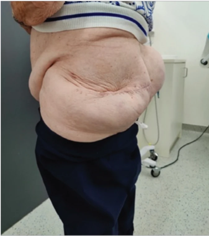
An 82-year-old obese (BMI 35.34) female patient presented with a progressively enlarging bilateral (left more than right) gluteal mass over the last 5 years. The patient had no further relevant personal or family medical history. Initially, when the masses were small, her general practitioner informed her that benign lipomas typically require no treatment. Therefore, she did not pursue further care. As the tumours grew, they began to impair her daily activities, prompting her to look for surgical advice (Fig. 1). Pre-operative imaging with magnetic resonance imaging (MRI) showed the presence of two large lipomas with a benign aspect of the gluteal region (Fig. 2). Due to functional impairment surgical excision was recommended for the largest lipoma, with plans to address the second lipoma after full recovery.
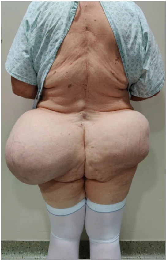
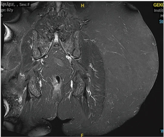
The patient underwent a rather significant surgical procedure, because of a delayed presentation to a medical doctor. The excision of the mass itself required an incision of 25 cm and was performed in an elliptical fashion with respect to the surrounding vascularization of the skin. The weight of the excised lipoma was 3,750 g, measuring 32.5 × 30.5 × 9.5 cm (Fig. 3, 4). Due to the development of seroma the hospital stay was extended and multiple visits in the outpatient clinic were needed. The wound had healed well on day 13, although fluid output remained at 150–200 cm3 per day. Three weeks after surgery, the sutures could be removed, however the patient continued to produce 160–200 cm of fluid daily, prompting sclerotherapy with doxycycline and removal of the Redon drain (Fig. 5). At 1, 2, and 3 months postoperative, the patient developed recurrent large seromas, each requiring drainage of 300–400 cm3 of fluid. The final drainage occurred at 5 months, with 80 cm3 removed. After this visit no further punctures were necessary. Although contralateral debulking was considered, the patient declined this procedure up till now. This was mainly due to her advanced age, less interference with daily life activities. To date, there has been no recurrence of the lipoma.
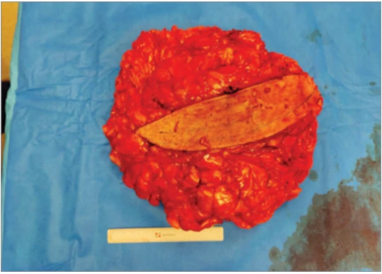
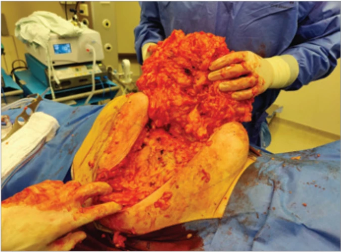
Histopathological analysis of the soft--tissue lesion revealed that most of the mass consisted of mature adipocytes with slight variation in size and nucleus, with no lipoblasts present in the lesion. These findings were consistent with the diagnosis of a benign giant lipoma with the possibility of being a low-grade lipomatous lesion such as an atypical spindle cell/pleomorphic lipomatous tumour (ASPLT). Eventually molecular testing by retinoblastoma 1 fluorescence in situ hybridization (Rb1-FISH) was preformed to exclude the possibility of an ASPLT.
Discussion
This case illustrates the consequences of prolonged conservative management of a giant lipoma, resulting in an unusually large, bilateral lesion requiring an extensive surgical procedure. To our knowledge, the combination of the atypical anatomical localization and the dimensions of the lesion has not been reported previously.
The etiological components of the extensive and sometimes rapid growth seen in giant lipomas, is still not completely understood [1,2]. Trauma-related rupture of fibrous septa and connections between the skin and deep fascia may contribute to rapid adipose proliferation [1,7]. However, there are endocrine, dysmetabolic and genetic factors playing a significant role as well [1,8]. In this case, the patient reported no trauma history to the gluteal region but had several risk factors, including advanced age and obesity [1,3].
Due to the continuous growth and size of the lesion in this patient, several other soft tissue tumours needed to be excluded. Lipomatous tumours range from benign lipomas to deep atypical lipomas (ATL), well-differentiated lipoma-like liposarcomas (WDL), and liposarcomas. This wide variety did not only create disagreement on the nomenclature of these different tumours, but controversy regarding the histopathological diagnosis as well [1]. The benign lipoma is the most common type of soft tissue tumour. Their peak incidence is between ages 40–60, though their exact incidence is unknown as most subcutaneous lipomas require no treatment and often go unreported. These tumors are typically well-circumscribed, lobulated, yellow masses with a thin fibrous capsule and no malignant potential [9,10].
Although ATL and WDL are histopathologically identical, they differ in recurrence rates, malignancy potential, and anatomical locations. ATL has a low local recurrence rate (20%), low risk of malignant transformation (< 5%) and presents on extremities and superficial trunk. WDL has a recurrence rate of up to 100%, a potential risk of malignancy of approximately 20% and arises from the retroperitoneum, mediastinum or paratesticular region [3,9]. Histopathologically, they are identical and are composed of mature adipocytes displaying more pleomorphism, nuclear atypia and hyperchromatic stroma cells compared to benign lipomas [3,9,11,12]. Due to the potential malignant qualities of ATL and WDL, there is a significant importance in differentiating these soft tissue tumours from a benign lipoma. While contrast-enhanced MRI, based on a scoring system created by Nagono et al., can differentiate them with high sensitivity (100%) and moderate specificity (71%), the gold standard to distinguish an ATL from a benign lipoma remains molecular testing via murine double minute-2 (MDM2) amplification on fluorescence in situ hybridization (FISH) analysis, achieving 92–100% specificity and 97–100% sensitivity [13].
Finally, there are liposarcomas. These malignant, severely aggressive soft tissue tumours can be divided into three different subtypes: dedifferentiated liposarcoma, myxoid liposarcoma and pleomorphic liposarcoma. Together they comprise up to 12.8% of all soft tissue sarcomas [14]. These tumors often present similarly to benign soft tissue masses as enlarging, painless lesions. MRI can aid in diagnosis by assessing the tumor’s fat content, with higher fat levels typically seen in benign lipomas and lower fat content in liposarcoma. However, the definitive diagnosis can only be established through tissue examination, making complete resection essential for an accurate diagnosis [3]. This was performed in our case.
Given the broad differential diagnosis of growing soft tissue tumors, early diagnosis and management are crucial. Criteria for surgical removal include tumors larger than 5 cm, involvement of the deep fascia, continuous growth, concerning features (such as firmness or irregularity), suspicious preoperative biopsy results, functional impairment, pain, or cosmetic concerns [3]. In this case, the lipoma exceeded 5 cm, caused functional and aesthetic issues, prompting the decision for surgical removal and histopathological analysis.
Surgical excision is the most common method of treatment, with liposuction also gaining interest as described by Silistreli et al. [15]. The surgical excision of giant lipomas is the superior method for assuring complete removal of the mass while ensuring the lowest rate of recurrences (< 5%) and damage to the surrounding tissue. Still temporary follow up of this region is recommend [15,16]. Surgery for giant benign lipomas is not always necessary. Treatment is often decided based on a number of factors with the most common being the aesthetic appearance of the lesion.
Conclusion
This case highlights the importance of early, accurate diagnosis and the risks of prolonged conservative management of soft tissue masses, which can lead to excessive growth and potential malignancy. The presence of giant lipomas presenting in the subcutaneous gluteal region is rare but not unheard of as described by Litchinko et al. [17].
MRI was performed to confirm the clinical suspicion of a benign giant lipoma and exclude other malignant soft tissue tumors. This was further validated postoperatively through microscopy and immunohistochemistry of the excised mass. Despite the lipoma’s excessive growth, complete excision and satisfactory reconstruction were achieved, meeting the patient’s aesthetic and functional goals.
The main goal of this case report is emphasizing the risk and consequences of conservative management of soft tissue masses. Misdiagnosing benign lipomas may result in overlooking malignant lipomatous tumors and requiring major surgery. Therefore, soft tissue lesions larger than 5 cm with continuous growth should be thoroughly examined with proper diagnostic testing to enable early intervention and avoid extensive surgery.
Roles of the authors
Ralph De Loecker, Ruben Van den Bossche, Maxime De Fré, Nicolas Vermeersch: investigation; clinical follow-up; assistance during surgery; writing – original draft, review & editing. Roanne Van Steen: investigation; clinical follow-up; writing – review & editing. Angelique Van Goethem: writing – review & editing; validation and verification of references. Thierry Tondu, Véronique Verhoeven, Filip Thiessen: surgery; clinical management and supervision of the patient during admission and follow-up; writing – original draft, review & editing; supervision.
Funding
None
Conflict of interest
No potential conflict of interest was reported by the authors.
Ethical approval
All applicable international, national, and/or institutional guidelines were followed, including the Declaration of Helsinki from the World Medical Association (WMA).
Informed consent
Informed consent was obtained from all individual participants included in the study.
Originality statement
This manuscript has not been published before, nor is it under consideration for publication elsewhere and publication of this manuscript has been approved by all (co-) authors.
Sources
1. Allen B., Rader C., Babigian A. Giant lipomas of the upper extremity. Can J Plast Surg. 2007, 15 (3): 141–144.
2. Aust MC., Spies M., Kall S., et al. Posttraumatic lipoma: fact or fiction? Skinmed. 2007, 6 (6): 266–270.
3. Johnson CN., Ha AS., Chen E., et al. Lipomatous soft-tissue tumors. J Am Acad Orthop Surg. 2018, 26 (22): 779–788.
4. Balach T., Stacy GS., Haydon RC. The clinical evaluation of soft tissue tumors. Radiol Clin North Am. 2011, 49 (6): 1185–1196.
5. Church DJ., Krumme J., Kotwal S. Evaluating soft-tissue lumps and bumps. Mo Med. 2017, 114 (4): 289.
6. Tedesco NS., Henshaw RM. Unplanned resection of sarcoma. J Am Acad Orthop Surg. 2016, 24 (3): 150–159.
7. Aust MC., Spies M., Kall S., et al. Posttraumatic lipoma: fact or fiction? Skinmed. 2007, 6 (6): 266–270.
8. Kubo T., Matsui Y., Naka N., et al. Specificity of fusion genes in adipocytic tumors. [online]. Available from: https: //ar.iiarjournals.org/content/30/2/661.
9. Goldblum JR., Folpe AL., Weiss SW. Enzinger & Weiss’s soft tissue tumors (7th edition). Elsevier. 2020.
10. Jo VY., Fletcher CDM. WHO classification of soft tissue tumours: an update based on the 2013 (4th edition). Pathology. 2014, 46 (2): 95–104.
11. Evans HL., Soule EH., Winkelmann RK. Atypical lipoma, atypical intramuscular lipoma, and well differentiated retroperitoneal liposarcoma: a reappraisal of 30 cases formerly classified as well differentiated liposarcoma. Cancer. 1979, 43 (2): 574–584.
12. Enzinger FM., Winslow DJ. Liposarcoma. A study of 103 cases. Virchows Arch Pathol Anat Physiol Klin Med. 1962, 335 : 367–388.
13. Weaver J., Downs-Kelly E., Goldblum JR., et al. Fluorescence in situ hybridization for MDM2 gene amplification as a diagnostic tool in lipomatous neoplasms. Mod Pathol. 2008, 21 (8): 943–949.
14. Amer KM., Congiusta DV., Thomson JE., et al. Epidemiology and survival of liposarcoma and its subtypes: a dual database analysis. J Clin Orthop Trauma. 2020, 11: S479–S484.
15. Silistreli ÖK., Durmuş EÜ., Ulusal BG., et al. What should be the treatment modality in giant cutaneous lipomas? Review of the literature and report of 4 cases. Br J Plast Surg. 2005, 58 (3): 394–398.
16. Allen B., Rader C., Babigian A. Giant lipomas of the upper extremity. Can J Plast Surg. 2007, 15 (3): 141–144.
17. Litchinko A., Cherbanyk F., Menth M., et al. Giant gluteal lipoma surgical management. BMJ Case Rep. 2019, 12 (8): e229842.
Labels
Plastic surgery Orthopaedics Burns medicine TraumatologyArticle was published in
Acta chirurgiae plasticae
2026 Issue 1-2
Most read in this issue
- A systematic review of sensory outcomes in nipple reinnervation after implant-based breast reconstruction
- Principles of nasal framework reconstruction for complex nasal defects: review
- Self-esteem and quality of sexual life in women undergoing breast reconstruction due to mastectomy: a cross-sectional study in a non-European setting
- Cutting-edge biotechnological strategies to enhance autologous fat graft survival