
Reconstructive challenges following Fournier’s gangrene: a case report
Authors:
K. Oben; A. Zarowska; L. De Roeck; I. Vrints
Authors‘ workplace:
Department of Plastic Surgery, Heilig Hart Ziekenhuis, Lier, Belgium
Published in:
ACTA CHIRURGIAE PLASTICAE, 68, 1-2, 2026, pp. 56-62
doi:
https://doi.org/10.48095/ccachp202656
Oben K, Zarowska A, De Roeck L et al. Reconstructive challenges following Fournier’s gangrene: a case report. Acta Chir Plast 2026; 68 (1–2): 56–62.
Introduction
Fournier’s gangrene (FG) is an acute necrotizing soft tissue infection (NSTI) of perineal, perianal, or genital areas. This disease is mostly seen in men, with a male-to-female ratio of 10 to 1 and an incidence of 1.6 cases per 100,000 men [1]. It often has a polymicrobial origin, with Streptococci and E. coli as frequent causative agents. Clinical features include sudden pain and swelling of the scrotum, fever and purulent wound discharge, and signs of septic shock may also be present [1,2]. The diagnosis of FG is mostly clinical. Early recognition and fluid resuscitation, extended surgical debridement and antibiotic therapy are the golden standard treatment for this disease [2].
In this report, we discuss the case of a 34-year-old man with painful scrotal swelling and fever presenting 48 h after a vasectomy. He was diagnosed with FG of both the scrotum and penile shaft. After infection control, the soft tissue reconstruction of his scrotal defect consisted of a bilateral fasciocutaneous superomedial thigh flap. The penile defect was reconstructed with MatriDerm skin substitution, covered with a full sheet split-thickness skin graft (STSG).
Case description
A 34-year-old man with no major clinical history presented to the emergency department two days after an elective vasectomy, performed under local anesthesia in an outpatient day-care setting. He complained of lower abdominal pain, scrotal swelling and fever up to 38.5 °C. The pain was aggravated by movement, radiates towards the groin, and is associated with nausea and vomiting. The immediate postoperative period was uneventful, aside from mild hematuria. During the night following discharge, the patient’s condition progressively deteriorated, with the development of oliguria and reduced intake, without significant weight loss or diarrhea. Flatulence remained normal and there were no signs of melena.
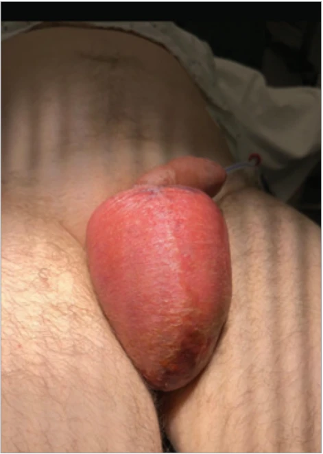
Upon arrival at the emergency department, the patient had already been seen by his general practitioner, and broad--spectrum antibiotics consisting of Ciprofloxacin 500 mg were already started. Physical examination showed a red and edematous scrotum and penile shaft, which were extremely painful on palpation (Fig. 1). There was an absence of soft--tissue crepitations or clear skin breaks.
Laboratory testing revealed leukocytosis (19 × 109/L), neutrophilia (16 × 109/L), an elevated CRP (C-Reactive Protein) (404 mg/L), as well as signs of acute renal failure (eGFR (estimated Glomerular Filtration Rate) 35 mL/min, creatinine 2.14 mg/dL). Additional ultrasound examination of the scrotum was performed, which showed normal vascularization. The provisional diagnosis was a septic shock caused by an underlying soft tissue infection of the genital area. Intravenous fluid suppletion with Plasmalyte was initiated and an exploratory surgical debridement of the scrotum was scheduled urgently. A small drain was left in place to ensure fluid drainage.
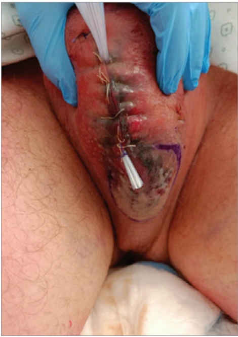
The antibiotic therapy was empirically switched to IV Amoxiclav and Amukin after peroperative collection of samples for microbial culture. Postoperatively, the patient was admitted to the intensive care unit (ICU). However, within 24 h, the patient deteriorated clinically with development of necrotic areas on the scrotum (Fig. 2).
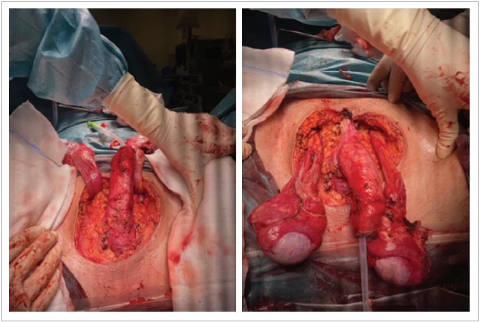
Broad surgical debridement of penile skin and scrotum were performed, until all necrotic tissue was excised (Fig. 3). Cultures and wound swabs came back positive for S. pyogenes and S. lugdunensis, necessitating an antibiotic switch to Piperacillin/Tazobactam and Clindamycin. Anatomopathological examination of the excised tissue showed edema and necrosis of the skin, subcutaneous tissue and dartos fascia in all resection pieces, consistent with necrotizing fasciitis.
Postoperative wound care consisted of daily Iso-Betadine dressing and frequent re-evaluation of the wound bed. Once no further spread of the infection was observed, additional negative pressure wound therapy (NPWT) was started to ensure granulation of the wound bed. NPWT was applied with continuous negative pressure set at 100 mmHg, delivered in 4 cycles over a total duration of 4 days. Based on the antibiogram, antibiotic therapy was de-escalated to intravenous amoxicillin-clavulanic acid (Augmentin) 2 g three times daily. Ten days after initial admission to the ICU, the patient could be transferred to the surgical nursing ward for continued treatment.
The extensive wound bed in the perineal and genital area after aggressive debridement necessitated a soft tissue reconstruction of the defect. A bilateral superomedial thigh perforator-based flap was chosen for the scrotal reconstruction, supplemented by the application of MatriDerm skin substitution and full sheet STSG to the penile shaft.
Doppler ultrasound at the level of the groin was used for preoperative indication of the perforating vessels from following three sources: external pudendal artery, anterior branch of the obturator artery, and medial circumflex femoral artery (Fig. 4). Granulation tissue was debrided to bleeding edges and was rinsed with Iso-Betadine. Hemostasis was performed to limit excessive blood loss.
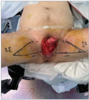
There are three important landmarks that border the flap. The first one is the line that reaches from the inferior part of the defect to the vertical line of the thigh, measuring 15 cm on the left and 16 cm on the right. The second line measures 8 cm bilaterally and connects the inguinal fold with the perineal region (not drawn). Finally, the most distal part of the first line can be connected with the inguinal line, with an angle depending on the degree of primary closure of the donor site (Fig. 4).
Afterwards, the flaps are dissected until sufficient mobilization is obtained. They are then rotated into the defect and the donorsites are closed primarily with Vicryl 1, Monocryl 3–0 and Stratafix 3–0. The flap is fixated using Vicryl 3–0 and Vicryl Rapide 4–0 (Fig. 5).
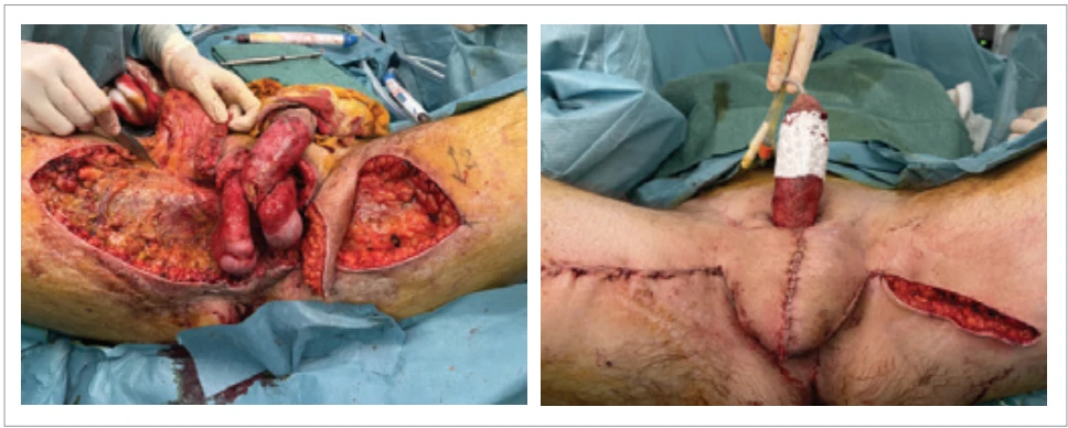
To reconstruct the penile defect, an electrical dermatome is used to obtain a STSG from the anterior right thigh. A full sheet graft is used to minimize the development of contractures and optimize erectile function postoperatively. Before applying this full sheet STSG, a MatriDerm skin substitute was hydrated with a saline solution and circularly applied around the penile shaft (Fig. 5). The graft is fixated using Vicryl Rapide 4–0 (Fig. 6). Negative pressure therapy is used to immobilize the graft and protect the wounds. Postoperatively, the patient could return to the nursing ward in good clinical condition.
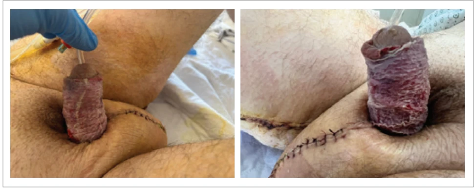
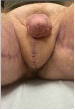
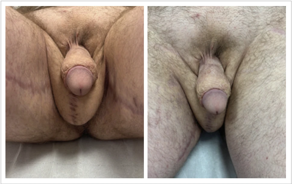
The patient is discharged 15 days after initial presentation at the emergency department. He is seen in the outpatient clinic with no signs of infection or flap failure 3 weeks postoperative (Fig. 7). Now, 18 months after reconstructive surgery, the patient reports full functional recovery (Fig. 8).
Discussion
Early recognition and prompt infection control are crucial for optimizing the prognosis of FG [2]. Following initial debridement, reconstruction is necessary. The choice for either minimally invasive procedures (primary closure, secondary healing, skin grafts, and local random pattern flaps) or more extensive procedures (axial pattern flaps or free flaps), depends on defect size, location, depth, and the availability of local tissue [3].
Primary closure is suitable only for very small defects, without skin tension after approximation of the wound edges [3,4]. Leaving the wound open to promote granulation tissue, known as healing by secondary intention, can be considered in patients with a contraindication for general anesthesia [3]. In this case, neither primary closure nor secondary healing were viable options for scrotal or penile reconstruction, due to the extensive tissue loss and expected poor functional and cosmetic outcomes.
Skin grafting, wherein healthy skin from a donor site is transplanted to cover up a defect elsewhere, is a widely used technique in FG management [3]. Grafts are categorized as split-thickness skin grafts (STSGs) or full-thickness skin grafts (FTSGs). STSGs offer advantages such as the simplicity of the procedure, shorter operative time, low donor site morbidity, and the ability to cover large defects [4]. Nonetheless, graft take can be compromised when the tunica vaginalis is lost. Other complications include vulnerability of the new scrotum to trauma, pain or discomfort and risk of infection due to the proximity of the perianal region. Covering defects exceeding 50% of the scrotal surface with a skin graft often leads to poor aesthetics, favoring flap-based reconstruction in such cases [4].
A scrotal advancement flap can address limited tissue loss if adjacent skin quality is sufficient and tension-free closure is achievable [4,5].
For more extensive defects, local fasciocutaneous flaps are preferred. These flaps provide a more sturdy and rigid protection to the testes, reduce contractures and are often considered aesthetically superior [4]. The Limberg flap, a random pattern flap, has been successfully used for larger perineal and scrotal defects. It is technically straightforward and allows tension-free closure, therefore minimizing the risk of developing necrosis, though achieving a natural scrotal appearance can be challenging [4].
Furthermore, axial pattern flaps like the anterolateral thigh flap, the groin flap, the pudendal thigh flap and the superomedial thigh flap have been discussed in literature for the reconstruction of defects after debridement of FG.
The anterolateral thigh flap, based on perforating branches of the descending branch of the lateral femoral circumflex artery, features a long pedicle (8–16 cm), allowing to cover extensive perianal, scrotal, or even abdominal defects [6]. Its distant donor site minimizes the risk of transferring infected tissue, and the flap can be sensate when incorporating the lateral femoral cutaneous nerve. Limitations include anatomical variability of the perforators, bulkiness, and potential complications with thermoregulation and spermatogenesis [7].
The “groin flap” and the superficial circumflex iliac perforator (SCIP) flap are both harvested from the inguinal region and ultimately derive their blood supply from the superior circumflex iliac artery (SCIA). However, they are distinct types of flaps. The classic groin flap is based on the deep branch of the SCIA, providing a larger fasciocutaneous territory but often requiring deeper dissection and potential thinning procedures. In contrast, the SCIP flap is a perforator variant, nourished by only a dominant perforator branch of the SCIA [8]. It has a shorter vascular pedicle with elastic skin, and is the thinnest flap in the perineal area (5–7 mm) with minimal subcutaneous fat, making it suitable for preserving spermatogenic function. However, it may lack sufficient tissue volume for extensive defects and offers only moderate aesthetic outcomes [7–9].
The pudendal thigh flap, supplied by a branch of the internal pudendal artery, is a simple, reliable option for scrotal reconstruction. Its texture closely resembles that of the natural scrotum, and it preserves spermatogenesis due to its limited thickness [10]. However, it is less suitable for large or deep defects, where a myocutaneous variant may be preferable [4].
For extensive defects, the superomedial thigh flap is an effective choice. This flap receives vascular supply from the external pudendal artery, obturator artery, and medial circumflex femoral artery [11]. It is a sensate flap with practical dissection and a short operative time, yielding favorable aesthetic outcomes, minimal functional loss and a low reported incidence of (partial) flap failure [4,5]. These considerations guided its use in the case of our 34-year-old patient, emphasizing the importance of selecting a fast and effective reconstructive method. Different reconstructive techniques were selected for the penile shaft and scrotum, according to their specific functional and aesthetic requirements. The penile shaft was reconstructed using a STSG, as it provides thin, pliable coverage without excessive bulk, thereby preserving penile contour and function [4]. The scrotal defect was reconstructed with a superomedial thigh flap, which was considered aesthetically superior to skin grafting because of its greater elasticity and a higher potential for reinnervation, with low donor-site morbidity and low risk of flap failure [4,5].
Despite Fournier following vasectomy being uncommon, it has already been documented in numerous other case reports with similar pathophysiology as this one. For example, a 33-year-old man who developed fatal FG within five days after vasectomy despite extensive surgical and medical treatment, highlighting the potential severity of FG [12]. Likewise, a 35-year-old men developed Fournier one week after vasectomy, which required repeated debridement and prolonged hospitalization before recovery [13]. These reports emphasize that, even in low-risk patients, clinicians should always be aware of potential serious pathology following routine vasectomy surgery.
Conclusion
The reconstruction of soft-tissue defects following FG can be challenging. Due to the often compromised general condition of affected patients, the chosen treatment strategy must prioritize rapidity and minimal procedural risk while also addressing aesthetic outcomes, functional restoration, and minimizing patient morbidity. We believe a superomedial thigh flap meets these requirements and should therefore be taken into consideration for the reconstruction of tissue defects following FG. Although uncommon, FG may occur as a severe complication following vasectomy, highlighting the need for early diagnosis and prompt multidisciplinary intervention, even in otherwise low-risk patients.
Roles of the authors
All authors contributed to the conception and design of the work. Clinical data were collected and interpreted by the treating surgeons. The reconstructive strategy and surgical techniques were performed and documented by the plastic surgery team. The first draft of the manuscript was written by the lead author, and all authors critically revised the manuscript for important intellectual content. All authors approved the final version and agreed to be accountable for all aspects of the work.
Disclosure of interest
The authors report no conflict of interest. The authors declare that this study has received no financial support. All procedures performed in this study involving human participants were in accordance with ethical standards of the institutional and/or national research committee and with the Helsinki declaration and its later amendments or comparable ethical standards.
Sources
1. Leslie SW., Foreman J., Rad J. Fournier gangrene. Treasure Island (FL): StatPearls Publishing, 2026.
2. Erickson BA., Flynn KJ. Management of necrotizing soft tissue infections (Fournier’s gangrene) and surgical reconstruction of debridement wound defects. Urol Clin North Am. 2022, 49 (3): 467–478.
3. Susini P., Marcaccini G., Efica J., et al. Fournier’s gangrene surgical reconstruction: a systematic review. J Clin Med. 2024, 13 (14): 4085.
4. Insua-Pereira I., Ferreira PC., Teixeira S., et al. Fournier’s gangrene: a review of reconstructive options. Cent European J Urol. 2020, 73 (1): 74–79.
5. Oufkir AA., Tazi MF., El Alami MN. The superomedial thigh flap in scrotal reconstruction: Technical steps to improve cosmetic results. Indian J Urol. 2013, 29 (4): 360–362.
6. Microsurgeon.org. Atlas of microsurgery techniques and principles. Available from: https: //www.microsurgeon.org/.
7. Maurya R., Mir MA., Mahajan S. Various options for scrotal reconstruction: a prospective observational study. Cureus. 2022, 14 (2): e22671.
8. Han HH., Lee JH., Kim SM., et al. Scrotal reconstruction using a superficial circumflex iliac artery perforator flap following Fournier’s gangrene. Int Wound J. 2016, 13 (5): 996–999.
9. Atik B., Tan O., Ceylan K., et al. Reconstruction of wide scrotal defect using superthin groin flap. Urology. 2006, 68 (2): 419–422.
10. Younes MT., Nawar AM., Shoulah AA. Outcome of pudendal thigh flap in scrotal reconstruction and its effect on testicular function. Egyptian J Surgery. 2019, 38 (3): 399–405.
11. Hirshowitz B., Moscona R., Kaufman T., et al. One-stage reconstruction of the scrotum following Fournier’s syndrome using a probable arterial flap. Plast Reconstr Surg. 1980, 66 (4): 608–612.
12. Viddeleer AC., Lycklama à Nijeholt GA. Lethal Fournier’s gangrene following vasectomy. J Urol. 1992, 147 (6): 1613–1614.
13. De Diego Rodríguez E., Correas Gomez MA., Martín García B., et al. Gangrena de Fournier postvasectomía [Fournier’s gangrene after vasectomy]. Arch Esp Urol. 2000, 53 (3): 275–278.
Labels
Plastic surgery Orthopaedics Burns medicine TraumatologyArticle was published in
Acta chirurgiae plasticae
2026 Issue 1-2
Most read in this issue
- A systematic review of sensory outcomes in nipple reinnervation after implant-based breast reconstruction
- Principles of nasal framework reconstruction for complex nasal defects: review
- Self-esteem and quality of sexual life in women undergoing breast reconstruction due to mastectomy: a cross-sectional study in a non-European setting
- Cutting-edge biotechnological strategies to enhance autologous fat graft survival