
Reconstruction of traumatic nasal tip defect using a composite graft: a case report
Authors:
K. Procházková 1,2; P. Kurial 1; P. Vodička 1; V. Mařík 1
Authors‘ workplace:
Department of Plastic Surgery, České Budějovice Hospital, České Budějovice, Czech Republic
1; Faculty of Health and Social Sciences, University of South Bohemia, České Budějovice, Czech Republic
2
Published in:
ACTA CHIRURGIAE PLASTICAE, 68, 1-2, 2026, pp. 46-50
doi:
https://doi.org/10.48095/ccachp202646
Procházková K, Kurial P, Vodička P et al. Reconstruction of traumatic nasal tip defect using a composite graft: a case report. Acta Chir Plast 2026; 68 (1–2): 46–50.
Introduction
A composite graft is a free graft composed of two or more tissue types – most commonly skin and cartilage – transplanted without a vascular pedicle. It is typically used for the reconstruction of small, well-vascularized defects, particularly in the nasal and auricular regions. The main advantage of a composite graft is that it is a single-stage procedure without additional scarring in the central face [1,2]. Like skin grafts, the composite graft relies on the wound bed to provide vascularisation to support the tissue [3]. However, the use of this technique may be associated with complications. These include in particular graft necrosis, scarring, infection, asymmetry, and others [1]. Therefore, it is necessary to follow several principles to minimize these risks. The survival of the composite graft is dependent on multiple factors, but the most important limitation is size [1,3–6]. Composite grafts used for nasal defect reconstruction are most commonly harvested from the ear; therefore, it is also necessary to carefully consider an appropriate reconstruction of the donor site, depending on the location used.
Description of the case
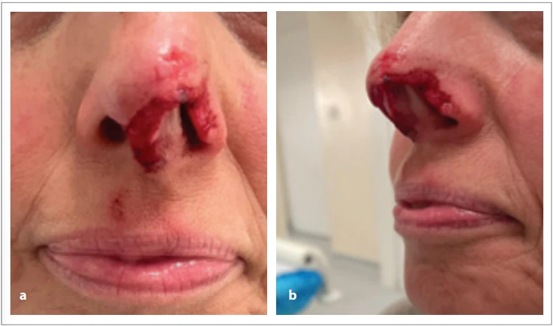
A nearly 50-year-old female patient sustained a dog bite injury in February 2025. The injury consisted of a defect of the acral portion of the left nasal ala, involving part of the nasal tip, with an additional defect in the columellar region (Fig. 1). The injury was caused by a properly vaccinated dog, and the patient had valid tetanus immunization. The patient was otherwise healthy, non-smoker, with no relevant medical history.
After multidisciplinary discussion and thorough evaluation of the defect, a composite graft was selected as the reconstructive method. The patient underwent regular wound dressing changes and was maintained on antibiotic therapy (Augmentin + Metronidazol) throughout the preoperative period. Surgical intervention was performed 8 days after the trauma. Fig. 2 shows crust formation in the area of the defect prior to surgery.

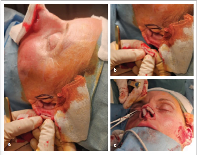
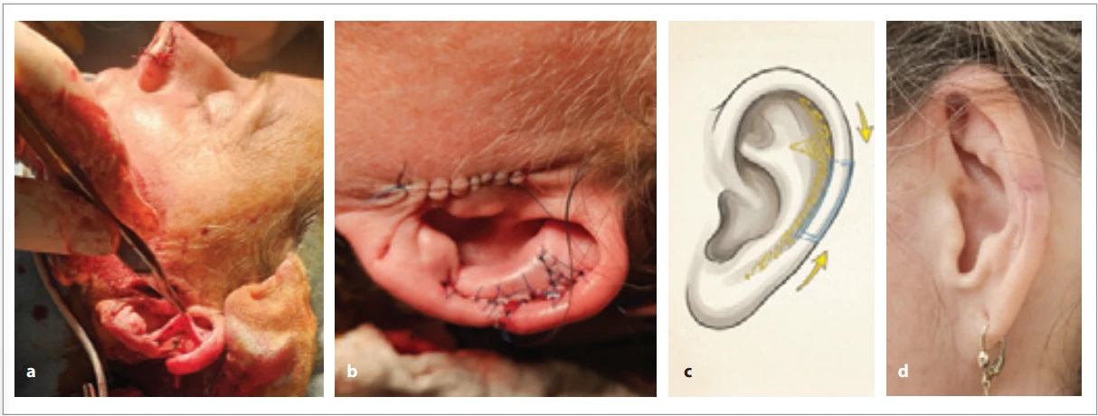
A composite graft was harvested from the patient’s left auricle. An initial meticulous, tissue-sparing debridement was performed to prevent enlargement of the defect. For reconstruction of the nasal ala, a 2 cm segment of the acral portion of the helix, including cartilage, subcutaneous tissue, and skin, was harvested (Fig. 3). The graft was then carefully sutured to the recipient bed using 5–0 Vicryl Rapid. The graft shape precisely matched the dimensions of the elongated defect (Fig. 3). For reconstruction of the columellar defect, a full--thickness skin graft was harvested from the area anterior to the left tragus and applied (Fig. 3, 4). Due to the limited amount of tissue available for helix reconstruction, a releasing incision was required, and a triangular piece of tissue was excised from the proximal part of the helix (Fig. 4).
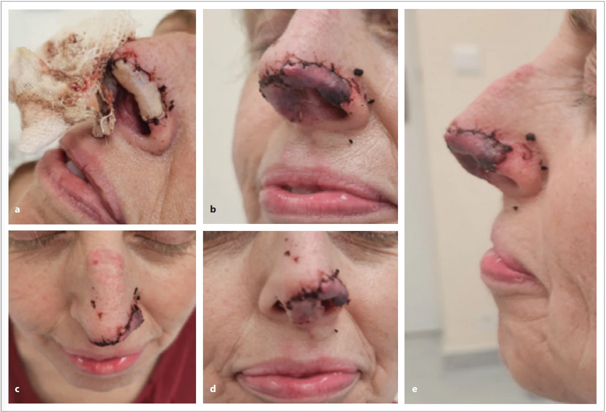
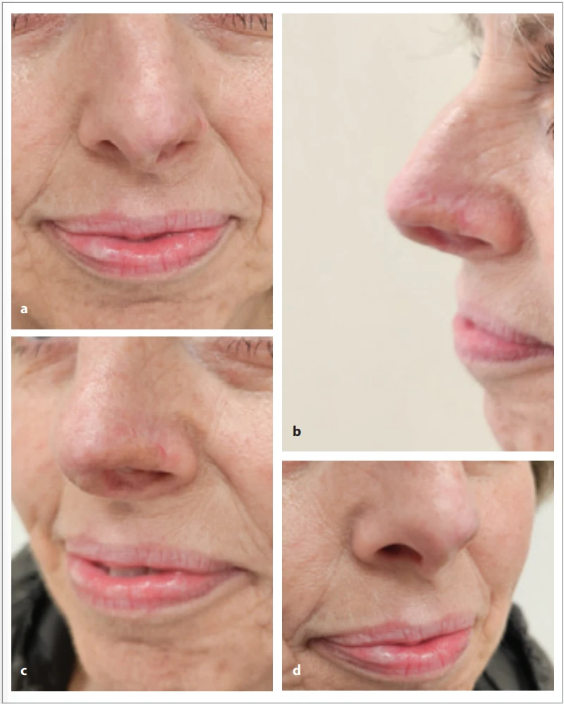
All areas were covered with fatty gauze moistened with saline and a small amount of Betadine. On the first postoperative day, the graft appeared pale (Fig. 5a). However, by the second postoperative day, the graft had become pink, and the patient was discharged for outpatient care (Fig. 5b–e). Three weeks postoperatively, the patient began scar treatment (moisturizing all treated areas). No further surgical procedures were required. The patient’s last follow-up was after 8 months (Fig. 6a–d).
Discussion
Traumatic defects of the nasal tip pose both functional and aesthetic challenges in reconstructive surgery. Several techniques can be considered for the reconstruction of nasal tip defects. In this case, a bilobed flap or a V–Y advancement flap was not feasible due to limited tissue mobility. A nasolabial flap could have been used [7] however, given the longitudinal acral nature of the defect, the aesthetic outcome would have been uncertain. For larger defects, a forehead flap represents an established option, typically performed as a two - to three--stage reconstruction [8]. Other reconstructive possibilities include a radial forearm flap, a reversed superficial temporal artery auricular flap, or a pedicled Washio retroauricular–temporal flap [9]. Another potential approach is the use of artificial dermis [10]. However, in our country, its application is not covered by insurance except in cases of burn injuries. Nevertheless, promising results have been reported, as demonstrated by the study You et al. According to this study, the multilayered application of Matriderm (an acellular collagen-elastin dermal substitute) in combination with a full-thickness skin graft (FTSG) represents a reliable method for covering nasal skin defects, particularly in the thick skin zone of the nasal tip and ala [10]. Among other surgical options for the treatment of defects of the acral part of the nose is the use of a composite graft [11]. In our case, a composite graft was considered the most appropriate option. However, each technique has its indications and limitations depending on defect size, location, and the desired aesthetic outcome. When multiple layers are involved in defect, the reconstruction requires combining procedures to replace each layer [12].
The maximum graft size is generally limited to 1.5–2 cm, and its shape is determined by the donor site, which provides limited plasticity [1,13,14]. Several studies support a limit of up to 1.5 cm [4,5]. According experience Chen et al., using composite grafts for partial and full-thickness ala defect, the best results occurred when the graft was 1 cm or less. Very few patients in their series with defect larger than 2 cm had good aesthetic outcomes [3]. On the contrary, some studies show that it is possible to use a larger size [1,6]. Gjorup et al. reported 18 patients with nasal defects, with an average maximum defect size of 13 mm (range, 6–25 mm). Based on the authors’ findings, defects up to 2 cm have a higher likelihood of successful graft take. The study concluded that composite grafts are particularly suitable for defects of the nasal ala and tip. Negative factors affecting healing included diabetes mellitus, smoking, and obesity. Reported complications included graft necrosis, scarring, and asymmetry. Overall outcomes were classified as good in 9 cases (50%), acceptable in 5 cases (28%), and poor in 4 cases (22%) [1]. The risk of graft necrosis is therefore relatively high.
The success of a composite graft relies on meticulous surgical technique, including precise suturing and tension--free adaptation to ensure optimal contact, as graft nourishment initially depends solely on diffusion. Because the graft is initially nourished solely by diffusion, it was highly advantageous in our case to use a thin, longitudinal graft that adhered along its entire length to the defect site, thereby significantly increasing the likelihood of complete graft survival. We also emphasize the importance of delaying the procedure after the injury, allowing the wound to partially heal and granulate before addressing only the essential areas. At the borderline size of 2 cm, the technique must be executed with extreme precision: the graft must lie completely flat, without any folds, particularly because the nose is one of the most prominent facial structures.
Conclusion
Use of a composite graft requires meticulous surgical technique. There is a relatively high risk of graft failure; therefore, extensive debridement that could enlarge the defect should be strictly avoided. It must always be kept in mind that graft tissue may detach, potentially resulting in an even larger defect. Although the technique itself is relatively straightforward, alternative reconstructive options remain available in the event of graft necrosis; however, these options are generally more invasive for the patient. Therefore, we recommend attempting a composite graft as the initial approach.
Roles of the authors
Pavel Kurial, Kristýna Procházková – authors, article compositors; Pavel Kurial, Kristýna Procházková – surgical team; Vladimír Mařík, Petr Vodička – review and editing.
Acknowledgements
This study was supported by the Research and Development Fund of Nemocnice České Budějovice – 2025_1 Application of Dermal Substitutes for the Reconstruction of Complex Tissue Defects.
Disclosure
All procedures performed in this study involving human participants were in accordance with ethical standards of the institutional and/or national research committee and with the Helsinki declaration and its later amendments or comparable ethical standards.
Conflicts of interest
The authors have no conflicts of interest to declare.
Sources
1. Gjorup CA., Moeller MP., Asklund C., et al. Versatility of composite grafts for nasal defects – a case series. Case Reports Plast Surg Hand Surg. 2022, 9 (1): 236–248.
2. Riopelle AM., Jeong D., Kaliszewski SL., et al. Superior antihelix composite graft for repair of nasal ala and lateral nasal tip defects: a retrospective case series. Dermatol Surg. 2023, 49 (5): 466–472.
3. Chen C., Patel R., Chi J. Comprehensive algorithm for nasal ala reconstruction: utility of the auricular composite graft. Surg J (N Y). 2018, 4 (2): e55–e61.
4. Harbison JM., Kriet JD., Humphrey CD. Improving outcomes for composite grafts in nasal reconstruction. Curr Opin Otolaryngol Head Neck Surg. 2012, 20 (4): 267–273.
5. Singh DJ., Bartlett SP. Nasal reconstruction: aesthetic and functional considerations for alar defects. Facial Plast Surg. 2003, 19 (1): 19–28.
6. Son D., Kwak M., Yun S., et al. Large auricular chondrocutaneous composite graft for nasal alar and columellar reconstruction. Arch Plast Surg. 2012, 39 (4): 323–328.
7. Salgarelli AC., Bellini P., Multinu A., et al. Reconstruction of nasal skin cancer defects with local flaps. J Skin Cancer. 2011, 2011 : 181093.
8. Dvořák Z., Cheimaris A., Knoz M., et al. Three-stage paramedian forehead flap reconstruction of the nose using the combination of composite septal pivot flap with the turbinate flap andL-septal cartilaginous graft – a case report. Acta Chir Plast. 2021, 63 (1): 6–13.
9. Morrison CM., Bond JS., Leonard AG. Nasal reconstruction using the Washio retroauricular temporal flap. Br J Plast Surg. 2003, 56 (3): 224–229.
10. You HJ., Choi YS., Kim DW. Use of a multilayered acellular dermal substitute with simultaneous full-thickness skin graft for the one-stage coverage of nasal skin defects. J Cosmet Dermatol. 2020, 19 (11): 3014–3019.
11. Ferreira S., Ayres Quaresma LE., Timóteo CA., et al. The primary closure approach of dog bite injuries of the nose. J Craniofac Surg. 2014, 25 (3): e216–218.
12. Driscoll BP., Baker SR. Reconstruction of nasal alar defects. Arch Facial Plast Surg. 2001, 3 (2): 91–99.
13. Teltzrow T., Arens A., Schwipper V. One-stage reconstruction of nasal defects: evaluation of the use of modified auricular composite grafts. Facial Plast Surg. 2011, 27 (3): 243–248.
14. Scheithauer MO., Rotter N., Lindemann J., et al. The auricle’s cavum conchae composite graft in nasal reconstruction. Am J Rhinol Allergy. 2013, 27 (2): e53–e57.
Labels
Plastic surgery Orthopaedics Burns medicine TraumatologyArticle was published in
Acta chirurgiae plasticae
2026 Issue 1-2
Most read in this issue
- A systematic review of sensory outcomes in nipple reinnervation after implant-based breast reconstruction
- Principles of nasal framework reconstruction for complex nasal defects: review
- Self-esteem and quality of sexual life in women undergoing breast reconstruction due to mastectomy: a cross-sectional study in a non-European setting
- Cutting-edge biotechnological strategies to enhance autologous fat graft survival