
Antegrade posterior interosseous artery flap for coverage of full-thickness elbow defects when radial forearm flap is not feasible
Authors:
W. Gutermann; N. Vermeersch; F. Thiessen; T. Tondu; M. De Fré
Authors‘ workplace:
Department of Plastic, Reconstructive and Aesthetic Surgery, Antwerp University Hospital, Antwerp, Belgium
Published in:
ACTA CHIRURGIAE PLASTICAE, 68, 1-2, 2026, pp. 74-75
Category:
doi:
https://doi.org/10.48095/ccachp202674
Gutermann W, Vermeersch N, Thiessen F et al. Antegrade posterior interosseous artery flap for coverage of full-thickness elbow defects when radial forearm flap is not feasible. Acta Chir Plast 2026; 68 (1–2): 74–75.
Dear Editor,
Reconstruction of soft-tissue defects around the elbow remains challenging, particularly in cases requiring well-vascularized tissue following repeated surgical procedures or chronic infection. The pedicled radial forearm flap is frequently used because of its reliable vascular anatomy and technical simplicity. However, this technique requires sacrifice of one of the major arterial axes of the forearm. In addition, the volar forearm soft tissue may not always be available for flap harvest due to previous surgery, scarring, skin grafting, trauma, or the need to preserve the volar aspect for potential future procedures. In such situations, alternative reconstructive options must be considered [1].
We would like to highlight the antegrade posterior interosseous artery (PIA) flap as a reliable and vessel-preserving reconstructive option when radial forearm flap harvest is undesirable or not feasible.
A 63-year-old female presented with a chronic and recalcitrant left olecranon bursitis persisting for approximately 6 months. She had previously undergone four surgical interventions performed by orthopedic and plastic surgery teams, including multiple wound debridements, vacuum-assisted closure therapy, and split-thickness skin grafting, all without durable resolution. Clinical examination revealed a persistent soft-tissue defect over the olecranon with chronically inflamed tissue. Plain radiographs did not demonstrate clear signs of osteomyelitis.
Definitive surgical management consisted of extensive debridement with excision of the entire pseudobursa, curettage of the underlying bone, and procurement of bone biopsies (Fig. 1A). Given the contaminated surgical field and repeated reconstructive failure, coverage with well-vascularized tissue was considered essential.
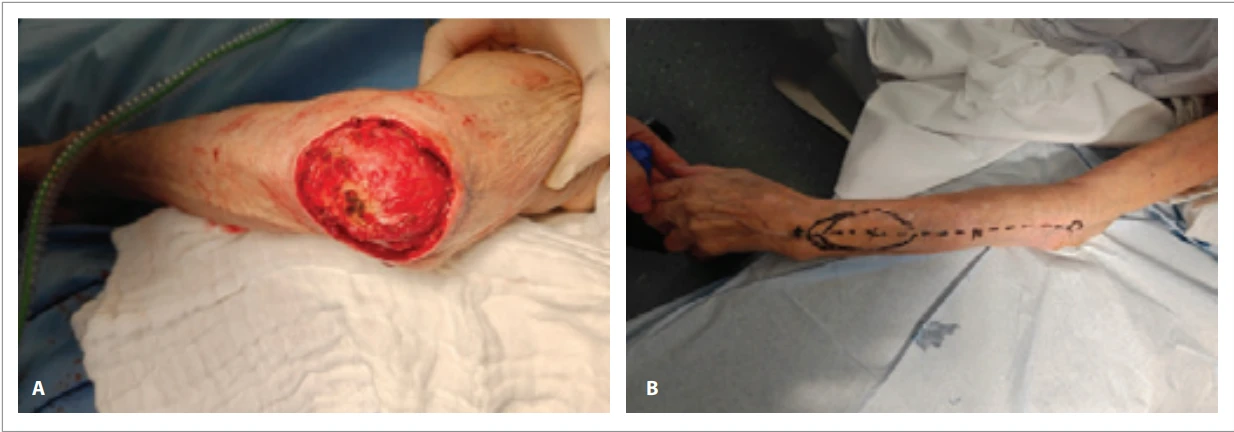
In our practice, pedicled radial forearm flaps are frequently used for such defects because of their robust perfusion and straightforward dissection. However, preoperative Doppler examination in this patient demonstrated two markedly gracile vessels at the wrist. Furthermore, the patient was scheduled to undergo a future volar corrective forearm osteotomy requiring plate osteosynthesis. A radial forearm flap would therefore have sacrificed a major arterial axis and potentially complicated future surgical access through previously grafted skin.
For these reasons, reconstruction was performed using an antegrade PIA flap (Fig. 1B). First described by Penteado et al. in 1986 [2] and introduced clinically by Zancolli and Angrigiani in 1988 [3], the PIA flap has been used predominantly in its retrograde form for coverage of the dorsal hand and wrist. The proximally based, antegrade variant for elbow coverage was reported subsequently but remains comparatively underused, although more recent work has confirmed its reliability [4,5]. In the present case, the antegrade PIA flap was not merely an alternative but represented the most appropriate reconstructive solution given the need to preserve the major forearm vessels and maintain the integrity of the volar forearm.
Flap elevation proved relatively straightforward. Careful dissection is required to identify and preserve the crossing branches of the posterior interosseous nerve as much as possible during flap harvest. When performed meticulously, the technique allows safe elevation with minimal donor-site morbidity (Fig. 2).
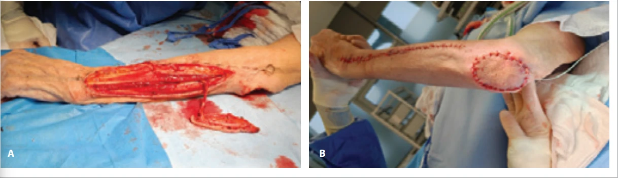
Postoperatively, the patient was discharged after one day with a protective elbow splint for 5 days. At 3 weeks of follow-up, all wounds had healed uneventfully, and the patient had regained full elbow function without limitations.
The antegrade PIA flap offers several advantages for elbow reconstruction. It preserves both the radial and ulnar arteries, provides thin and pliable tissue suitable for the olecranon region, and avoids donor-site morbidity associated with radial artery sacrifice [4,5]. Additionally, preservation of the volar forearm may be beneficial in patients requiring future surgical procedures in this area. This case illustrates that the antegrade PIA flap should be considered in the reconstructive algorithm for elbow defects when preservation of the radial and ulnar arteries is desirable.
Roles of the authors
All authors have been actively involved in the planning, preparation, analysis and interpretation of the findings, enactment and processing of the article with the same contribution.
Disclosure of conflict of interest
The authors declare no conflicts of interest in relation to this work.
Funding
This work received no specific grant from any funding agency in the public, commercial, or not-for-profit sectors.
Ethical approval and informed consent
Written informed consent was obtained from the patient for publication of this case and any accompanying images.
Sources
1. Adkinson JM., Chung KC. Flap reconstruction of the elbow and forearm: a case-based approach. Hand Clin. 2014, 30 (2): 153–163. v.
2. Penteado CV., Masquelet AC., Chevrel JP. The anatomic basis of the fascio-cutaneous flap of the posterior interosseous artery. Surg Radiol Anat. 1986, 8 (4): 209–215.
3. Zancolli EA., Angrigiani C. Posterior interosseous island forearm flap. J Hand Surg Br. 1988, 13 (2): 130–135.
4. Robinson LP., Usmani RH., Hazel A., et al. Use of the antegrade posterior interosseous artery flap for coverage of complex elbow wounds. Plast Reconstr Surg. 2021, 148 (6): 1316–1319.
5. Zaidenberg EE., Zancolli P., Farias Cisneros E., et al. Antegrade posterior interosseous flap for nonhealing wounds of the elbow: anatomical and clinical study. Plast Reconstr Surg Glob Open. 2018, 6 (11): e1959.
Labels
Plastic surgery Orthopaedics Burns medicine TraumatologyArticle was published in
Acta chirurgiae plasticae
2026 Issue 1-2
Most read in this issue
- A systematic review of sensory outcomes in nipple reinnervation after implant-based breast reconstruction
- Principles of nasal framework reconstruction for complex nasal defects: review
- Self-esteem and quality of sexual life in women undergoing breast reconstruction due to mastectomy: a cross-sectional study in a non-European setting
- Cutting-edge biotechnological strategies to enhance autologous fat graft survival