
Painful neuroma pathophysiology review: double crush syndrome due to coexistent proximal neural lesion?
Authors:
V. Mačionis
Authors‘ workplace:
Independent researcher, Vilnius, Lithuania
Published in:
ACTA CHIRURGIAE PLASTICAE, 68, 1-2, 2026, pp. 26-33
doi:
https://doi.org/10.48095/ccachp202626
Mačionis V. Painful neuroma pathophysiology review: double crush syndrome due to coexistent proximal neural lesion? Acta Chir Plast 2026; 68 (1–2): 26–33.
Introduction
The development of post-traumatic neuroma as a pseudotumor produced by the sprouting axons of the severed nerve stump has been well explored and can be considered a normal reaction to nerve injury [1]. Clinically important are only painful (symptomatic) neuromas because they cause significant functional impairment [2]. Despite advances in treatment and research of painful neuromas, their pathogenesis remains largely unexplained. Possible local and central mechanisms of sensitization that accompany painful neuromas have been discussed in many reviews [3–5], most of which have not concluded why neuroma pain develops only in some but not in all patients and not in all multiple injured nerves. These are essential unanswered questions that make the current understanding of symptomatic neuroma mysterious.
The etiopathogenetic conundrum of painful neuromas has recently been explained by the possibility of sensitization due to a coexistent proximal neural lesion (PNL), in particular neural entrapment [6] (see the subsection below for details). However, the introduced concept has not explained numerous specific features of painful neuromas [3]. To fill this knowledge gap, this paper reviews the clinical, epidemiological, and histochemical characteristics of symptomatic neuromas and provides literature--based interpretations of these features regarding the sensitizing neuroinflammatory effects of a possible co-occurring PNL.
Evaluation of the topic
Methods
For the narrative review, the Google Scholar and PubMed databases were selectively searched for relevant literature on painful neuroma characteristics and their association with neural lesions proximal to the neuroma. The search terms included various combinations and derivations of the following key words: amputation, animals, axon, clinical, compression, diagnosis, digital, double crush, entrapment, experimental, extremity, fibrosis, hand, histochemistry, histology, imaging, injury, lesion, myelination, nerve, neural, neuroma, neuron, neuropathy, ongoing activity, pain, painful, symptoms, pathophysiology, proximal, recurrence, review, scar, sensitization, sprouting, surgery, syndrome, Tinel’s sign, trauma, treatment. The references of the articles of interest were further scrutinized for pertinent publications. A total of 229 articles were analyzed, 108 of which were included in this review. Most recent papers and those containing well-referenced relevant discussions were preferred to cite.
Interpretive question-driven literature review
PNL as a possible cause of sensitization in neuroma pain
Neuroma pain has been associated with the development of sensitization (sensory hypersensitivity) [3–5]. In neurophysiological terms, sensitization is due to ongoing activity (continuous spontaneous firing) of afferent neurons and a reduction in their excitation threshold [7], which can result in allodynia, hyperalgesia, and persistent spontaneous pain. Importantly, for sensitization to persist, continuous local irritation and consequent local inflammation of neural fibers are necessary; otherwise, ongoing activity and axonal mechanosensitivity fade away [8,9]. The pathophysiological activity of neuromas normally decreases with time [10], as is usually the case in the healing of all tissues. Furthermore, it has been experimentally proven that there is no substantial spontaneous electrical activity in neuromas [11]. Thus, it is unclear how sensitization can be maintained by painful neuromas.
Axonal sprouts within neuromas seem to be naturally sensitive to pressure [12], and the decrease in their mechanical sensitivity with neuroma age [13] may be due to their encapsulation by a fibrotic matrix [1] that probably serves as protection against painful external pressure (Fig. 1A). However, such protection may be insufficient, and even the internal pressure of the neuroma can produce a noxious stimulus if the nerve trunk is sensitized by a coexistent proximal compressive neural lesion (Fig. 1B). This hypothesis is based on research indicating that proximal compressive neural lesions can cause peripheral and central sensitization, which, in turn, can account for allodynic pain in many usually non--painful peripheral conditions [6]. In this regard, a PNL is primarily understood as intermittent neural compression in the proximity of the dorsal root ganglion (DRG) and spinal plexuses. The closer the lesion is to the neuron body of the DRG, the more likely the development of peripheral sensitization is. Notably, sensory root demyelination by chronic compression may cause ephapse formation and ephaptic transmission (cross-talk between axons), resulting in tactile allodynia [14]. It must be stressed that the concept of PNL also extends to lesions of the central nervous system (CNS) and consequent central sensitization [6].
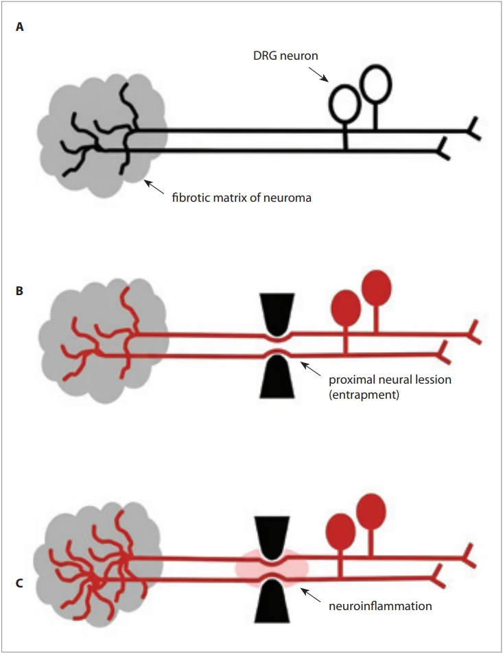
It has been shown that compressed nerves become susceptible to another lesion, which is the pathophysiological basis of double crush syndrome [15]. Two heterotopic nerve trunk lesions produce worse symptoms than a single lesion [15]. This implies that asymptomatic neural compression can become symptomatic if another lesion develops on the same neural axis. According to this concept, a painful neuroma may be a variant of the double crush phenomenon because any neuroma can be regarded as compressive entrapment of the sprouting axons of the nerve stump [16].
Painful neuromas tend to be larger in sensory than in mixed nerves
A recent prospective study of 50 painful neuromas concluded that purely sensory nerves form relatively larger symptomatic neuromas and that symptomatic neuromas grow over time [17]. These findings can be explained by enhanced and continuing axonal sprouting due to the sensitizing neuroinflammatory effects of coexisting PNL (Fig. 1C).It has been directly shown in rats that nerve compression results in axonal sprouting both distal and proximal to the compression site [18]. Several studies have found that spinal cord injury in animals results in sprouting of the central axons of undamaged primary sensory neurons into laminae II–V of the dorsal horns [19]; that is, lesions of the second-order neuron, i.e., PNLs [6], stimulate sprouting of the first-order neuron. Furthermore, transection of the mouse L3 spinal nerve results in collateral sprouting of the neighboring undamaged L4 spinal nerve [20]. In addition, axon sprouting can be enhanced by damaging a longer length of the nerve trunk [21], which is in direct agreement with the concept of double crush injury, which, in turn, alludes to PNL, as discussed above. Finally, it should be noted that, while both sensory and motor neurons can sprout, only sensory neurons can undergo sensitization, as implied by its definition alone.
Painful neuromas tend to recur and are more likely to develop in preexisting neuropathy
It is well known that symptomatic neuromas are prone to recur after surgical interventions, especially after simple excisions [22–26]. Furthermore, while some surgical techniques can provide improvement of neuroma symptoms in up to 92% of cases [27], many patients have insufficient pain relief after neuroma surgery [2,26,28–31].
The concept of PNL offers a reasonable explanation for painful neuroma recurrence, in particular in terms of double crush syndrome as a multifocal neuropathy [32]. It has previously been noted that cutting neuropathic nerves results in pain exacerbation and that this may be due to DRG and/or CNS involvement [33]. Obviously, pain recurs because a new neuroma forms and the cause of preexisting nerve sensitization persists [33], which can be due to PNL. Furthermore, trauma-induced neuroinflammation may spread upwards along nerves [34], which can produce neuropathy due to intra-/perineural edema and fibrosis with resultant sensitization of the involved neurons.
Perineural edema identified by magnetic resonance imaging (MRI) in oncological amputees correlates with the occurrence of painful neuromas [35]. This may be an indication of neuropathy resulting from cancer and its treatment [36]. In a series of 60 oncological amputees, 71.8% of symptomatic neuromas were found out of 78 neuromas identified by MRI [35]. For comparison, average rates of traumatic painful neuromas reported by systematic meta-analyses are 13% in upper extremity amputees [37] and 15–19% in lower extremity amputees [38,39]. Notably, post-mastectomy pain in breast cancer patients occurs in more than 50% of cases [40,41], which has been shown to be likely caused by intercostal neuromas [42] that may be sensitized due to pre - and/or post-mastectomy adjuvant treatments [43].
The susceptibility to the formation and recurrence of painful neuromas of the superficial sensory branch of the radial nerve (SSBRN) at the wrist [28] can be attributed to a tethering of this nerve in the forearm [44]. This factor, along with post-traumatic inflammation and immobility, can cause neural entrapment, which is thought to exacerbate symptoms of SSBRN neuromas [45]. The recurrence rate of radial sensory nerve neuromas is higher in patients with multiple neuromas [28], which can be attributed to a more extensive PNL. By the same token, post-traumatic digital nerve entrapment [46] may cause digital neuroma pain. Notably, digital nerve repair has not been found to diminish the incidence of painful neuromas [47].
Proximal Tinel’s sign in painful neuromas may indicate a PNL
Tinel’s sign proximal to a neuroma, possibly indicating backward regeneration of injured axons, has been advised as a diagnostic test for painful neuromas [48,49]. However, as experimentally shown, the presumably backward regenerating excessive axons in the short segments proximal to the neuroma should normally be present in all neuroma cases [50]. Then, why should the proximal Tinel’s sign be present only in symptomatic neuromas? A feasible explanation is that Tinel’s sign proximal to a neuroma indicates neural sensitization due to a proximal axonal lesion.
Painful neuromas tend to occur more frequently in mobile nerves
While injured, immobile nerves are likely to heal without neuropathic consequences, mobile entrapped nerves continue to receive noxious stimuli due to motion, which maintains ongoing activity (i.e., sensitization) of the involved nerves. The intermittence of compressive injury of entrapped mobile nerves can act as irritation rather than complete cessation of axonal viability. This can explain why post-traumatic painful neuromas mostly occur in the extremities and are relatively rare elsewhere [22,51]. Comparative rates are unknown, but the absolute numbers are impressive. For example, one of the most extensive systematic reviews found 393 head and neck, 2,082 upper extremity, and 2,531 lower extremity symptomatic neuromas out of the total of 5,562 reported neuromas [51]. In the head, however, the ratio between reported symptomatic neuromas of the very mobile lingual and the almost immobile alveolar nerves was 5.35 to 1 [51].
These statistics account well for the higher prevalence of entrapment neuropathies of more mobile nerves at more mobile sites in the upper extremity [52,53]. For example, the incidence of carpal tunnel, ulnar neuropathies, and radial neuropathies in males has been reported as 104.8, 36.0, and 8.5, respectively, per 100,000 person-years [53]. The much higher prevalence of spinal radiculopathies (ranging, e. g., in females from 63 to 580 per 100,000 person-years) [54] is explainable by the proximity of entrapment to the DRG and the proneness of spinal nerves to overload injury.
Similar injuries do not always result in painful neuromas
As has been shown by one imaging study [55], macroscopically detectable neuromas do not always develop after nerve injury, probably because of successful regeneration of the damaged fibers into muscle [1] and/or skin [56]. The varying occurrence of neuroma formation, however, does not explain the varying occurrence of painful neuromas after similar injuries, in particular in cases of injury to multiple nerves.
While multiple symptomatic neuromas can occur sporadically, as can be seen from patient-to-neuroma number ratios in the relevant studies, single traumatic painful neuromas are most often reported [17,22,23,57–59]. A good example could be a study of bilateral upper limb amputees, in which symptoms of neuroma were reported in 26.2% of 172 patients, and only 9.3% (8 patients) of them had bilateral symptomatic neuromas [60]. Painful neuromas of both digital nerves of the same digit also seem to be rare [57].
The varying occurrence of compressive PNL [52–54] could explain the varying rates of painful neuromas [17,22,37–39,51,57–60] as well as the inconsistent results of their treatment [2,22–26,28–31,61]. The rarity of multiple painful neuromas is explainable by the fact that compressive neural lesions are usually not uniform across the nerve cross-section. Nerve compression affects nerve fibers differently depending on their location [62] and type [63], which can also be seen from, e. g., the varying distribution of digital paresthesia in carpal tunnel syndrome [64]. Relevantly, in bilateral upper limb amputees, phantom limb pain, which is frequently accompanied by symptomatic neuroma [65,66], does not always affect both sides [67]. This may be due to the asymmetric occurrence of PNL.
Painful neuromas are more likely after severe injuries
Nerve injury in digital amputation results in symptomatic neuroma significantly more often than in non-amputation digital injuries (7.8% compared to 1%, respectively) [68]. It has also been found that traumatic painful neuromas occur more often in digital avulsion injuries [57]. In a study of 32 lower limb amputees, multiple symptomatic neuromas were identified in 67% of cases [69], which is well above the average of 19% and the maximum of 49% [39]. Symptomatic neuromas after landmine injuries of the lower extremities also develop more frequently compared to other injuries (57.5% vs. 37.8%) [70]. These correlations may be due to a higher probability of concomitant and extensive proximal neural damage in more severe injuries.
Indirect conclusions about the influence of injury severity on PNL occurrence and consequent painful neuroma development can also be made from relevant studies of persistent pain. Severity of injury is a known risk factor for chronic pain development [71]. Neuropathic post-amputation pain occurs more often in proximal than in distal limb amputees [72], which is explainable by the proximity of the injury site to the DRG in proximal amputations. Also, injury severity-related factors such as pain, loss of extremity parts, and motor-sensory disturbances can change posture, gait, and manual behavior, resulting in the compression of nerve trunks at entrapment-prone locations [73,74].
Local factors cannot explain neuroma pain
Although the injury severity may influence the development of painful neuromas, no research could be found to explain how local changes in symptomatic neuromas could be caused by the neuroma itself. While scarring around a neuroma may restrict nerve mobility and cause pain, ultrasound and MRI of symptomatic and asymptomatic neuromas have found no considerable differences between them in terms of local fibrosis [75,76]. Also, neuroma pain does not depend on neuroma size [17,76], which suggests additional causes of nerve irritation.
Immunohistochemical features of symptomatic neuromas are explainable by PNLs
Fascicular architecture
A recent study found a smaller number of organized fascicles in human painful neuromas than in non-symptomatic ones [77]. Here, again, the stimulatory effect of PNL on axonal sprouting needs to be considered (Fig. 1C), as discussed in the above subsection “Painful neuromas tend to be larger in sensory than in mixed nerves”. In general, neuromas are disorganized axon bundles in connective tissue that also include normal axonal patterns [1]. Naturally, PNL--enhanced sprouting can increase the ratio of disorganized to normal axonal patterns.
Ion channels
Voltage-gated sodium channels PN1 (also known as Nav1.7) and PN3 (synonymous with Nav1.8) are more highly expressed in painful than in non-painful neuromas [78]. Sodium channels Nav1.8 and Nav1.9 have been found to be expressed in human lingual nerve neuromas, and higher expression of these channels correlates with neuropathic pain [79]. These findings are compatible with the pathophysiology of PNL because relevant research indicates that sodium channel density increases along axons in chronic neural compressive injury [80].
The protein ankyrin-G has been found to be expressed in both painful and non--painful neuromas, but higher expression has been observed in painful neuromas [81]. This is in line with the fact that ankyrin-G is required for the transport of sodium channels into axons from the axon initial segment [82] located near the neuronal body, and this process, as indicated above, can be more intense because of the sensitizing neuroinflammatory action of PNLs.
Neuropeptides
Neuroma pain has been associated with increased levels of neuropeptides, such as calcitonin gene-related peptide, substance P [3], and nerve growth factor (NGF) [83]. The reason for neuropeptide appearance in neuromas can be linked to their release following proximal neural stimulation [84], which can occur in compressive PNLs. For example, neuronal NGF levels increase after experimental chronic nerve constriction injury [85]. Of note, increased NGF content has been found both in painful neuromas and in the nerve segments proximal to the neuromas, but only in nerve fibers and not in other neuroma cells [83]. Also of relevance, neuropeptide-containing nerves have been observed in painful scars [86], and abnormal scarring may be caused by continued neuropeptide release by sensory terminals [87], which in turn may be due to the stimulatory effects of a neural lesion proximal to the scar site.
Myofibroblasts
Alpha-smooth muscle actin (a-SMA), a contractile protein of myofibroblasts [88], has been found to be well expressed in human traumatic painful neuromas, but only slightly in non-painful neuromas [89]. Similar results have been reported in rats [90]. Myofibroblasts were detected in human upper extremity painful neuromas, with their numbers decreasing over time [91]. The discovery of a-SMA and myofibroblasts in symptomatic neuromas has become the basis of the currently popular theory that neuroma pain is a result of internal compression produced by myofibroblast contraction [5,16]. However, myofibroblast numbers are significantly reduced after tissue healing is complete [92]. Why, then, should myofibroblasts be present and still contracting in healed nerve stumps?
The PNL as a cause of ongoing neural activity explains this puzzle. It has been shown that direct contact between fibroblasts and neurons is necessary for fibroblast differentiation into myofibroblasts and for the induction of collagen gel contraction [93]. This suggests that myofibroblasts in painful neuromas develop and contract because of neuropeptide release stimulated by PNL, as discussed in the preceding subsection.
Cannabinoid CB2 receptors
Cannabinoid CB2 receptors have been shown to be expressed in human painful neuromas of the injured brachial plexus, as well as in normal brachial plexus nerves [94]. CB2 receptors are present in various peripheral tissues [95], including sensory neurons [96], and are known for their antinociceptive/analgesic properties [96]. Their levels increase after inflammation and neural injury [97]. Regarding the PNL, cannabinoid receptor expression after proximal nerve constriction injury has been observed both in the DRG and the spinal cord of rats [98].
Unmyelinated fibers
In three clinical cases of painful neuromas, unmyelinated fibers were found to be much more abundant than myelinated fibers, the ratio being up to 20 : 1, respectively [99]. Unfortunately, this study comes without a controlled exploration of non-painful neuromas. Demyelination seems to be a normal process in neuroma development [100]. On the other hand, the abundance of unmyelinated fibers in painful neuromas may be related to the fact that proximal neural compression results in demyelination [63,101] and induces unmyelinated fiber regeneration [63].
The success of conservative and surgical treatments may indicate an improvement in PNL
Conservative modalities have been recommended for the initial treatment of painful neuromas [102]. Their effectiveness could be explained by the relief of pressure on nerve trunks [103]. As mentioned above, trauma can change postural and manual behavior, which can lead to nerve entrapment [73,74]. A study by Fisher and Boswick [104] reported minimal dressings and early postoperative use of the hand in 100 patients with 144 hand digit amputations, which resulted in only 4 (2.8%) painful neuromas (the relevant average rate being 13%) [37]. This is in accord with the fact that motion facilitates nerve gliding [105] and thus can prevent proximal fibrous compressive neuropathy.
All surgical treatments of neuromas involve intentional or non-intentional neurolysis of the affected nerve stump. This diminishes, at least in part, the possible double crush lesion of the nerve and thus may produce pain relief. The success of the techniques that prevent the formation of a neuroma as such can similarly be explained. This includes a significant reduction in recurrence rates of painful neuromas with the use of novel muscle reinnervation techniques [106,107].
Discussion
This review suggests that many features of painful neuromas can be consequences rather than causes of sensitization in neuroma pain. The rarity of multiple traumatic painful neuromas suggests that peripheral rather than central sensitization drives neuroma pain.
Frequent misdiagnosis of painful neuropathic conditions [108] suggests that missing the PNL in traumatic painful neuromas is very likely. Diagnosing a pre-existing PNL may be difficult because it can remain asymptomatic even after another lesion of the same neural axis occurs, as implied by the double crush phenomenon. On the other hand, significant neural damage, such as complete transection and a consequent neuroma, may mask a clinically less manifest PNL. In practice, the extraneuronal causes of neuroma pain could be revealed by a targeted search for proximal neuropathy signs. An objective verification of PNL can be a reduction of nerve conduction velocity proximal to the neuroma. Unfortunately, electrodiagnostics is not routinely performed in painful neuroma evaluation.
The concept of PNL-driven neuroma pain suggests that experimental neuroma research should involve comparisons between animal groups with and without chronic constriction injury of the nerve trunk proximal to the neuroma. It can be postulated that chronic constriction injury would be associated with changes in the histochemistry and microstructure of the neuroma and that concurrent electrophysiologic explorations would reveal mechanical sensitivity of sensory nerve fibers ending in the experimental neuroma.
Conclusion
Many features of painful neuromas, in particular frequent recurrence and incomplete improvement after surgical treatment, suggest that not only local factors are involved in the pathogenesis of this condition. While there are no clinical studies to show a direct association of neuroma pain with proximal neural damage, the available literature indicates that neural lesions proximal to the painful neuroma could account for its specific features. PNL-focused approaches in diagnostics and research of symptomatic neuromas may advance the understanding of its pathophysiology and eventually provide new treatments.
Roles of the authors
Valdas Mačionis is the single author of all contributions to the manuscript, including conceptualization, the review outline, manuscript draft, writing, and design and preparation of the figures.
Disclosure
The author has no conflicts of interest to disclose. The author declares that this study has received no financial support.
Sources
1. Oliveira KMC., Pindur L., Han Z., et al. Time course of traumatic neuroma development. PLoS One. 2018, 13 (7): e0200548.
2. Dahlin E., Gudinge H., Dahlin LB., et al. Neuromas cause severe residual problems at long-term despite surgery. Sci Rep. 2023, 13 (1): 15693.
3. Hwang CD., Hoftiezer YAJ., Raasveld FV., et al. Biology and pathophysiology of symptomatic neuromas. Pain. 2024, 165 (3): 550–564.
4. Krauss EM., Mackinnon SE. Definition, etiology, and epidemiology of symptomatic neuroma. In: Eberlin KR., Ducic I., Moore A., et al. (eds). Contemporary neuroma management. Springer International Publishing. 2024, 3–15.
5. Scott BB., Winograd JM. Peripheral sensitization: peripheral mechanisms of neuroma and neuropathic pain. In: Eberlin KR., Ducic I., Moore A., et al. (eds). Contemporary neuroma management. Springer International Publishing. 2024, 17–25.
6. Macionis V. Chronic pain and local pain in usually painless conditions including neuroma may be due to compressive proximal neural lesion. Front Pain Res (Lausanne). 2023, 4 : 1037376.
7. Bove GM., Dilley A. The conundrum of sensitization when recording from nociceptors. J Neurosci Methods. 2010, 188 (2): 213–218.
8. Satkeviciute I., Goodwin G., Bove GM., et al. Time course of ongoing activity during neuritis and following axonal transport disruption. J Neurophysiol. 2018, 119 (5): 1993–2000.
9. Bove GM., Ransil BJ., Lin HC., et al. Inflammation induces ectopic mechanical sensitivity in axons of nociceptors innervating deep tissues. J Neurophysiol. 2003, 90 (3): 1949–1955.
10. Deininger S., Schumacher J., Blechschmidt A., et al. Nerve injury converts Schwann cells in a long-term repair-like state in human neuroma tissue. Exp Neurol. 2024, 382 : 114981.
11. Burchiel KJ., Russell LC. Has the amount of spontaneous electrical activity in experimental neuromas been overestimated? Somatosens Res. 1987, 5 (1): 63–75.
12. Gorodetskaya N., Grossmann L., Constantin C., et al. Functional properties of cutaneous A - and C-fibers 1–15 months after a nerve lesion. J Neurophysiol. 2009, 102 (6): 3129–3141.
13. Meyer RA., Raja SN., Campbell JN., et al. Neural activity originating from a neuroma in the baboon. Brain Res. 1985, 325 (1–2): 255–260.
14. Ding YQ., Qi JG. Sensory root demyelination: transforming touch into pain. Glia. 2022, 70 (3): 397–413.
15. Dellon AL., Mackinnon SE. Chronic nerve compression model for the double crush hypothesis. Ann Plast Surg. 1991, 26 (3): 259–264.
16. Foltán R., Klíma K., Spacková J., et al. Mechanism of traumatic neuroma development. Med Hypotheses. 2008, 71 (4): 572–576.
17. Weigel DT., Raasveld FV., Liu WC., et al. Neuroma-to-nerve ratio: does size matter? Neurosurgery. 2025, 96 (3): 545–554.
18. Hayashi A., Pannucci C., Moradzadeh A., et al. Axotomy or compression is required for axonal sprouting following end-to-side neurorrhaphy. Exp Neurol. 2008, 211 (2): 539–550.
19. Hagg T. Collateral sprouting as a target for improved function after spinal cord injury. J Neurotrauma. 2006, 23 (3–4): 281–294.
20. Lemaitre D., Court FA. New insights on the molecular mechanisms of collateral sprouting after peripheral nerve injury. Neural Regen Res. 2021, 16 (9): 1760–1761.
21. Xu QG., Midha R., Martinez JA., et al. Facilitated sprouting in a peripheral nerve injury. Neuroscience. 2008, 152 (4): 877–887.
22. Wolvetang NHA., Lans J., Verhiel SHWL., et al. Surgery for symptomatic neuroma: anatomic distribution and predictors of secondary surgery. Plast Reconstr Surg. 2019, 143 (6): 1762–1771.
23. Pet MA., Ko JH., Friedly JL., et al. Traction neurectomy for treatment of painful residual limb neuroma in lower extremity amputees. J Orthop Trauma. 2015, 29 (9): e321–325.
24. Guse DM., Moran SL. Outcomes of the surgical treatment of peripheral neuromas of the hand and forearm: a 25-year comparative outcome study. Ann Plast Surg. 2013, 71 (6): 654–658.
25. Tupper JW., Booth DM. Treatment of painful neuromas of sensory nerves in the hand: a comparison of traditional and newer methods. J Hand Surg Am. 1976, 1 (2): 144–151.
26. Raasveld FV., Nieuwdorp NJ., Der Avoort DJJCV., et al. Long-term outcomes of symptomatic neuromas in the wrist, hand, and digits: a prospective cohort study. J Plast Reconstr Aesthet Surg. 2026, 116 : 34–45.
27. Langeveld M., Hundepool CA., Duraku LS., et al. Surgical treatment of peripheral nerve neuromas: a systematic review and meta-analysis. Plast Reconstr Surg. 2022, 150 (4): 823e–834e.
28. Gottlieb RW., Westenberg RF., Chen NC., et al. Long-term outcomes after surgical treatment of radial sensory nerve neuromas: patient-reported outcomes and rate of secondary surgery. Plast Reconstr Surg. 2021, 147 (1): 101–111.
29. Anantavorasakul N., Lans J., Macken AA., et al. Surgery for lower extremity symptomatic neuroma: long-term outcomes. J Plast Reconstr Aesthet Surg. 2020, 73 (8): 1456–1464.
30. Dahlin E., Zimmerman M., Nyman E. Patient reported symptoms and disabilities before and after neuroma surgery: a register-based study. Sci Rep. 2023, 13 (1): 17226.
31. Stokvis A., Coert JH., van Neck JW. Insufficient pain relief after surgical neuroma treatment: prognostic factors and central sensitisation. J Plast Reconstr Aesthet Surg. 2010, 63 (9): 1538–1543.
32. Cohen BH., Gaspar MP., Daniels AH., et al. Multifocal neuropathy: expanding the scope of double crush syndrome. J Hand Surg Am. 2016, 41 (12): 1171–1175.
33. Guerrero-Moreno A., Fakih D., Parsadaniantz SM., et al. How does chronic dry eye shape peripheral and central nociceptive systems? Neural Regen Res. 2021, 16 (2): 306–307.
34. Devor M., Tal M. Nerve resection for the treatment of chronic neuropathic pain. Pain. 2014, 155 (6): 1053–1054.
35. van Vliet E., Raasveld FV., Liu WC., et al. Evaluation of MRI features of neuromas in oncological amputees, and the relation to pain. Skeletal Radiol. 2025, 54 (5): 979–991.
36. Yoon SY., Oh J. Neuropathic cancer pain: prevalence, pathophysiology, and management. Korean J Intern Med. 2018, 33 (6): 1058–1069.
37. Chaker SC., Hung YC., Saad M., et al. Systematic review and meta-analysis of global neuroma incidence in upper extremity amputees. Ann Plast Surg. 2024, 92 (1): 80–85.
38. List EB., Krijgh DD., Martin E., et al. Prevalence of residual limb pain and symptomatic neuromas after lower extremity amputation: a systematic review and meta-analysis. Pain. 2021, 162 (7): 1906–1913.
39. Huang YJ., Assi PE., Drolet BC., et al. A systematic review and meta-analysis on the incidence of patients with lower-limb amputations who developed symptomatic neuromata in the residual limb. Ann Plast Surg. 2022, 88 (5): 574–580.
40. Tasmuth T., von Smitten K., Hietanen P., et al. Pain and other symptoms after different treatment modalities of breast cancer. Ann Oncol. 1995, 6 (5): 453–459.
41. Alves Nogueira Fabro E., Bergmann A., do Amaral E Silva B., et al. Post-mastectomy pain syndrome: incidence and risks. Breast. 2012, 21 (3): 321–325.
42. Broyles JM., Tuffaha SH., Williams EH., et al. Pain after breast surgery: etiology, diagnosis, and definitive management. Microsurgery. 2016, 36 (7): 535–538.
43. Kerr AJ., Dodwell D., McGale P., et al. Adjuvant and neoadjuvant breast cancer treatments: a systematic review of their effects on mortality. Cancer Treat Rev. 2022, 105 : 102375.
44. Dellon AL., Mackinnon SE. Susceptibility of the superficial sensory branch of the radial nerve to form painful neuromas. J Hand Surg Br. 1984, 9 (1): 42–45.
45. Dellon AL., Mackinnon SE. Radial sensory nerve entrapment in the forearm. J Hand Surg Am. 1986, 11 (2): 199–205.
46. Dobyns JH. Digital nerve compression. Hand Clin. 1992, 8 (2): 359–367.
47. Dunlop RLE., Wormald JCR., Jain A. Outcome of surgical repair of adult digital nerve injury: a systematic review. BMJ Open. 2019, 9 (3): e025443.
48. Ray WZ., Mackinnon SE. Nerve problems in the lower extremity. Foot Ankle Clin. 2011, 16 (2): 243–254.
49. Mackinnon SE. Evaluation and treatment of the painful neuroma. Tech Hand Up Extrem Surg. 1997, 1 (3): 195–212.
50. Ko JH., Kim PS., O’Shaughnessy KD., et al. A quantitative evaluation of gross versus histologic neuroma formation in a rabbit forelimb amputation model: potential implications for the operative treatment and study of neuromas. J Brachial Plex Peripher Nerve Inj. 2011, 6 : 8.
51. Remy K., Raasveld FV., Saqr H., et al. The neuroma map: a systematic review of the anatomic distribution, etiologies, and surgical treatment of painful traumatic neuromas. Surgery. 2024, 176 (4): 1239–1246.
52. Latinovic R., Gulliford MC., Hughes RAC. Incidence of common compressive neuropathies in primary care. J Neurol Neurosurg Psychiatry. 2006, 77 (2): 263–265.
53. Hulkkonen S., Lampainen K., Auvinen J., et al. Incidence and operations of median, ulnar and radial entrapment neuropathies in Finland: a nationwide register study. J Hand Surg Eur Vol. 2020, 45 (3): 226–230.
54. Mansfield M., Smith T., Spahr N., et al. Cervical spine radiculopathy epidemiology: a systematic review. Musculoskeletal Care. 2020, 18 (4): 555–567.
55. Fournier-Farley C., Boudier-Revéret M., Gagnon DH. Residual limb neuropathic pain association with neuroma, prosthetic, function, and participation outcomes in individuals living with a transtibial amputation: an exploratory study. J Rehabil Med. 2025, 57: jrm40551.
56. Mackinnon SE., Dellon AL., Hudson AR., et al. Alteration of neuroma formation by manipulation of its microenvironment. Plast Reconstr Surg. 1985, 76 (3): 345–353.
57. Vlot MA., Wilkens SC., Chen NC., et al. Symptomatic neuroma following initial amputation for traumatic digital amputation. J Hand Surg Am. 2018, 43 (1): 86.e1–86.e8.
58. Koch H., Haas F., Hubmer M., et al. Treatment of painful neuroma by resection and nerve stump transplantation into a vein. Ann Plast Surg. 2003, 51 (1): 45–50.
59. Lemaire-Petit A., Milaire A., Amar S., et al. Surgical treatment of painful neuroma in amputated and non-amputated patients: does the level of neurotomy affect clinical outcomes? Eur J Trauma Emerg Surg. 2024, 50 (4): 1799–1806.
60. Soroush M., Modirian E., Soroush M., et al. Neuroma in bilateral upper limb amputation. Orthopedics. 2008, 31 (12): orthosupersite.com/view.asp?rID=32929.
61. Poppler LH., Parikh RP., Bichanich MJ., et al. Surgical interventions for the treatment of painful neuroma: a comparative meta-analysis. Pain. 2018, 159 (2): 214–223.
62. Aguayo A., Nair CP., Midgley R. Experimental progressive compression neuropathy in the rabbit. Histologic and electrophysiologic studies. Arch Neurol. 1971, 24 (4): 358–364.
63. Mackinnon SE., Dellon AL., Hudson AR., et al. Chronic human nerve compression – a histological assessment. Neuropathol Appl Neurobiol. 1986, 12 (6): 547–565.
64. Gomes I., Becker J., Ehlers JA., et al. Prediction of the neurophysiological diagnosis of carpal tunnel syndrome from the demographic and clinical data. Clin Neurophysiol. 2006, 117 (5): 964–971.
65. Penna A., Konstantatos AH., Cranwell W., et al. Incidence and associations of painful neuroma in a contemporary cohort of lower-limb amputees. ANZ J Surg. 2018, 88 (5): 491–496.
66. Buch NS., Qerama E., Brix Finnerup N., et al. Neuromas and postamputation pain. Pain. 2020, 161 (1): 147–155.
67. Modirian E., Shojaei H., Soroush MR., et al. Phantom pain in bilateral upper limb amputation. Disabil Rehabil. 2009, 31 (22): 1878–1881.
68. Van der Avoort DJJC., Hovius SER., Selles RW., et al. The incidence of symptomatic neuroma in amputation and neurorrhaphy patients. J Plast Reconstr Aesthet Surg. 2013, 66 (10): 1330–1334.
69. Nigam M., Webb A., Harbour P., et al. Symptomatic neuromas in lower extremity amputees: implications for pre-emptive-targeted muscle reinnervation. Plast Reconstr Surg Glob Open. 2019, 7 (8 Suppl): 80–81.
70. Örücü Atar M., Demir Y., Kılınç Kamacı G., et al. Neuroma prevalence and neuroma-associated factors in patients with traumatic lower extremity amputation. GMJ. 2022, 64 (1): 54–59.
71. Holmes A., Williamson O., Hogg M., et al. Predictors of pain 12 months after serious injury. Pain Med. 2010, 11 (11): 1599–1611.
72. Lans J., Groot OQ., Hazewinkel MHJ., et al. Factors related to neuropathic pain following lower extremity amputation. Plast Reconstr Surg. 2022, 150 (2): 446–455.
73. Mackinnon SE., Novak CB. Clinical commentary: pathogenesis of cumulative trauma disorder. J Hand Surg Am. 1994, 19 (5): 873–883.
74. Levine NA., Rigby BR. Thoracic outlet syndrome: biomechanical and exercise considerations. Healthcare. 2018, 6 (2): 68.
75. Chung BM., Lee GY., Kim WT., et al. MRI features of symptomatic amputation neuromas. Eur Radiol. 2021, 31 (10): 7684–7695.
76. O’Reilly MAR., O’Reilly PMR., Sheahan JN., et al. Neuromas as the cause of pain in the residual limbs of amputees. An ultrasound study. Clin Radiol. 2016, 71 (10): 1068.e1–1068.e6.
77. Pardo LA., Thomas C., Schilling AF., et al. Preserved fascicular architecture predicts neuroma pain: a morphometric study. Acta Neuropathol Commun. 2025, 13 (1): 248.
78. Kretschmer T., Happel LT., England JD., et al. Accumulation of PN1 and PN3 sodium channels in painful human neuroma-evidence from immunocytochemistry. Acta Neurochir. 2002, 144 (8): 803–810.
79. Bird EV., Christmas CR., Loescher AR., et al. Correlation of Nav1.8 and Nav1.9 sodium channel expression with neuropathic pain in human subjects with lingual nerve neuromas. Mol Pain. 2013, 9 : 52.
80. Gold MS., Weinreich D., Kim CS., et al. Redistribution of Na (V) 1.8 in uninjured axons enables neuropathic pain. J Neurosci. 2003, 23 (1): 158–166.
81. Kretschmer T., Nguyen DH., Beuerman RW., et al. Painful neuromas: a potential role for a structural transmembrane protein, ankyrin G. J Neurosurg. 2002, 97 (6): 1424–1431.
82. Barry J., Gu Y., Jukkola P., et al. Ankyrin-G directly binds to kinesin-1 to transport voltage-gated Na+ channels into axons. Dev Cell. 2014, 28 (2): 117–131.
83. Atherton DD., Taherzadeh O., Facer P., et al. The potential role of nerve growth factor (NGF) in painful neuromas and the mechanism of pain relief by their relocation to muscle. J Hand Surg Br. 2006, 31 (6): 652–656.
84. Brain SD. Sensory neuropeptides: their role in inflammation and wound healing. Immunopharmacology. 1997, 37 (2–3): 133–152.
85. Reis C., Chambel S., Ferreira A., et al. Involvement of nerve growth factor (NGF) in chronic neuropathic pain – a systematic review. Rev Neurosci. 2023, 34 (1): 75–84.
86. Crowe R., Parkhouse N., McGrouther D., et al. Neuropeptide-containing nerves in painful hypertrophic human scar tissue. Br J Dermatol. 1994, 130 (4): 444–452.
87. Lebonvallet N., Laverdet B., Misery L., et al. New insights into the roles of myofibroblasts and innervation during skin healing and innovative therapies to improve scar innervation. Exp Dermatol. 2018, 27 (9): 950–958.
88. Hinz B., Celetta G., Tomasek JJ., et al. Alpha-smooth muscle actin expression upregulates fibroblast contractile activity. Mol Biol Cell. 2001, 12 (9): 2730–2741.
89. Yan H., Gao W., Pan Z., et al. The expression of a-SMA in the painful traumatic neuroma: potential role in the pathobiology of neuropathic pain. J Neurotrauma. 2012, 29 (18): 2791–2797.
90. Weng W., Zhao B., Lin D., et al. Significance of alpha smooth muscle actin expression in traumatic painful neuromas: a pilot study in rats. Sci Rep. 2016, 6 : 23828.
91. Badalamente MA., Hurst LC., Ellstein J., et al. The pathobiology of human neuromas: an electron microscopic and biochemical study. J Hand Surg Br. 1985, 10 (1): 49–53.
92. Merkt W., Zhou Y., Han H., et al. Myofibroblast fate plasticity in tissue repair and fibrosis: deactivation, apoptosis, senescence and reprogramming. Wound Repair Regen. 2021, 29 (4): 678–691.
93. Fujiwara T., Kubo T., Kanazawa S., et al. Direct contact of fibroblasts with neuronal processes promotes differentiation to myofibroblasts and induces contraction of collagen matrix in vitro. Wound Repair Regen. 2013, 21 (4): 588–594.
94. Anand U., Otto WR., Sanchez-Herrera D., et al. Cannabinoid receptor CB2 localisation and agonist-mediated inhibition of capsaicin responses in human sensory neurons. Pain. 2008, 138 (3): 667–680.
95. Maccarrone M., Bab I., Bíró T., et al. Endocannabinoid signaling at the periphery: 50 years after THC. Trends Pharmacol Sci. 2015, 36 (5): 277–296.
96. Atwood BK., Mackie K. CB2: a cannabinoid receptor with an identity crisis. Br J Pharmacol. 2010, 160 (3): 467–479.
97. Bie B., Wu J., Foss JF., et al. An overview of the cannabinoid type 2 receptor system and its therapeutic potential. Curr Opin Anaesthesiol. 2018, 31 (4): 407–414.
98. Wotherspoon G., Fox A., McIntyre P., et al. Peripheral nerve injury induces cannabinoid receptor 2 protein expression in rat sensory neurons. Neuroscience. 2005, 135 (1): 235–245.
99. Cravioto H., Battista A. Clinical and ultrastructural study of painful neuroma. Neurosurgery. 1981, 8 (2): 181–190.
100. Fried K., Govrin-Lippmann R., Rosenthal F., et al. Ultrastructure of afferent axon endings in a neuroma. J Neurocytol. 1991, 20 (8): 682–701.
101. Gupta R., Rowshan K., Chao T., et al. Chronic nerve compression induces local demyelination and remyelination in a rat model of carpal tunnel syndrome. Exp Neurol. 2004, 187 (2): 500–508.
102. Nath RK., Mackinnon SE. Management of neuromas in the hand. Hand Clin. 1996, 12 (4): 745–756.
103. Novak CB., Collins ED., Mackinnon SE. Outcome following conservative management of thoracic outlet syndrome. J Hand Surg Am. 1995, 20 (4): 542–548.
104. Fisher GT., Boswick JA. Neuroma formation following digital amputations. J Trauma. 1983, 23 (2): 136–142.
105. Luu D., Seto R., Deoraj K. Exercise rehabilitation for neurogenic thoracic outlet syndrome: a scoping review. J Can Chiropr Assoc. 2022, 66 (1): 43–60.
106. Mauch JT., Kao DS., Friedly JL., et al. Targeted muscle reinnervation and regenerative peripheral nerve interfaces for pain prophylaxis and treatment: a systematic review. PM R. 2023, 15 (11): 1457–1465.
107. Dumanian GA., Potter BK., Mioton LM., et al. Targeted muscle reinnervation treats neuroma and phantom pain in major limb amputees: a randomized clinical trial. Ann Surg. 2019, 270 (2): 238–246.
108. Hendler N. Why chronic pain patients are misdiagnosed 40 to 80% of the time? J Rec Adv Pain. 2016, 2 (3): 94–98.
Labels
Plastic surgery Orthopaedics Burns medicine TraumatologyArticle was published in
Acta chirurgiae plasticae

2026 Issue 1-2
Most read in this issue
- A systematic review of sensory outcomes in nipple reinnervation after implant-based breast reconstruction
- Principles of nasal framework reconstruction for complex nasal defects: review
- Self-esteem and quality of sexual life in women undergoing breast reconstruction due to mastectomy: a cross-sectional study in a non-European setting
- Cutting-edge biotechnological strategies to enhance autologous fat graft survival