
Principles of nasal framework reconstruction for complex nasal defects: review
Authors:
Z. Dvořák 1,2; M. Kubát 1; A. Berkeš 1; J. Menoušek 1; R. Pink 3; T. Kubek 3; I. Polovko 1
Authors‘ workplace:
Department of Plastic and Aesthetic Surgery, St. Anne’s University Hospital and Faculty of Medicine, Masaryk University, Brno, Czech Republic
1; Department of Oral and Maxillofacial Surgery, University Hospital Olomouc and Faculty of Medicine, Palacký University Olomouc, Czech Republic
2; Department of Surgical Oncology, Masaryk Memorial Cancer Institute, Brno, Czech Republic
3
Published in:
ACTA CHIRURGIAE PLASTICAE, 68, 1-2, 2026, pp. 34-41
doi:
https://doi.org/10.48095/ccachp202634
Dvořák Z, Kubát M, Berkeš A et al. Principles of nasal framework reconstruction for complex nasal defects: review. Acta Chir Plast 2026; 68 (1–2): 34–41.
Background
In the past, nose reconstrucion technique typically focused on the reconstruction of the outer cutaneous envelope. Of the skeltal parts, only the central support was usually reconstructed, omitting the lateral nasal walls. Due to the postoperative edema and temporary tissue stiffness that initially masked structural deficiencies, this approach often led to aesthetically acceptable results shortly after surgery. Over time, however, the progressing healing and scarring led to irreversible deformities and soft-tissue retraction. The six fundamental principles of nasal reconstruction are summarized in Tab. 1.
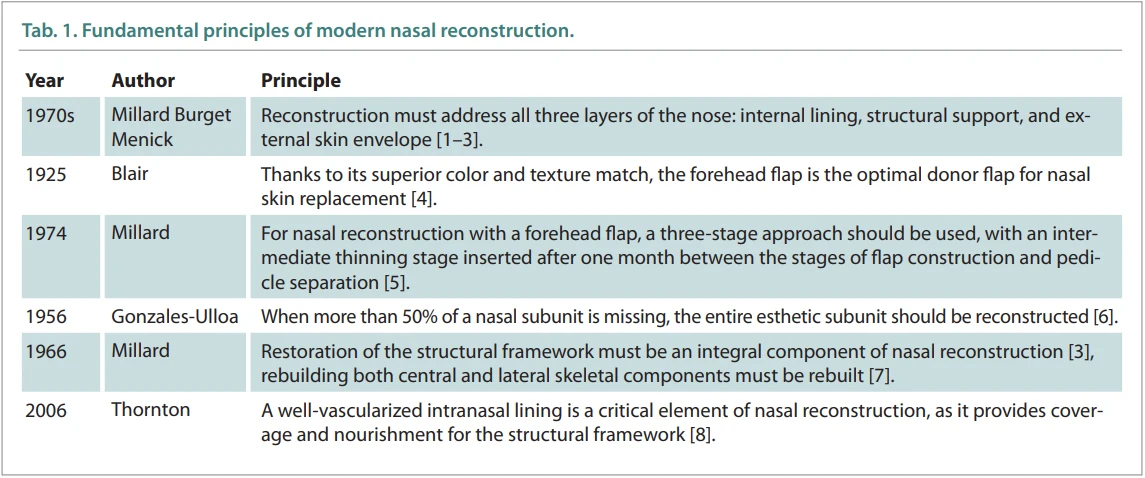
First, the well-vascularized internal lining is reconstructed. Subsequently, the structural framework (composed of cartilage grafts harvested from the auricular conchal bowl, rib cartilage, or nasal septum, or non-vascularized bone grafts taken from the outer parietal cortex or rib) is positioned and secured onto this lining. To allow a proper integration of the structural framework, it needs to be covered with a well-vascularized external skin/soft tissue envelope [1].
The shape of the nose is primarily determined by the bone, cartilage, and compact fibroadipose tissues. The skin and lining, on the other hand, possess only minimal rigidity and tend to simply drape over the supporting structures. The newly reconstructed nose must be designed and positioned proportionally to the face, and the rebuilt framework must provide long-term stability. Reconstruction of the skeletal support is, therefore, an essential component of nasal reconstruction, ensuring the durability and structural integrity of the outcome over time [3].
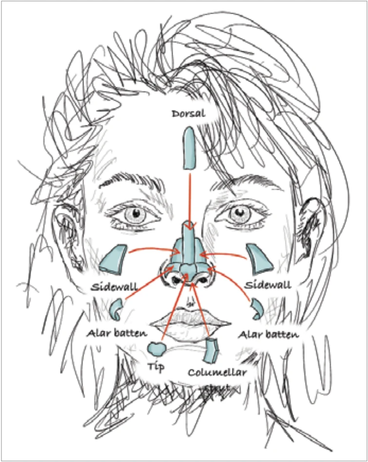
During nasal reconstruction, the complete nasal framework needs to be created in both the vertical and horizontal dimensions. The cartilage must be implanted extra-anatomically into the alar region because without such reinforcement, soft-tissue contraction and retraction are bound to occur. It is essential to recognize that the structural framework of the reconstructed nose must be stronger and more robust than the native nasal skeleton (Fig. 1) [1].
Methods
A literature search was performed using the Discovery system of the Masaryk University Medical Faculty Library, employing the key words ALGORITHM and NASAL RECONSTRUCTION. This search yielded 34 publications. Additionally, cross-referencing was performed using reference lists of selected articles, and relevant monographs on nasal reconstruction were reviewed. Thirteen articles published between 1979 and 2021 were added in this way [9–21]. Papers with high risk of bias, institutional case summaries without methodological innovation, and works not bringing new options for reconstruction of the supporting framework were excluded. Ultimately, 37 works were selected as sources for this review.
Timing of nasal framework implantation
The primary reconstruction surgery is the ideal time for creating the nasal framework. In cases involving double--layered flaps or when bulky soft tissue obstructs the nasal cavity or nostril, the framework is, however, implanted during the second stage of reconstruction along with the thinning of both internal and external layers. Typically, only intranasal mucosal flaps allow immediate primary implantation of the framework. For other flap types – local or free – the supporting layer is inserted only with thinning of the soft-tissue envelope [22].
Secondary implantation of the framework is no longer commonly performed due to the technical difficulty and suboptimal, compromised outcomes. The design of the nasal skeleton generally mirrors the intact contralateral structures [23]. Alternatively, the dimensions of the reconstructed nose may also be defined based on anthropometric measurements [24,25].
Materials and tissues for skeletal reconstruction
The choice of material is less important than the shape and structural design of the framework; the material must primarily provide sufficient rigidity. Still, however, alloplastic materials should generally be avoided because of the risk of extrusion, which may jeopardize the entire reconstruction.
The center of the septal cartilage can be used to harvest a 2–3 × 2–3 cm graft that is 2–3 mm strong and is suitable for multiple applications. The septal cartilage is firm, offers excellent support, and is useful for reconstructing the lateral nasal walls. Harvesting is performed similarly to open rhinoplasty. Importantly, a 1 cm L-strut always needs to be preserved to maintain nasal projection [26].
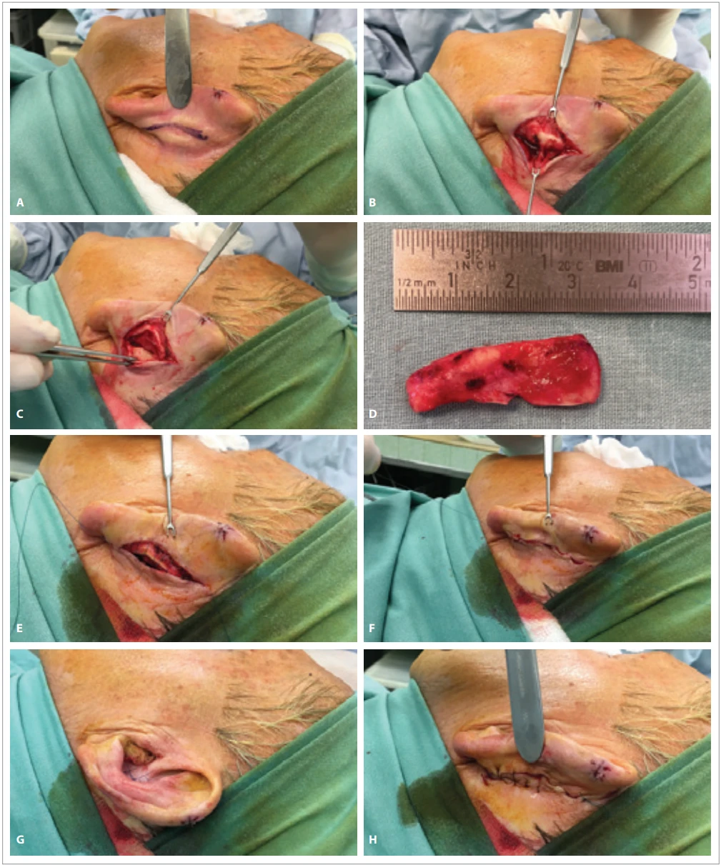
Auricular (conchal) cartilage is ideal for shaping the nasal tip and alae. It can be precisely contoured with sutures, but it does not provide adequate support for broader surfaces. Its harvest technique is shown in Fig. 2.
Costal cartilage is traditionally harvested from the submammary region at the synchondrosis of the 6th–8th ribs. Although the 9th and 10th floating ribs offer ideal curvature and length, the disadvantages of their use include prolonged donor-site pain and a higher scar visibility. When harvesting, a combined osteochondral graft (50% bone + 50% cartilage) is advantageous. Redundant material (if any) can then be stored subcutaneously in the scalp or within the thoracic subcutaneous pocket for potential secondary use [1,2,27,28].
Fixation of graft components during both primary and secondary construction is performed using Polydioxanone (PDS) or non-absorbable sutures. During secondary procedures, pockets preformed in the primary stage may be utilized. Creating a stable nasal framework requires reconstruction of the central support, while the lateral components may be formed from slightly more pliable materials [8].
It is essential to distinguish onlay grafts – used when residual structural support is present to smooth or enhance the projection – and cantilever grafts or L-grafts, which are used in the absence of such support. The latter are proximally secured to the nasal bones with screws or plates, or braced against them. The central nasal framework can be reconstructed using a composite septal pivot flap as well.
Reconstruction of the central nasal framework
The knowledge of the technical parameters of nasal bones (as described by Sullivan [29]) is crucial for anchoring graft materials to these bones. The nasal bones are approximately 12 mm wide at their free end, being widest at the frontonasal suture (14 mm) and narrowest at the nasofrontal angle (10 mm).
The thickness of the nasal bones decreases in the cranio-caudal direction: in men, it is approx. 6 mm in the upper part, decreasing to 3–4 mm at 5–10 mm below the nasofrontal angle. This thickness is still sufficient for screw fixation. Below that point, however, the application of osteosynthetic materials is not recommended due to the further decrease in thickness.
The four most widely used methods of central framework reconstruction include the L-graft, hinged septal flap, septal pivot flap, and cantilever graft.
L-graft
This graft consists of a long arm placed against the root of the nose and a short arm supported by the anterior nasal spine. It can be made of bone or cartilage. Ever since described by Gillies in 1920, it has been widely used (along with its modification). For example, Chait et al. (1980) used an osteochondral graft from the 5th rib. The downside of L-graft lies in the lateral instability of the result [30].
Hinged septal flap
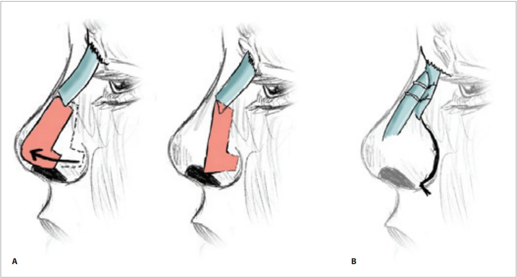
The principle of this technique lies in the rotation of an L-shaped composite flap from the septum around a cranial pivot point (Fig. 3) [31]. The flap is vascularized by the anterior medial nasal branch of the anterior ethmoidal artery [32].
Septal pivot flap
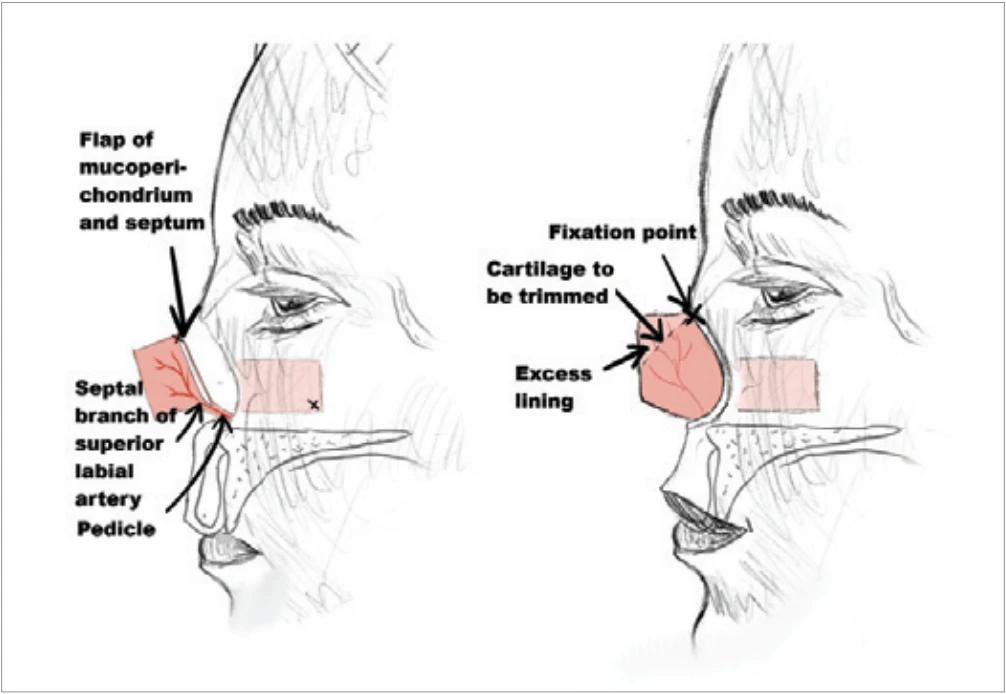
In 1989, Burget and Menick [33] described rotation of the entire septum as a composite flap supplied by the septal branch of the superior labial artery. This was then followed by dorsal augmentation using a cantilever graft. The basic principle of this method is illustrated in Fig. 4.
Cantilever graft
Principally, this technique employs a long bone graft fixed to the nasal bones (which may sometimes require reduction; Fig. 4) [16]. The original idea by Converse and Millard was subsequently developed by other authors, who most commonly use an osteocartilaginous rib graft [30,34]. The rib bone is secured to the nasal bones, while cartilage is used to reconstruct the cartilaginous framework of the nose. Structural stability is typically achieved within 3 weeks.
Neu [35] combined a cantilever graft with an L-shaped cartilage strut to provide both the dorsal and tip support. In his technique, the cranial portion was fashioned from the calvarial bone, whereas the caudal L-shaped segment was created from the septal or auricular cartilage.
Thomassin et al. [36] recommended constructing the L-graft from the calvarial bone – which is membranous in origin and, therefore, highly resistant to resorption – and fixing it using miniscrews and miniplates.
Shubailat used an osteocartilaginous cantilever graft (approximately 70% bone to 30% cartilage) harvested from the 10th rib. At 8–10 mm caudally to the nasal bones, the graft was perforated with a 1.5 mm drill and secured using 2 mm screws. In a cohort of 48 patients, the author reported excellent long-term stability without graft resorption [37].
Brackley and Jones [38] described a technique using a periosteal flap rotated from the forehead to reconstruct the central portion of the missing intranasal lining. On top of the flap, they placed auricular conchal grafts from both ears, and the structure was covered with a paramedian forehead flap. Conchal cartilage grafts were also used by Pirsig [39] for the reconstruction of the anterior septal segment in cases of full-thickness septal loss or saddle-nose deformity. During harvesting, the authors preserved perichondrium on one side, doubled the graft over it, and sutured it. This method employing an anterolateral approach led to the acquisition of a 2.5–3 cm graft demonstrating long-term stability without signs of resorption.
Reconstruction of the lateral nasal framework
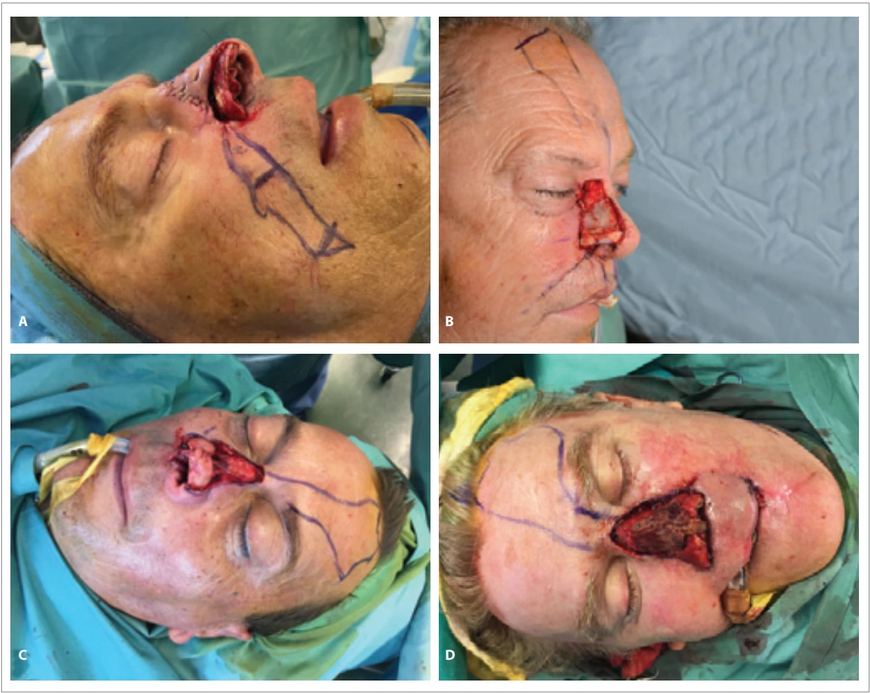
The bony portion of the lateral walls can be fashioned not only from the calvarial bone [36], but (much more easily) from the rib bone, which can be split along one edge and opened to create a stable pyramidal construct [40]. Upper lateral cartilages are best reconstructed using septal cartilage or split rib cartilage, while the alar subunit is best supported by the auricular conchal cartilage, as practically all authors agree [1,23,27,41]. Examples of nasal skeleton reconstruction for nasal defects of different sizes are shown in Fig. 5.
Discussion
The entire nasal skeleton must be reconstructed using well-vascularized tissue.
When reconstructing the intranasal lining, one should always aim to select the thinnest possible flap to prevent airway obstruction after an adequate structural framework is placed. In patients with extensive soft-tissue loss, microvascular free-tissue transfer is often the only viable option [42,43] but even here, the thinnest available flap should be opted for.
In 1974, Millard introduced a three--stage concept for nasal reconstruction using the forehead flap [5], inserting a procedure at 3 weeks after the initial transfer. During that procedure, the thick dorsal skin was thinned (by removal of the frontalis muscle) and the flap was then re-inset. This approach was later modified by Burget and Menick, who advocated against any initial thinning of the distal flap segments to prevent compromising the vascularity of the flap’s peripheral portions and to perform any thinning exclusively during the second surgical stage, 3–4 weeks after the transfer [44].
Even a thin, well-vascularized soft-tissue envelope is able to nourish the implanted cartilaginous and bone grafts enough to prevent their extrusion or resorption. At the same time, using such a thin layer avoids obstruction of the nasal airway and allows clear expression of the aesthetic contours of the reconstructed nasal framework, thereby preserving the natural appearance of the nose.
The question of using alloplastic materials to replace the nasal framework appears repeatedly in the literature. In cases of total nasal loss, Bikhazi [45] used Vitalium or titanium mesh for dorsal support, which was then covered with an expanded forehead flap; the nasal tip was reinforced with conchal cartilage. The fact that no bony graft needs to be harvested, thus preventing any potential donor site morbidity is a clear benefit of this approach. On the other hand, disadvantages include a higher risk of infection and implant extrusion, as well as interference with radiography and CT imaging (which is especially relevant in oncologic patients).
Romo et al. [46] reported outcomes from a group of 187 nasal framework reconstructions using porous polyethylene implants (MEDPOR, Stryker, Minessota, USA), followed up for 6 to 42 months. They observed a 2.6% rate of infection requiring implant removal. Niechajev [47], using Medpor in 23 patients (primarily) with saddle-nose deformities, recorded two complications – one extrusion and one infection [47]. On the other hand, Burget and Menick consistently advise avoiding alloplastic materials in nasal reconstruction [2,27].
Conclusion
The stability of the reconstructed nose is directly proportional to the successful restoration of all nasal layers. Reconstruction of a well-vascularized intranasal lining and its overlay with a well-nourished forehead flap permits the implantation of avascular bone and cartilage grafts in complex nasal defects. A complete structural framework ensures long-term durability of the reconstruction, preventing collapse of the reconstructed nose and maintaining patency as well as functional integrity of the upper airway. Thus, the use of contemporary reconstructive principles allows excellent functional and aesthetic outcomes in nasal reconstruction.
Roles of the authors
Martin Kubát, Jan Menoušek, Ievgenij Polovko conducted the literature search, designed the flowchart and analysed the collected data. Andrej Berkeš drew the pictures. Zdeněk Dvořák and Ievgenij Polovko wrote the manuscript with the input from all authors. Richard Pink and Tomáš Kubek conceived the study and revised it critically for important intellectual content. All the authors approved the version to be published.
Disclosure
The authors have no conflicts of interest to disclose.
Funding
Supported by the internal grant projects of Faculty of Medicine, Masaryk University in Brno - MUNI/A/1754/2024 and MUNI/A/1820/2025.
Sources
1. Baker SR. Principles of nasal reconstruction. Springer. 2011.
2. Menick FJ. Aesthetic nasal reconstruction. In: Neligan PC. (ed). Plastic surgery. Elsevier. 2013, 134–186.
3. Millard DR. Total reconstructive rhinoplasty and a missing link. Plast Reconstr Surg. 1966, 37 (3): 167–183.
4. Dvořák Z., Novák P., Výška T., et al. Reconstruction of defects with forehead flap. Acta Chir Plast. 2015, 57 (3–4): 46–48.
5. Millard DR. Reconstructive rhinoplasty for the lower half of a nose. Plast Reconstr Surg. 1974, 53 (2): 133–139.
6. Gonzalez-Ulloa M. Restoration of the face covering by means of selected skin in regional aesthetic units. Br J Plast Surg. 1956, 9 (3): 212–221.
7. Burget GC., Menick FJ. The subunit principle in nasal reconstruction. Plast Reconstr Surg. 1985, 76 (2): 239–247.
8. Thornton JF., Griffin JR. Nasal reconstruction. Sel Read Plast Surg. 2006, 10 (12): 1–39.
9. Parrett BM., Pribaz JJ. An algorithm for treatment of nasal defects. Clin Plast Surg. 2009, 36 (3): 407–420.
10. Hesamirostami M., Sarparast L., Radfar A., et al. Choosing appropriate technique for nasal reconstruction in challenging cases of panfacial burn: treatment algorithm. J Burn Care Res. 2021, 42 (6): 1215–1226.
11. Chen C., Patel R., Chi J. Comprehensive algorithm for nasal ala reconstruction: utility of the auricular composite graft. Surg J (N Y). 2018, 4 (2): e55–e61.
12. Guo L., Pribaz JR., Pribaz JJ. Nasal reconstruction with local flaps: a simple algorithm for management of small defects. Plast Reconstr Surg. 2008, 122 (5): 130e–139e.
13. Yong JS., Christophel JJ., Park SS. Repair of intermediate-size nasal defects: a working algorithm. JAMA Otolaryngol Head Neck Surg. 2014, 140 (11): 1027–1033.
14. Halani SH., Ma C., Pierce J., et al. Nasal reconstruction after Mohs cancer resection: lessons learned from 2553 consecutive cases. Plast Reconstr Surg. 2021, 148 (1): 171–182.
15. Kim YJ., Cho HH., Kim SO., et al. Reconstruction algorithm for nasal basal cell carcinoma with skin involvement only: analysis of 221 cases repaired by minor surgery. Clin Exp Dermatol. 2015, 40 (7): 728–734.
16. Manson PN., Hoopes JE., Chambers RG., et al. Algorithm for nasal reconstruction. Am J Surg. 1979, 138 (4): 528–532.
17. Moolenburgh SE., McLennan L., Levendag PC., et al. Nasal reconstruction after malignant tumor resection: an algorithm for treatment. Plast Reconstr Surg. 2010, 126 (1): 97–105.
18. Bayramiçli M. A new classification system and an algorithm for the reconstruction of nasal defects. J Plast Reconstr Aesthetic Surg. 2006, 59 (11): 1222–1232.
19. Almeyda R., van der Eerden P., Vuyk H. Skin graft survival on subcutaneous hinge flaps: an algorithm for nasal reconstruction. Laryngoscope. 2013, 123 (3): 605–612.
20. Konofaos P., Alvarez S., McKinnie JE., et al. Nasal reconstruction: a simplified approach based on 419 operated cases. Aesthetic Plast Surg. 2015, 39 (1): 91–99.
21. Yoon T., Benito-Ruiz J., García-Díez E., et al. Our algorithm for nasal reconstruction. J Plast Reconstr Aesthetic Surg. 2006, 59 (3): 239–247.
22. Dvořák Z., Kubát M., Berkeš A., et al. Possibilities of intranasal reconstruction in complex nasal defects. Acta Chir Plast. 2025, 67 (1): 27–41.
23. Menick FJ. Aesthetic nasal reconstruction: principles and practice. Aesthetic Nasal Reconstruction Press. 2017.
24. Farkas LG. Anthropometry of the head and face in medicine. Elsevier. 1981.
25. Farkas LG., Kolar JC., Munro IR. Geography of the nose: a morphometric study. Aesthetic Plast Surg. 1986, 10 (4): 191–223.
26. Dvořák Z., Cheimaris A., Knoz M., et al. Three-stage paramedian forehead flap reconstruction of the nose using the combination of composite septal pivot flap with the turbinate flap and L-septal cartilaginous graft – a case report. Acta Chir Plast. 2021, 63 (1): 6–13.
27. Burget GC. Aesthetic reconstruction of the nose. In: Mathes SJ. (ed). Plastic surgery. 2: The Head and Neck. Saunders 2006, Pt 1 : 573–648.
28. Baker SR. Local flaps in facial reconstruction. Elsevier/Saunders. 2014.
29. Sullivan PK., Varma M., Rozzelle AA. Optimizing bone-graft nasal reconstruction: a study of nasal bone shape and thickness. Plast Reconstr Surg. 1996, 97 (2): 327–335.
30. Chait LA., Becker H., Cort A. The versatile costal osteochondrial graft in nasal reconstruction. Br J Plast Surg. 1980, 33 (2): 179–184.
31. Millard DR. Hemirhinoplasty. Plast Reconstr Surg. 1967, 40 (5): 440–445.
32. MacArthur FJD., McGarry GW. The arterial supply of the nasal cavity. Eur Arch Otorhinolaryngol. 2017, 274 (2): 809–815.
33. Burget GC., Menick FJ. Nasal support and lining: the marriage of beauty and blood supply. Plast Reconstr Surg. 1989, 84 (2): 189–202.
34. Daniel RK. Rhinoplasty and rib grafts: evolving a flexible operative technique. Plast Reconstr Surg. 1994, 94 (5): 597–609.
35. Neu BR. Segmental bone and cartilage reconstruction of major nasal dorsal defects. Plast Reconstr Surg. 2000, 106 (1): 160–170.
36. Thomassin JM., Paris J., Richard-Vitton T. Management and aesthetic results of support grafts in saddle nose surgery. Aesthetic Plast Surg. 2001, 25 (5): 332–337.
37. Shubailat GF. Cantilever rib grafting in salvage rhinoplasty. Aesthetic Plast Surg. 2003, 27 (4): 281–285.
38. Brackley PTH., Jones NS. The use of a periosteal/forehead flap with sandwiched conchal cartilage graft: a novel approach for nasal reconstruction in the absence of a nasal septum. Plast Reconstr Surg. 2002, 110 (3): 831–835.
39. Pirsig W., Kern EB., Verse T. Reconstruction of anterior nasal septum: back-to-back autogenous ear cartilage graft. Laryngoscope. 2004, 114 (4): 627–638.
40. Dvořák Z., Kubek T., Pink R., et al. Moderní principy rekonstrukce nosu. Otorinolaryngol Foniatr. 2018, 67 (4): 100–106.
41. Millard DR. Reconstructive rhinoplasty for the lower two-thirds of the nose. Plast Reconstr Surg. 1976, 57 (6): 722–728.
42. Veselý J., Dvořák Z., Binková H., et al. Rekonstrukce pooperačních defektů v dutině ústní a hltanu. In: Smilek P., Plzák J., Klozar J., et al. (eds). Karcinomy dutiny ústní a hltanu. Medicína hlavy a krku. Tobiáš 2015, 294–312.
43. Binková H., Hložek J., Kostřica R., et al. Volné laloky v rekonstrukci rozsáhlých defektů po onkochirurgických výkonech v oblasti hlavy a krku. Otorinolaryngol Foniatr. 2014, 63 (2): 75–81.
44. Menick FJ. A 10-year experience in nasal reconstruction with the three-stage forehead flap. Plast Reconstr Surg. 2002, 109 (6): 1839–1855.
45. Bikhazi NB., Chow AW., Maas CS. Nasal reconstruction using a combination of alloplastic materials and autogenous tissues: a surgical alternative. Laryngoscope. 1997, 107 (8): 1086–1093.
46. Romo T., Sclafani APOD., Sabini P. Use of porous high-density polyethylene in revision rhinoplasty and in the platyrrhine nose. Aesthetic Plast Surg. 1998, 22 (3): 211–221.
47. Niechajev I. Porous polyethylene implants for nasal reconstruction: clinical and histologic studies. Aesthetic Plast Surg. 1999, 23 (6): 395–402.
Labels
Plastic surgery Orthopaedics Burns medicine TraumatologyArticle was published in
Acta chirurgiae plasticae
2026 Issue 1-2
Most read in this issue
- A systematic review of sensory outcomes in nipple reinnervation after implant-based breast reconstruction
- Principles of nasal framework reconstruction for complex nasal defects: review
- Self-esteem and quality of sexual life in women undergoing breast reconstruction due to mastectomy: a cross-sectional study in a non-European setting
- Cutting-edge biotechnological strategies to enhance autologous fat graft survival