
Results of the study of mucosal immunity indices in patients with cancer of the oral cavity and oropharynx during radiotherapy or chemoradiotherapy therapy and immunotherapy with α/β-defensins
Authors:
H. A. Hirna 1,2; D. V. Maltsev 3; M. M. Rozhko 2; I. D. Kostyshyn 1
Authors‘ workplace:
Department of Oncology, Educational and Scientifi c Institute of Postgraduate Education, Ivano-Frankivsk National Medical University, Ministry of Health of Ukraine, Ivano-Frankivsk, Ukraine
1; Department of Dentistry, Educational and Scientifi c Institute of Postgraduate Education, Ivano-Frankivsk National Medical University, Ministry of Health of Ukraine, Ivano-Frankivsk, Ukraine
2; Laboratory of Immunology and Molecular Biology, Institute of Experimental and Clinical Medicine, O. O. Bogomolets National Medical University, Kyiv, Ukraine
3
Published in:
Klin Onkol 2023; 36(2): 112-123
Category:
Original Articles
doi:
https://doi.org/10.48095/ccko2023112
Overview
Background: The aim of the study was to investigate the concentration of interferon (INF) -a, INF - g, interleukin (IL) -6, and secretory IgA (sIgA) in saliva during various regimens of antitumour treatment and immunotherapy (IT) with a/b-defensins in patients with cancer of the oral cavity and oropharynx in order to find ways to increase the effectiveness and improvement of the tolerability of antitumour treatment on the base of the identification of biomarkers for the evaluation of the antitumour effect and the prediction of complications. Materials and methods: We have studied the changes in the immunity indices of 105 patients who were diagnosed with squamous cell carcinoma of the oral cavity or oropharynx for the first time. The patients received radiotherapy (RT) or chemoradiotherapy and IT with a/b-defensins in different doses (40 and 60 mg) at the 1st phase of the special treatment. Results: A determined drop in the concentration of INF-a after cytostatic treatment, and the additional use of IT with a/b-defensins in different doses do not produce the protective effect on the production of INF-a. Regarding INF - g, a more than two-fold decrease in the concentration of INF - g in the saliva of patients in group receiving a double dose of an immunotherapeutic agent along with radiation therapy (RT) was noted, which may indicate an adjuvant effect of a/b-defensins in relation to RT, enhancing its antitumour influence, and thereby ensuring the regression of neoplasia. In case of an increased dose of a/b-defensins use during RT, there was found immunomodulatory effect in relation to IL-6. In the group of patients who received RT and a higher dose of the immune agent, the “scissors phenomenon” was noted – a simultaneous decrease in the concentration of INF - g and an increase in the concentration of sIgA in saliva, which, taking into account the reduced risk of mucositis and better regression of the tumour, shows the meaningful adjuvant and immunomodulating effects of a/b-defensin therapy in the study group. Conclusion: High-dose IT with a/b-defensins against the background of cytostatic therapy in patients with cancer of the oral cavity and oropharynx potentially leads to an adjuvant and immunomodulatory effect with a decrease in the concentration of INF - g and a parallel increase in the concentration of sIgA in saliva, i.e., reconstruction of the immune response from Th1 - to Th2-profile – the profile associated with the tumour regression. With the development of the radio-induced mucositis in these patients, a decrease in concentration of sIgA in saliva with a tendency to a progressive decrease of this index with the increase of mucositis severity was noted. The data obtained allow us to consider INF - g and sIgA as biomarkers of the effectiveness of traditional anticancer therapy during the use of a/b-defensins, and sIgA as a biomarker of the risk of developing radio-induced mucositis in patients with cancer of the oral cavity and oropharynx, which should be verified in further clinical studies with better design.
Keywords:
oral cavity – immunotherapy – Oropharynx – cancer – regression – interferon-α – interferon- – secretory IgA
Introduction
Due to the latest advances in immuno-oncology, it has been determined that the local antitumour immune response is an important factor in controlling the growth of the tumour in the human body [1]. Therefore, the study of indices of local immunity turns out to be an attractive prospect for finding the effective biomarkers for assessing the severity of the patients’ condition and predicting the course of an oncological disease. In case of cancer of the oral cavity and oropharynx, it is advisable to study indices of mucosal immunity of the oropharyngeal system [2].
Interferon (INF) -a and INF - g play a key role in the antitumour immune response of the human body [3]. They are produced mostly by Th1-cells in the area of inflammation, both in response to tumour antigens and to the action of gamma-rays during therapy, and affect the generation and properties of immune and malignant cells [4]. The biological effects of interleukin (IL) -6 are diverse, but the important thing is that this cytokine can be involved into the pathogenesis of the tumourigenic process, increasing inflammation and angiogenesis, which sometimes contributes to the progression of the tumour process and metastasis. IL-6 can participate in the formation of tumour resistance to chemotherapy (CHT), which allows some researchers to consider this cytokine as a factor in enhanced tumour growth [5]. As it is known, the dominant class of immunoglobulins in saliva is secretory IgA (sIgA) – an effector of humoral immunity of the mucous membrane of the oral cavity which plays an important role in its protection against infectious and tumourigenic factors [6]. It is believed that the level of sIgA in saliva is a sensitive index of immune-mediated diseases, and its diagnostic value in cancer of the oral cavity and oropharynx should be studied in more details in the future [7].
An increased level of defensins is found in the saliva of patients with oral cavity cancer compared to healthy individuals [8,9]. As it is known, defensins are the immune peptides that have antimicrobial and cytotoxic properties [10], and their potential influence on the development of the malignant process is promising for further scientific study. An immunotherapeutic agent containing natural a/b-defensins may be a useful tool in conventional radiotherapy (RT) and chemoradiotherapy (CHRT) due to its immunomodulatory effect and potential ability to enhance the efficacy of immune peptide antitumour therapy. It seems appropriate to study the effect of a medicine based on a/b-defensins on the indices of local immunity in patients with cancer of the oral cavity and oropharynx during RT or CHRT treatment, in particular, on the concentration of INF-a, INF - g, IL-6, sIgA in saliva, which are important indices of mucosal immune protection of the human body.
The aim of the work is to study the concentration of INF-a, INF - g, IL-6, sIgA in saliva during different regimens of anticancer treatment and immunotherapy (IT) with a/b-defensins in patients with cancer of the oral cavity and oropharynx in order to find the ways to increase the effectiveness and improve the tolerability of anticancer treatment due to the identification of biomarkers for the evaluation of the antitumour effect and the prediction of complications.
The tasks of the study are: 1) to study the concentrations of INF-a, INF - g, IL-6, sIgA in saliva during various regimens of antitumour treatment and IT with a/b-defensins; 2) to study the concentrations of INF-a, INF - g, IL-6, sIgA in saliva during the development of chemoradiotherapy-induced mucositis of the oral mucous membrane; 3) to evaluate the possibility of using the studied laboratory indices as biomarkers of the effectiveness of the treatment and prevention of complications.
The endpoints of the study are: 1) differences in the concentration of INF-a, INF - g, IL-6, sIgA in saliva in different groups of patients with different modes of cytostatic therapy and IT; 2) development of mucositis induced by RT or CHRT.
The null hypothesis: the indices of mucosal immunity may reflect the use of both RT or CHRT and IT with a/b-defensins, and may also be associated with tumour regression in response to the treatment and the development of mucositis as a complication of RT or CHRT, which may be used to evaluate the effectiveness and tolerability of the applied therapeutic strategies and predict the further course of the disease.
Materials and methods
This is a prospective, single-center, non-randomized, controlled, comparative study. The study of changes in indices of humoral immunity was performed including 105 patients who were diagnosed with squamous cell cancer of the oral cavity or oropharynx for the first time.
In the study, the enrollment of patients and the distribution between groups was carried out depending on the indicated method of treatment (RT or CHRT) at the department of head and neck tumours of Communal Non-Profit Enterprise “Precarpathian Clinical Oncology Center of the Ivano-Frankivsk Regional Council” during the years 2017–2021.
Thus, the research groups were: I (RT-IT), II (CHRT-IT) and III (RT-2IT); the comparison groups were: IV (RT) and V (CHRT). Twenty-five patients of group I (RT-IT) received RT and IT with a/b-defensins at the 1st phase of special treatment in a total dose of 40 mg per course. Twenty patients of group II (CHRT-IT) had RT with intra-arterial cisplatin potentiation and IT at a dose of 40 mg. Group III (RT-2IT) included 20 patients who received RT and the immune agent of a/b-defensins in a total dose of 60 mg.
According to the localization, in our study, the percentages of patients with oropharyngeal cancer and oral cavity cancer were 36% and 64%, respectively (Tab. 1). The groups were compared by tumour localization using the chi-square test (P > 0.05).

According to the international classification of TNM-8 (AJCC, 2017), there were 94 (89.5%) patients in an extensive-degree of the disease (stage III and IV), and 11 (10.5%) patients had stages I–II (Tab. 2). Stage III of the disease was most common in each of the groups: 9 (36%) patients were in group I and most of them had localization in the oropharynx, 13 (65%) patients were in group II, including 9 (45%) with tongue cancer, 8 (40%) patients were in group III (half of them had cancer of the oral cavity), 10 (50%) patients were in group IV and 11 (55%) patients were in group V. Nine (36%, 45%, 45%) patients in groups I, III, IV, and 6 (30%) and 7 (35%) patients in groups II and V had stage IV disease. According to the analysis performed, no differences were found between the groups according to the stage of tumour disease (P > 0.419); the groups were compared by this characteristic.
![Disposition of patients with cancer of the oral cavity and oropharynx according to the international TNM-8
classifi cation [37].](https://pl-master.mdcdn.cz/media/cache/media_object_image_large/media/image_pdf/8ed3dcc0f96ef18ef800d7f73f0af3ce.png)
In 82 (78.1%) patients, the spread of the tumour to the regional lymph nodes was confirmed by a puncture biopsy of the lymph node, ultrasound, multi-slice spiral computed tomography or magnetic resonance imaging (MRI) examinations. The majority of patients with N1 were in group II – 14 (70%) patients, and the least in group IV – 7 (35%) patients. Regional metastasis of cancer of the oral cavity and oropharynx, which was interpreted as N2, occurred most often in group IV – 8 (40%) patients, and the least in group II – 4 (20%) patients. In groups I and III, there was one patient with N3, and 2 (10%) patients in group IV. Having performed the analysis according to the chi-square test, it was determined that the groups were compared according to the rate of metastasis to the regional lymph nodes (P > 0.442).
RT with ionizing radiation was performed using the device Cobalt-60 at a dose of 2–2.5 Gy per session up to the total dose (TD) of 36–40 Gy. CHT treatment consisted of retrograde intra-arterial (through the superficial temporal artery on one or both sides or the external carotid artery) potentiation with cisplatin. The scheme of its introduction was as follows: single dose (SD) 20 mg/m2 within 5 days from the day of the beginning of RT [11]. IT with a/b-defensins was performed according to two schemes, the difference between them was the total dose received by the patients. Patients of groups I (RT-IT) and II (CHRT-IT) were intramuscularly injected an immune preparation at a dose of 2.0 mL twice a day 2 days before the start of the special treatment for 5 days and in the following 10 days during the treatment once a day, with TD of 40 mg per course. Patients of group III (RT-2IT) were intramuscularly administered a larger dose of the immune agent, 60 mg per course, according to the scheme of 2.0 mL twice a day 2 days before the start of the special treatment for 10 days and once a day in the following 10 days during the treatment [12].
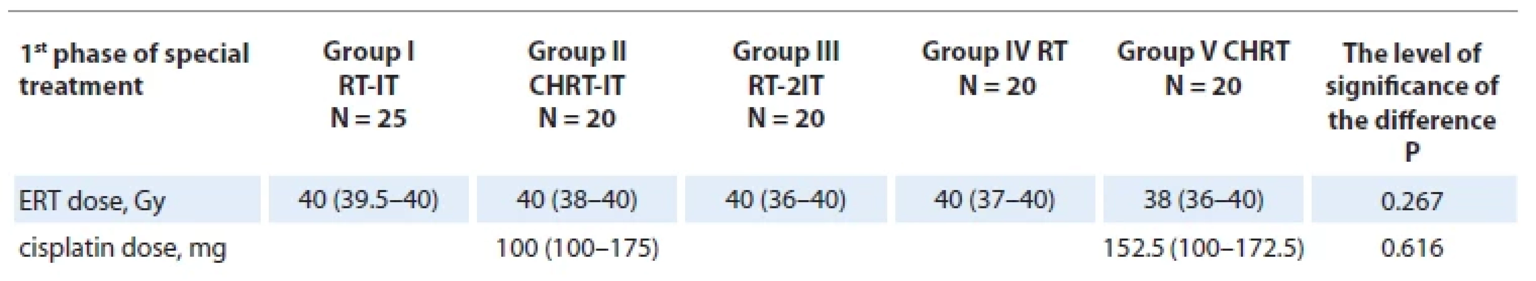
All patients with different doses of RT and cytostatics received the planned treatment at the 1st phase, the median values of which are given in Tab. 3.
At the end of the 1st phase of special treatment, we have recorded the presence of one or another degree of mucositis in the oral cavity or oropharynx (Graph 1). Thus, in 12% of patients of group I, 25% of group II and 5% of group III, the phenomena of mucositis were not observed until the end of the treatment; degree I mucositis was present in 44% patients of group I, 10% patients of group II, 35% patients of group III and 10% patients in group IV. There were no such patients in the comparison group. It is worth noting that in groups IV and V, there were 75% and 45% of patients with degree III mucositis; in groups I, II and III, the ratios of such patients were significantly lower 4%, 15%, and 10%, respectively, which clinically attests to high-quality use of IT with a/b-defensins in the treatment. This means that in the groups where the combination of treatment included IT with a/b-defensins, there was a greater proportion of patients with degree II mucositis, but less with degree III mucositis (Graph 1). The data of all groups were statistically significant (P < 0.001).

According to the data of clinical examination, ultrasound examination, CT and MRI, the size of the tumour was assessed and compared with the initial data of the patients, and tumour regression was recorded at the end of the 1st phase of special treatment.
After completion of the treatment, tumour regression was > 50% in 40% of patients of group I, and in 63% in groups II and III. Also, only in these groups receiving special treatment and IT, tumour regression was > 75% (Graph 2). The median value of the regression index for groups I, II and III is the same – 50%, and the interquartile ranges differ slightly: 37.5–70% for group I, 40–57.5% for group II and 40–55% for group III. In groups IV and V, the median value with interquartile ranges was slightly lower – 30% (22.5–40%) and 40% (30–50%), respectively. According to these data, reliable indices of tumour regression are between groups I, II, III and IV (P < 0.004). The best results are in the group having received RT and IT. The results of groups IV and V are significantly different in comparison, and the lowest average index is in group IV, having received only RT (Graph 2).

Research methodology
Before the initiation of special treatment, saliva was sampled from patients to determine the initial levels of INF-a, INF - g, IL-6, and sIgA. Re-sampling of biomaterial was carried out on the day after the end of IT in research groups (I and II), on average after a dose of 20 Gy, which was also an index for saliva sampling in the comparison groups (IV and V). In group III, where the scheme of IT was different, the collection of biomaterial was done after 26 Gy.
The saliva collection procedure was performed in the morning, on an empty stomach, before oral hygiene and without the use of stimuli for salivation. The amount of collected saliva was approximately 5–10 mL. Saliva samples were stored in a refrigeration system (freezer) at a temperature of about −20 °C until analytical procedures were carried out at O.O. Bogomolets National Medical University, Scientific-Research Institute of Experimental and Clinical Medicine. Immunological studies of oral fluid were performed using an Epics XL flow cytofluorimeter with the use of “Vector-Best” reagents and an ELISA Kit (FINETEST).
Statistical analysis of the results was performed using the Statistical software package EZR v. 1.54 (graphical user interface for R statistical software version 4.0.3, R Foundation for Statistical Computing, Vienna, Austria).
The distribution of quantitative indices was tested for normality using the Shapiro-Wilk test. In case of normal distribution, quantitative indices are expressed through the mean value ± standard deviation (SD), and in case of a non-normal distribution – through the median and the interquartile range (QI–QIII). Most of the parameters were not subjected to the normal distribution law, therefore, non-parametric criteria were used: Kruskal-Wallis test, for pairwise comparisons, Dunn or Mann–Whitney test was used, taking into account the Bonferroni corrections. The chi-square test was used to compare the qualitative characteristics, and the posterior comparison was carried out according to Fisher’s exact test, taking into account the Bonferroni correction. The differences in groups were indicated as P-value with an indication of the level of significance. The data were considered to be different at P < 0.05.
Results
We have performed evaluation of the concentrations of INF-a, INF - g, IL-6, and sIgA in the saliva of patients with cancer of the oral cavity and oropharynx of the studied groups receiving various regimens of cytostatic treatment with/without IT with a/b-defensins.
Since the mean values turned out to be unrepresentative due to the non-standard distribution of the variants of a number of values, we have determined the median value of the indices and the corresponding interquartile intervals (Tab. 4)

As can be seen from Tab. 4, RT and CHRT lead to almost complete inhibition of INF-a production by the oral mucous membrane. An increase in the concentration of INF-a in groups II, III, and V before the start of the treatment can be considered as the manifestation of immunoreactivity – the response of the immune system to the tumour. The drop in these concentrations indicates the well-known immunosuppressive effect of RT and CHRT. Concerning INF-a production, additional use of IT with a/b-defensins in different doses does not have a protective effect. We’ve tested whether the addition of the immune agent a/b-defensins could preserve INF-a production during cytostatic treatment, potentially indicating preservation of the antitumour immune response despite the immunosuppressive effects of the therapeutic interventions. However, the results obtained do not allow to confirm the null hypothesis.
As for INF - g, there were no significant differences in the concentrations in saliva before and after cytostatic therapy in groups I, II, IV, and V, but there was a more than two-fold drop in the concentration of INF - g in the saliva of patients in group III receiving an increased dose of the immunotherapeutic agent along with RT. Since the production of INF - g is a consequence of the implementation of a specific cytotoxic cellular immune response against tumour antigens, the preservation of the secretion of this cytokine may indicate the preservation of the intensity of the cellular immune reaction in response to malignant cells. A sharp decrease in the concentration of INF - g in the saliva of group III patients may indicate either that an increased dose of a/b-defensin exerts an adjuvant effect upon RT, enhancing its antitumour effect and thereby ensuring the regression of neoplasia, or tumour growth and/or increased immunosuppression. The better indices of tumour regression after the 1st phase of treatment (Graph 1) in the observation group III confirm the above mentioned assumptions regarding the adjuvant effect of the immunotherapeutic agent based on a/b-defensins. Since mucositis of the mucous membrane of the oral cavity, provoked by special treatment, was less common in this group, it could be assumed that due to the adjuvant effect of a/b-defensins and weakening of the cytotoxic immune antitumour response due to tumour regression, it was possible to reduce the risk of inflammatory complications in the oral cavity. Since this group had better results of tumour treatment, i.e., more pronounced tumour regression, it can be assumed that the decrease in INF - g production is not a consequence of an increase in radiation-induced immunosuppression, but of a decrease in antigenic irritation of the immune system due to a reduction in the size of the tumour because of an increase in the clinical effectiveness of antitumour therapy.
For the most part, there was no significant dynamics of IL-6 concentration in saliva before and after therapy in different observation groups (Tab. 4). It is worth noting a certain increase in the concentration of IL-6 in the saliva of the studied groups, which could indicate the risk of tumour progression. However, increasing the dose of a/b-defensins eliminated this effect in the observation group III, which may indicate dose-dependent differences in the immunomodulatory effects of a/b-defensins in patients with cancer of the oral cavity and oropharynx during cytostatic therapy.
Regarding the concentration of sIgA in saliva, no significant differences were found in patients before and after therapy in all observation groups, except for group III, where an increase in the concentration of sIgA in saliva was determined, which indicated an increase in humoral local mucosal immunity and, possibly, potentiation of local antitumour resistance (Tab. 4). As it is known, the cytotoxic cellular immune response characterized by INF - g indicates the exacerbation of the tumour process, and its reduction with the parallel development of the humoral response, including the production of sIgA, may indicate the remission of the malignant tumour. In group III, the “scissors phenomenon” was recorded – a simultaneous decrease in the concentration of INF - g and an increase in the concentration of sIgA in saliva, which, taking into account the reduced risk of mucositis and better tumour regression, indicates the pronounced adjuvant and immunomodulatory effects of therapy with a/b-defensins in the patients of the study groups. Therefore, the additional use of a/b-defensins in an increased dose (60 mg) has clinical significance in terms of treatment results and prevention of complications in patients with cancer of the oral cavity and oropharynx during conventional anticancer therapy. However, a smaller dose (40 mg) of the medicine with a/b-defensins did not demonstrate similar adjuvant and immunomodulatory effects, which indicates a dose-dependent manner of their implementation in the observation groups.
During the analysis, there was found no connection between the level of the analyzed indices after treatment and the severity of mucositis at the end of the treatment (P > 0.05 for all indices), but a certain trend was noted. The study of indices of local mucosal immunity in groups of patients with cancer of the oral cavity and oropharynx who had different degrees of mucositis at the end of treatment indicates a progressive decrease in the concentration of sIgA with the increasing severity of mucositis, which indicates the potential possibility of using sIgA as a marker of the risk of developing mucositis (Tab. 5).
When analyzing the changes (Tab. 6), i.e., the difference in indices before and after the treatment (dX = Xi after – Xi before, i.e. a decrease in the index (with the sign −), and an increase in the index (with the sign +), depending on the severity of mucositis at the time of treatment completion, no connection was found between the change in the level of the analyzed indices and the severity of mucositis (P > 0.05 for all indices). Graph 3 shows the median value, interquartile range, minimum and maximum value, and revealed a trend towards a decrease in the concentration of sIgA in saliva with increasing severity of mucositis at the end of the treatment (P = 0.016 according to the Jonckheere-Terpstra criterion).


As it is shown in Tab. 4–6, the IT with a/b-defensins in an increased dose due to the effect of immunomodulation including a simultaneous decrease in INF - g production and an increase in sIgA production, i.e., a restructuring of the immune response from the Th1 - -pathway to the Th2-pathway, was associated with a reduced risk of severe mucositis during conventional anticancer therapy. The concentration of INF-a and IL-6 in saliva did not change with different severity of mucositis, and this does not allow this index to be considered an informative marker for predicting this complication in patients of the studied groups.
Discussion
The local effect of gamma rays on the tumour modifies its microenvironment and promotes the generation of pro-inflammatory cytokines, including the most important anti-proliferative agents – interferons (IFN-a, IFN - g) promoting a persistent immune response against the tumour [4]. IFN-a is important for the activation of the innate and adaptive immune antitumour response, which is regulated by T-cells [13]. Induction of interferons has been found to be important in tumour shrinkage in response to RT in several studies [14,15]. However, despite the expected increase in the production of interferons in response to external beam RT, it was found that in patients with cancer of the oral cavity and oropharynx at the salivary level, there is a several-fold decrease in the level of antibodies to INF-a, which paradoxically has a negative influence on local antitumour protection [16]. Therefore, the results of research regarding the influence of locally produced INF-a in cancer of the oral cavity and oropharynx are ambiguous, since this cytokine can both inhibit and stimulate tumour growth depending on specific conditions. In this study, it was shown that the concentration of INF-a in saliva in the observation groups after the course of therapy is quite low, although it was slightly increased in some groups before the start of treatment, which could be considered as a manifestation of immunoreactivity. We’ve made an attempt to find an agent influencing the production of IFN-a in saliva to potentiate the antitumour response mediated by interferons, however, as it turned out, a/b-defensins do not contribute to the increased synthesis of this cytokine or to the preservation of such synthesis during RT and CHRT exposure in patients with cancer of the oral cavity and oropharynx.
IFN - g is a strong activator of NK-cells and CD8+ cytotoxic T-cells, and it has pleiotropic effects in the tumour microenvironment [17]. Radiotherapy causes inflammation in which IFN - g definitely plays an important role. During the tumour process, the production of IFN - g usually increases, which can mediate an effective antitumour response, but to achieve this effect, a balance of positive and negative effects of IFN - g is necessary, taking into account many other factors [18]. The results of one of the studies confirm that RT dose-dependently increases the concentration of IFN - g in blood serum in 88% of patients with squamous cell carcinoma of the esophagus who were completely cured [19]. But in patients with oral cavity cancer; however, this theory is not absolutely confirmed, and there is currently no accurate information about the role of this cytokine in the development of oral cavity or oropharyngeal cancer. In one of the studies, it was determined that the production of IFN - g by malignant cells is significantly reduced after RT [20], and in another trial, it was determined that after CHRT treatment, there is an increased synthesis of IFN - g in the mucous membrane of patients with cancer of the oral cavity and oropharynx in comparison with the condition before the treatment [16]. In this study, some decrease in the concentrations of INF - g under the influence of RT and CHRT, and an increase in this reaction with an increase in the dose of an immunotherapeutic agent based on a/b-defensins, were revealed. Since this phenomenon was associated with tumour regression, we’ve considered the decrease in the concentration of INF - g when increasing the dose of a/b-defensins not as a result of drug-induced immunosuppression, but as a sign of the adjuvant effect of a/b-defensins, that is, their ability to enhance the effectiveness of cytostatic therapy. The decrease in the production of INF - g in this case could be explained by the weakening of the antigenic irritation of the immune system due to the reduction in the size of the tumour. However, no data were obtained that could recommend the use of INF - g as a marker of the risk of mucositis as a result of anticancer therapy.
According to the research results, significant difference in the levels of IL-6 in cancer patients were recorded. Thus, in healthy individuals, IL-6 concentration levels range from 1.4 ± 0.9 to 47.46±18.74 pg/mL. Most investigations studied this cytokine in patients with oral cavity cancer without taking into account the factors affecting its production, primarily – inflammation of the mucous membrane provoked by CHRT [21]. However, the results of many studies confirm that in patients with cancer of the oral cavity and oropharynx before the treatment, the concentration of IL-6 is increased 16–22-fold (on the average – 137 pg/mL for an exophytic tumour and 186 pg/mL for an endophytic tumour) compared to healthy individuals [22]. It has been proven that their expression of the IL-6 gene in endophytic tumour is higher than in exophytic one [23]. Therefore, this index is used as a tumour marker for early detection of cancer and monitoring the effectiveness of its treatment [24]. In this study, the median concentration of IL-6 was small, corresponding to the range of values from 3.78–5.39 pg/mL, which does not correspond to the results of other studies. There are reasons to believe that IT with a/b-defensins in an increased dose reduces the concentration of IL-6 in saliva, which could be explained by the adjuvant effect of immunotherapeutic interventions.
The results of previous studies indicate different concentrations of sIgA in saliva in patients with cancer of the oral cavity and oropharynx [25–27]. When using different methods of measurement, the levels of sIgA in cancer patients are significantly lower [28] than in healthy people (they are in the range 4–40 μg/mL) [7]. The results of one study showed that sIgA concentrations measured by nephelometry were 17.0 ± 10.4 mg/dL in healthy individuals and 7.2 ± 5.0 mg/dL in patients with cancer of the oral cavity and oropharynx, and when using radial immunodiffusion measurement methods, they were 13.7 ± 9.1 mg/dL and 5.6 ± 4.2 mg/dL for healthy people and patients with cancer of the oral cavity and oropharynx, respectively [29,30]. No correlation was found between sIgA concentration and patient-related parameters such as clinical stage, histological tumour type, and the presence of lymph node metastases before the treatment [29,31].
Some authors have hypothesized that the reduced sIgA level may be associated with the increased mortality in cancer patients due to the attenuation of the humoral immune response against the tumour [32]. Moreover, a decrease in the concentration of sIgA increases the risk of inflammatory complications in the mucous membrane, including mucositis [33,34]. A significant decrease in the concentration of sIgA was found in sick children who suffered from a malignant disease, and low values were associated not only with the disease, but also with the development of mucositis induced by CHRT [35]. The phenomenon of a decrease in the concentration of sIgA in patients with cancer of the oral cavity and oropharynx and deepening of the sIgA deficiency in the development of mucositis is considered as an unfavourable prognostic factor. This study demonstrated an increase in the concentration of sIgA in saliva with the addition of a/b-defensins in an increased dose, which was associated with more pronounced tumour regression during the performance of the conventional anticancer treatment. With the increase of inflammatory phenomena in the mucous membrane of the oral cavity, the deficiency of sIgA in saliva deepened, which corresponds to the results of other studies in this area (Tab. 4,5). Therefore, the result in the observation group III, which was achieved under the influence of a/b-defensins – an increase in the concentration of sIgA in saliva – indicates a positive prognosis in patients with cancer of the oral cavity and oropharynx. The study of sIgA as a biomarker in oncology patients is promising for further scientific research, taking into account the possibility of correcting the concentration of sIgA in saliva during adjuvant IT, which can improve the prognosis by influencing both the development of local complications of RT and the further course of oncological disease.

Although there have been numerous attempts to evaluate sIgA, INF-a, INF - g as biological markers in oral cavity and oropharyngeal cancer, no convincing evidence has been found for the routine use of these indices in clinical practice, despite the encouraging results of individual studies. The results of this work strengthen the evidence base for the feasibility of assessing local mucosal immunity in patients with cancer of the oral cavity and oropharynx undergoing various RT and CHT regimens. In particular, it has been shown that these indices can indicate both tumour regression under the influence of cytostatic therapy and the immunosuppressive effect of these treatment methods, as well as indicate the risk of developing mucositis, which needs to be clarified in further studies with a larger number of participants and a better design. It also seems obvious that the use of high-dose IT with a/b-defensins exerts an immunomodulatory effect on local mucosal immunity within the studied indices; this could be associated with an improvement of the effectiveness and tolerability of RT for patients with cancer of the oral cavity and oropharynx. These adjuvant and tolerogenic effects of IT with a/b-defensins should be investigated in additional studies to find reliable evidence for the use of this immunotherapeutic strategy in oncology clinical practice. The data obtained are consistent with the results of our previous study regarding the effect of IT with a/b-defensins on indices of systemic immunity in patients with cancer of the oral cavity and oropharynx [36]. There was achieved a pronounced dose-dependent decrease in the relative number of lymphocytes, a probable decrease in the absolute number of CD3+ of T-cells simultaneously with an increase in the relative number of natural killer cells, as well as a decrease in the absolute number of NKT--cells in the blood. There were noted adjuvant and immunomodulatory effects of a/b-defensins with restructuring of the subpopulation composition of lymphocytes with an increase in the relative amount of natural killers in the blood having pronounced antitumour activity.
Conclusions
High-dose IT with a/b-defensins against the background of conventional cytostatic therapy in patients with cancer of the oral cavity and oropharynx leads to the adjuvant and immunomodulatory effects with a decrease in the concentration of INF - g and a parallel increase in the concentration of sIgA in saliva, i.e., restructuring of the immune response from Th1 - to the Th2-profile associated with tumour regression.
During the development of radiation-induced mucositis in patients with cancer of the oral cavity and oropharynx undergoing conventional cytostatic therapy, a decrease in the concentration of sIgA in saliva was noted with a tendency to a progressive decrease of this index with increasing severity of mucositis.
The data obtained allow us to consider INF - g and sIgA as the potential biomarkers of the effectiveness of conventional anticancer therapy during the use of a/b-defensins, and sIgA as a biomarker of the risk of developing radiation-induced mucositis in patients with cancer of the oral cavity and oropharynx, which should be tested in further clinical studies with a better design.
Disclosure
This work is a fragment of the scientific-research work “Clinical effectiveness of comprehensive treatment of diseases of the hard tissues of the teeth and periodontium in the population of environmentally unfavorable regions”, state registration number 0118U004144.
Ethical aspects
All patients were informed about the planned treatment and their written consent was obtained. The study was performed in accordance with the principles of the Declaration of Helsinki and the Ethics Committee of the Ivano-Frankivsk National Medical University (research protocol № 94/17 dated November 16, 2017).
Halyna Anatoliyivna Hirna
Department of Oncology
Ivano-Frankivsk National Medical
University
Halytska Street 2
Ivano-Frankivsk
Ukraine
e-mail: halynagіrna@gmail.com, ggyrna@ifnmu.edu.ua;
Submitted/Obdrženo: 2. 8. 2022
Accepted/Přijato: 30. 8. 2022
The authors declare that they have no potential confl icts of interest concerning drugs, products, or services used in the study.
Autoři deklarují, že v souvislosti s předmětem studie nemají žádné komerční zájmy.
The Editorial Board declares that the manuscript met the ICMJE recommendation for biomedical papers.
Redakční rada potvrzuje, že rukopis práce splnil ICMJE kritéria pro publikace zasílané do bi omedicínských časopisů.
Klin Onkol 2023; 36(2): 112 – 123
Sources
1. Kotani Y, Shinkai S, Okamatsu H et al. Oral intake of Lactobacillus pentosus strain b240 accelerates salivary immunoglobulin A secretion in the elderly: a randomized, placebo-controlled, double-blind trial. Immun Ageing 2010; 7 : 11. doi: 10.1186/1742-4933-7-11.
2. Diesch T, Filippi C, Fritschi N et al. Cytokines in saliva as biomarkers of oral and systemic oncological or infectious diseases: a systematic review. Cytokine 2021; 143 : 155506. doi: 10.1016/j.cyto.2021.155506.
3. Dunn GP, Koebel CM, Schreiber RD. Interferons, immunity and cancer immunoediting. Nat Rev Immunol 2006; 6 (11): 836–848. doi: 10.1038/nri1961.
4. Lugade AA, Moran JP, Gerber SA et al. Local radiation therapy of B16 melanoma tumors increases the generation of tumor antigen-specific effector cells that traffic to the tumor. J Immunol 2005; 174 (12): 7516–7523. doi: 10.4049/jimmunol.174.12.7516.
5. Miron N, Miron MM, Milea VG et al. Proinflammatory cytokines: an insight into pancreatic oncogenesis. Roum Arch Microbiol Immunol 2010; 69 (4): 183–189.
6. Lynge Pedersen AM, Belstrоm D. The role of natural salivary defences in maintaining a healthy oral microbiota. J Dent 2019; 80 (1): S3–S12. doi: 10.1016/j.jdent.2018.08.010.
7. Sun H, Chen Y, Zou X et al. Salivary secretory immunoglobulin (SIgA) and lysozyme in malignant tumor patients. Biomed Res Int 2016; 2016 : 8701423. doi: 10.1155/2016/8701423.
8. Mizukawa N, Sugiyama K, Fukunaga J et al. Defensin-1, a peptide detected in the saliva of oral squamous cell carcinoma patients. Anticancer Res 1998; 18 (6B): 4645–4649.
9. Musella M, Galassi C, Manduca N et al. The yin and yang of type I IFNs in cancer promotion and immune activation. Biology (Basel) 2021; 10 (9): 856. doi: 10.3390/biology10090856.
10. Lehrer RI, Ganz T, Selsted ME. Defensins: endogenous antibiotic peptides of animal cells. Cell 1991; 64 (2): 229–230. doi: 10.1016/0092-8674 (91) 90632-9.
11. Method of chemotherapeutic potentiation during radiation therapy of patients with oral cancer: Patent 142686 Ukraine № u201911428; stated 25. 11. 2019; published 25. 06. 2020, Biul. № 12. 3 p.
12. Hirna HA, Maltsev DV, Natrus LV et al. Study of the immunomodulating influence of preparation alpha/beta-defensins on chemo/radiotherapy of patients with oral and oropharyngeal cancer. Fiziol Zh 2021; 67 (4): 86–96. doi: 10.15407/fz67.04.086.
13. Arnold KM, Flynn NJ, Raben A et al. The impact of radiation on the tumor microenvironment: effect of dose and fractionation schedules. Cancer Growth Metastasis 2018; 11 : 1179064418761639. doi: 10.1177/1179064418761 639.
14. Burnette BC, Liang H, Lee Y et al. The efficacy of radiotherapy relies upon induction of type I interferon-dependent innate and adaptive immunity. Cancer Res 2011; 71 (7): 2488–2496. doi: 10.1158/0008-5472.CAN-10 - 2820.
15. Deng L, Liang H, Xu M et al. STING-dependent cytosolic DNA sensing promotes radiation-induced type I interferon-dependent antitumor immunity in immunogenic tumors. Immunity 2014; 41 (5): 843–852. doi: 10.1016/j.immuni.2014.10.019.
16. Hirna H, Kostyshyn I, Rozhko M et al. Analysis of immune changes and their role in the development of oral and oropharyngeal cancer. Georgian Med News 2021; 310 : 29–35.
17. Agarwal A, Agrawal U, Verma S et al. Serum Th1 and Th2 cytokine balance in patients of superficial transitional cell carcinoma of bladder pre - and post-intravesical combination immunotherapy. Immunopharmacol Immunotoxicol 2010; 32 (2): 348–356. doi: 10.3109/08923970903300151.
18. Sologuren I, Rodríguez-Gallego C, Lara PC. Immune effects of high dose radiation treatment: implications of ionizing radiation on the development of bystander and abscopal effects. Transl Cancer Res 2014; 3 (1): 18–31. doi: 10.3978/j.issn.2218-676X.2014.02.05.
19. Ma JL, Jin L, Li YD et al. The intensity of radiotherapy-elicited immune response is associated with esophageal cancer clearance. J Immunol Res 2014; 2014 : 794249. doi: 10.1155/2014/794249.
20. Germano G, Allavena P, Mantovani A. Cytokines as a key component of cancer-related inflammation. Cytokine 2008; 43 (3): 374–379. doi: 10.1016/j.cyto.2008. 07.014.
21. Cheng YS, Rees T, Wright J. A review of research on salivary biomarkers for oral cancer detection. Clin Transl Med 2014; 3 (1): 3. doi: 10.1186/2001-1326-3-3.
22. Korostoff A, Reder L, Masood R et al. The role of salivary cytokine biomarkers in tongue cancer invasion and mortality. Oral Oncol 2011; 47 (4): 282–287. doi: 10.1016/j.oraloncology.2011.02.006.
23. Jimi E, Furuta H, Matsuo K et al. The cellular and molecular mechanisms of bone invasion by oral squamous cell carcinoma. Oral Dis 2011; 17 (5): 462–468. doi: 10.1111/j.1601-0825.2010.01781.x.
24. Mundy-Bosse BL, Young GS, Bauer T et al. Distinct myeloid suppressor cell subsets correlate with plasma IL-6 and IL-10 and reduced interferon-alpha signaling in CD4+ T cells from patients with GI malignancy. Cancer Immunol Immunother 2011; 60 (9): 1269–1279. doi: 10.1007/s00262-011-1029-z.
25. Richards TM, Hurley T, Grove L et al. The effect of parotid gland-sparing intensity-modulated radiotherapy on salivary composition, flow rate and xerostomia measures. Oral Dis 2017; 23 (7): 990–1000. doi: 10.1111/odi.12686.
26. Jasim HH. Effects of X-radiation on the salivary composition. Eur J Pharm Med Res 2017; 4 : 110–114.
27. Ajila V, Shetty V, Babu S et al. Immunoglobulin A in oral potentially malignant disorders and oral squamous cell carcinoma. J Med Sci 2017; 37 (5): 195–200. doi: 10.4103/jmedsci.jmedsci_29_17.
28. Zhang S, Zhang X, Yin K et al. Variation and significance of secretory immunoglobulin A, interleukin 6 and dendritic cells in oral cancer. Oncol Lett 2017; 13 (4): 2297–2303. doi: 10.3892/ol.2017.5703.
29. Shpitzer T, Bahar G, Feinmesser R et al. A comprehensive salivary analysis for oral cancer diagnosis. J Cancer Res Clin Oncol 2007; 133 (9): 613–617. doi: 10.1007/s00432-007-0207-z.
30. Brandtzaeg P. Role of secretory antibodies in the defence against infections. Int J Med Microbiol 2003; 293 (1): 3–15. doi: 10.1078/1438-4221-00241.
31. De Souza RM, Lehn CN, Porto Denardin OV. Serum and salivary immunoglobulin A levels in patients with cancer of the mouth and oropharynx. Rev Assoc Med Bras (1992) 2003; 49 (1): 40–44.
32. Phillips AC, Carroll D, Drayson MT et al. Salivary immunoglobulin A secretion rate is negatively associated with cancer mortality: the west of Scotland twenty-07 study. PLoS One 2015; 10 (12): e0145083. doi: 10.1371/journal.pone.0145083.
33. Pels EJ. Oral mucositis and saliva IgA, IgG and IgM concentration during anti-tumor treatment in children suffering from acute lymphoblastic leukemia. Adv Clin Exp Med 2017; 26 (9): 1351–1358. doi: 10.17219/acem/64940.
34. Proc P, Szczepańska J, Zubowska M et al. Salivary immunoglobulin A level during steroids and chemotherapy treatment administered in remission induction phase among pediatric patients with acute lymphoblastic leukemia. Medicine (Baltimore) 2020; 99 (42): e22802. doi: 10.1097/MD.0000000000022 802.
35. Karolewska E, Konopka T, Pupek M et al. Antibacterial potential of saliva in children with leukaemia. Oral Surg Oral Med Oral Pathol Oral Radiol Endod 2008; 105 (6): 739–744. doi: 10.1016/j.tripleo.2007.10.010.
36. Hirna HA. Analysis of the results of radiotherapy and chemoradiotherapy on the background of immunotherapy of patients with cancer of the oral cavity and oropharynx. Klin Onkol 2022; 35 (3): 222–231. doi: 10.48095/ccko2022222.
37. Sobin LH, Gospodarowicz MK, Christian Wittekind C (eds). International Union Against Cancer (UICC): TNM Classification of Malignant Tumours. 8th ed. Oxford: Wiley-Blackwell (2017).
Labels
Paediatric clinical oncology Surgery Clinical oncologyArticle was published in
Clinical Oncology
2023 Issue 2
Most read in this issue
- Liver adenomatosis mimics metastatic liver involvement on FDG-PET/ CT
- Ixazomib – lenalidomide – dexamethason in heavily pretreated multiple myeloma patients – case reports
- Selected trends in head and neck cancer epidemiology in Slovakia – an international comparison
- Biomarkers as prognostic and predictive factors in patients with hepatocellular carcinoma undergoing radiological oncological interventions