
Anthropometric study of the ear in the Vietnamese adult population
Authors:
N. Hong Ha 1,2; T. T. Thanh Huyen 1,2; D. T. Viet Ha 1
Authors‘ workplace:
Department of Maxillofacial, Plastic and Aesthetic Surgery, Viet Duc University Hospital, Hanoi, Vietnam
1; University of Medicine & Pharmacy, Vietnam National University, Hanoi, Vietnam
2
Published in:
ACTA CHIRURGIAE PLASTICAE, 67, 3, 2025, pp. 167-171
doi:
https://doi.org/10.48095/ccachp2025167
Introduction
Regarding facial aesthetics, people may not initially think about the ears. However, if the ears’ size, shape, or symmetry is not typical, they can become a noticeable feature of a person’s appearance. Congenital malformations of the pinna (microtia, aural atresia, anotia) associated with syndromes such as Down’s syndrome or acquired deformities due to trauma can cause dissatisfaction with one’s appearance and psychological/psychosocial disturbances [1,2].
Therefore, the desire to surgically correct external ears is present, and it is crucial for surgeons to have an understanding of ear norms and to perform ear reconstructive surgeries. Anthropometric data on the ear can also improve the design of ergonomic devices, such as hearing aids that adapt to the shape of the ear. It is worth noting that a British design that fits 90% of the British population fits only 84% of the Swedish population, 35% of the Sri Lankan population, and even only 13% of the Vietnamese population, indicating significant morphological differences between ethnicities [3].
Prof. Farkas and his pioneering extensive work of craniofacial anthropometry have pointed out that various anthropometric investigations of the external ear from different regions of the world display important variability, dependent on factors (age, gender, and ethnic background) [4–10]. Our study is the first to provide data about the Vietnamese ear.
Material and methods
The study employed a cross-sectional design and included a sample of 2,000 Vietnamese participants aged between 18 and 25 years. The cohort consisted of 1,000 females and 1,000 males. None of the participants had any history of ear malformations, trauma, or surgery. The study’s purpose was explained to the subjects, and their cooperation was voluntary.
The participants were asked to sit upright on a chair with their Frankfurt horizontal plane parallel to the floor and turned 90 degrees to the camera (NIKON 700D/Nikkor AF-S 28-105 mm/f3,5-4,5, Tokyo, Japan). A distance of 1.5 meters was maintained between the camera and the chair, ensuring uniform units on the DENTAVN program. A ruler was placed 3 cm above the head as a reference point (Fig. 1).
They were asked to remove any ear accessories, and the left ear was measured using the DENTAVN program. The measurements were rounded to two decimal places, and all pictures and measurements were taken by the same researcher.
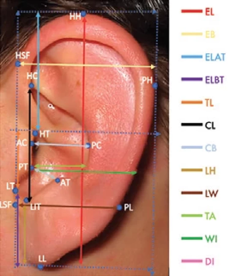
To establish anatomical landmarks for measurements, 14 points were defined, and a line between the highest point of the helix (HSF) and the lobule-skin-face intersection (LSF) was used as the orientation line for all other measurement lines, which were either parallel or at right angles. The measurements of the intertragic notch, width of the intertragic incisura (WI) and depth of the intertragic incisura (DI) were exceptions, and were perpendicular to each other (Fig. 2).

Statistical analyses
IBM SPSS® Statistics Version 20.0 (Armonk, NY, USA) was used for statistical analysis.
A P-value < 0.05 was considered to indicate statistical significance.
Results
The mean, standard deviation, and P-value of each measurement for the left ears of both genders are displayed in Tab. 1.
Our study found significant differences in the morphology of the left ear between males and females. Out of the 13 measurements taken, 10 were significantly larger in male participants than in female participants, with a confidence level of 99% (P < 0.01). These measurements included ear length, ear breadth, ear length above and below tragus, tragus length, concha length, lobule height, distance from tragus to antihelix and to helix, and width of the intertragic incisura. Therefore, our research clearly supports the existence of sexual dimorphism in ear morphology.
However, there were no significant differences in concha breadth and lobule width (P > 0.05) between male and female participants.
We also discovered that only the depth of the intertragic incisura was more significant in female subjects than in males (P < 0.01, confidence level 99%).


Discussion
The ear is a vital organ not only for hearing but also for aesthetics. Individuals with congenital malformations or acquired ear defects may face discrimination and cultural stigmas. The stigmatized person tends to seek anonymity and wishes to blend in with society by appearing “normal” [11–13]. The dissatisfaction with their own appearance and belief that others are staring or talking about their visible deformity results in decreased self-confidence, which, in turn, affects social life and leisure activities negatively [2]. However, successful reconstructive procedures extend benefits beyond aesthetics, exerting positive psychological and psychosocial effects. Patients report a noticeable boost in their self-confidence and an overall improvement in their social life [2]. Furthermore, a normal ear can provide the necessary support for glasses.
However, it should be remembered that for surgical reconstruction of the ear, surgeons must rely on anthropometric data, and it is known that measurements differ between genders as well as ethnicity. Each anthropological study used a different method to measure the ear, including measurements on the ear using a calliper, 3D-computerized digitizer or 3D-surface scan, and measurements on pictures taken with a camera in a computer program [1,6,14–17]. Direct measurements on the ear require many researchers and a significant amount of time, resulting in few variables being included in a study and a high margin of error among investigators. In recent decades, advanced 3D technologies have become popular. The MicroScribe digitizer captures landmarks with a stylus tip attached to a movable, articulated sensor arm to collect 2D linear measurement and 3D coordinate data [18,19]. It is faster than a conventional calliper, and deviations as small as 0.1 mm have been reported [20,21].
Another technology is a 3D scanner that emits lasers or visible light to scan surfaces. The data obtained can be imported into another software for accurate measurements [17,19]. However, both MicroScribe and 3D surface scanners are costly, with the latter ranging between $5,000 and $50,000 [22]. These costs are not suitable for Vietnam’s economy. Considering the limitations associated with the measurement techniques discussed earlier, we opted to utilize a digital camera to capture images for our measurements due to its numerous benefits. Even with many study participants, a single researcher efficiently conducts a large volume of measurements using computer software. This method results in precise data that are conveniently processed and stored on the computer. Last but not least, it is a cost-effective approach with significant practical applications.
Our data reveal sexual dimorphism with statistical significance (P < 0,01), as measured outcomes were greater in males for 12 of 13 variables. Notably, men’s ear length and width are larger, consistent with other research findings [5,7,10,14]. Interestingly, the depth of the intertragic incisura (DI) was greater in females by 0.21 mm, corresponding to 1/10 of the standard deviation (this value corresponds to 1/10 of the standard deviation).
Ethnic disparities in ear dimensions have been substantiated in prior research (Tab. 2) [4–10]. The results of our study indicate that Vietnamese ears are smaller than those of Caucasians, as well as Chinese and Japanese populations [4,9]. Cultural practices, such as ear pulling and stretching, prevalent in China and Japan, potentially account for the observed larger ear dimensions in these populations, as large ears are associated with good fortune, wealth, blessings, long life, and high social status. Compared to Ahmed’s data from Saudi Arabia, our findings show that the ear length of an Arab woman is longer than that of a Vietnamese woman, but the width is smaller [10]. The smaller width of Arab ears could be due to the tradition and culture of women wearing large and heavy ear jewelry. However, the ear length of Arab men is shorter than that of Vietnamese men [10]. Similarly, Indian ears exhibit smaller dimensions than Vietnamese ears in both genders [7].
Our research included a large sample of 2,000 subjects, but we only gathered data on the left ear, leading to questions about significant asymmetry. Previous anthropometric studies of the ear by Ahmed, Jappati, Murgod, and Purkait have all reported measurements of bilateral asymmetry [7,10,15,23]. While Ahmed and Murgod reported significant asymmetry, the data from Japatti and Purkait did not reach statistical significance [7,10,15,23]. On the other hand, Alexander’s research demonstrated symmetry, particularly in height and width [5]. Given these findings, it would be of great interest to conduct further research on the right ear to determine whether symmetry or asymmetry exists.
Conclusion
Our study provides comprehensive anthropometric data on the left ear of Vietnamese individuals aged 18–25 years. We used an established photographic analysis and found significant sexual dimorphism, with measurements being higher in males for 10 out of 13 variables. Furthermore, our findings highlight distinctive ear dimensions among Vietnamese individuals compared to other ethnic groups. However, since we only studied the left ear, further research should investigate the dimensions of the right ear and as assess symmetry between both sides.
Roles of authors
Nguyen Hong Ha – conception and design, analysis and interpretation of data, writing publication, critical revision of publication, approval of final publication, supervision, resources; Dang Thi Viet Ha – data acquisition, approval of final publication, technical, administrative, or material support; Thi Thanh Huyen – analysis and interpretation of data, writing publication, approval of final publication.
Disclosure
The authors have no conflicts of interest to disclose. The authors declare that this study has received no financial support. All procedures performed in this study involving human participants were in accordance with ethical standards of the institutional and/or national research committee and with the Helsinki declaration and its later amendments or comparable ethical standards.
Sources
1. Sforza C., Dellavia C., Tartaglia GM., et al. Morphometry of the ear in Down’s syndrome subjects. A three-dimensional computerised assessment. Int J Oral Maxillofac Surg. 2005, 34 (5): 480–486.
2. Horlock N., Vögelin E., Bradbury ET., et al. Psychosocial outcome of patients after ear reconstruction: a retrospective study of 62 patients. Ann Plast Surg. 2005, 54 (5): 517–524.
3. Abeysekera JDA., Shahnavaz H. Body size variability between people in developed and developing countries and its impact on the use of imported goods. Int J Indust Ergonom. 1989, 4 (2): 139–149.
4. Farkas LG. Anthropometry of the normal and defective ear. Clin Plast Surg. 1990, 17 (2): 213–221.
5. Alexander KS., Stott DJ., Sivakumar B., et al. A morphometric study of the human ear. J Plast Reconstr Aesthet Surg. 2011, 64 (1): 41–47.
6. Bozkir MG., Karakaş P., Yavuz M., et al. Morphometry of the external ear in our adult population. Aesthetic Plast Surg. 2006, 30 (1): 81–85.
7. Japatti SR., Engineer PJ., Reddy BM., et al. Anthropometric assessment of the normal adult human ear. Ann Maxillofac Surg. 2018, 8 (1): 42–50.
8. Niemitz C., Nibbrig M., Zacher V. Human ears grow throughout the entire lifetime according to complicated and sexually dimorphic patterns – conclusions from a cross-sectional analysis. Anthropol Anz. 2007, 65 (4): 391–413.
9. Zhao S., Li D., Liu Z., et al. Anthropometric growth study of the ear in a Chinese population. J Plast Reconstr Aesthet Surg. 2018, 71 (4): 518–523.
10. Ahmed AA., Omer N. Estimation of sex from the anthropometric ear measurements of a Sudanese population. Leg Med (Tokyo). 2015, 17 (5): 313–319.
11. Bradbury E. The psychology of aesthetic plastic surgery. Aesthetic Plast Surg. 1994, 18 (3): 301–305.
12. Macgregor FC. The place of the patient in society. Aesthetic Plast Surg. 1981, 5 (1): 19–26.
13. Richardson SA., Goodman N., Hastorf AH., et al. Cultural uniformity in reaction to physical disabilities. Am Sociol Rev. 1961, 26 (2): 241–247.
14. Brucker MJ., Patel J., Sullivan PK., et al. A morphometric study of the external ear: age - and sex-related differences. Plast Reconstr Surg. 2003, 112 (2): 647–652.
16. Prasetyo AT., Putri IL. Anthropometric study of human ear: a baseline data for ear reconstruction. J Craniofac Surg. 2022, 33 (4): 1245–1249.
17. Wang D., Jiang H., Pan B., et al. Standardised measurement of the auricle: a method of high-precision and reliability based on 3D scanning and Mimics software. Exp Ther Med. 2019, 18 (6): 4575–4582.
18. Seguchi N., Dudzik B., Murphy MM., et al. Chapter one – introduction. In: Seguchi N., Dudzik B., (eds). 3D data acquisition for bioarchaeology, forensic anthropology, and archaeology. Academic Press 2019 : 1–16.
19. Waltenberger L., Rebay-Salisbury K., Mitteroecker P. Three-dimensional surface scanning methods in osteology: a topographical and geometric morphometric comparison. Am J Phys Anthropol. 2021, 174 (4): 846–858.
20. Boldt F., Weinzierl C., Hertrich K., et al. Comparison of the spatial landmark scatter of various 3D digitalisation methods. J Orofac Orthop. 2009, 70 (3): 247–263.
21. Stephen AJ., Wegscheider PK., Nelson AJ., et al. Quantifying the precision and accuracy of the MicroScribe G2X three-dimensional digitiser. Dig Appl Archaeol Cultur Herit. 2015, 2 (1): 28–33.
22. Kuzminsky SC., Gardiner MS. Three-dimensional laser scanning: potential uses for museum conservation and scientific research. J Archaeolog Sci. 2012, 39 (8): 2744–2751.
23. Purkait R., Singh P. Anthropometry of the normal human auricle: a study of adult Indian men. Aesthet Plast Surg. 2007, 31 (4): 372–379.
Nguyen Hong Ha
Department of Maxillofacial, Plastic and Aesthetic Surgery
Viet-Duc University Hospital
40 Trang Thi, Hanoi, Vietnam
nhadr4@gmail.com
Submitted: 12.3.2025
Accepted: 25.10.2025
Labels
Plastic surgery Orthopaedics Burns medicine TraumatologyArticle was published in
Acta chirurgiae plasticae
2025 Issue 3
Most read in this issue
- Overall and area-specific tactile recovery following different methods of surgical reinnervation in post-mastectomy breast reconstruction – a systematic review and meta-analysis
- Brachymetacarpia – our experience with internal device for distraction osteogenesis in adolescent patients
- The importance of sentinel node biopsy and examination in malignant melanoma of the head and neck
- Case report of severe complications after gel injection breast augmentation – treatment and use of hemostatic net for effective management