-
Medical journals
- Career
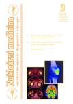
18F-FDG PET/CT pattern of Erdheim-Chester disease – a group of Czech patients
Authors: Zdeněk Řehák 1,2; Renata Koukalová 1; Jiří Vašina 1; Václav Ptáčník 3; Petr Szturz 4; Josef Karban 5; Jindřich Polívka 5; Zdeněk Adam 4
Authors‘ workplace: Oddělení nukleární medicíny, MOÚ Brno, ČR 1; Regionální centrum aplikované molekulární onkologie, MOÚ Brno, ČR 2; Ústav nukleární medicíny, 1. lékařské fakulty UK a Všeobecné fakultní nemocnice v Praze, ČR 3; Interní hematologická a onkologická klinika, LF MU a FN Brno, ČR 4; I. interní klinika – klinika hematologie, 1. lékařské fakulty UK a Všeobecné fakultní nemocnice v Praze, ČR 5
Published in: NuklMed 2018;7:50-52
Category: Original Article
Overview
Introduction:
Erdheim-Chester disease (ECD) is a rare unit of histiocytic diseases. The goal of our work was to assess 18F-FDG PET/CT presentation of this disease in patients from the Czech Republic.
Methods:
We analyzed overall 44 18F-FDG PET/CT examinations in 6 patients with this disease. We assessed 18F-FDG accumulation in staging examinations of these 6 patients at usual localizations, i.e. bones, brain, orbit, paranasal sinuses, periaortal space, heart, lungs, perirenal space and skin.
Results:
Bone 18F-FDG accumulation was detected in all patients; in 5 mostly in lower extremities. Maxillar sinuses were involved in 5/6 patients. Vascular and perirenal involvement was detected in 4/6 patients. Two patients had involved skin and hypophysis, one patient also orbits and heart. Lung involvement was not detected in any patient.
Conclusions:
18F-FDG avid involvement of skeleton was the main and regular characteristic of PET/CT presentation of Erdheim-Chester disease. Also other localizations of 18F-FDG avid involvement (cardiovascular, CNS, paranasal sinuses, orbitis, skin, perirenal space) confirm known observations in ECD.
Key Words:
18F-FDG, PET/CT, Erdheim-Chester disease
Sources
- Chester W. Uber Lipoidgranulomatose. Virchows Arch 1930;279 : 561–602
- Mazor RD, Manevich-Mazor M, Shoenfeld Y. Erdheim-Chester Disease: a comprehensive review of the literature. Orhanet Journal of Rare Diseases 2013;8 : 137 doi.org/10.1186/1750-1172-8-137
- Szturz P, Adam Z, Koukalová R et al. Erdheimova-Chesterova nemoc v obrazech. Vnitřní lékařství 2010;56(Suppl 2):2S170-178
- Arnaud L, Hervier B, Neel A et al. CNS involvement and treatment with interferon-alpha are independent prognostic factors in Erdheim-Chester disease: a multicenter survival analysis of 53 patients. Blood 2011;117 : 2778–2782
- Haroche J, Arnaud L, Amoura Z. Erdheim–Chester disease. Current opinion in rheumatology 2012;24 : 53-59
- Estrada-Veras JI, O’Brien KJ, Boyd LC et al. The clinical spectrum of Erdheim-Chester disease: an observational cohort study. Blood Advances 2017 : 1:357-366
- Ohara Y, Kato S, Yamashita D et al. An autopsy case report: Differences in radiological images correlate with histology in Erdheim–Chester disease. Pathology International 2018; 68 : 374–381
- Loeffler AG, Memoli VA. Myocardial involvement in Erdheim-Chester disease. Arch Pathol Lab Med 2004;128 : 682–685
- Berti A, Ferrarini M, Ferrero E et al. Cardiovascular manifestations of Erdheim-Chester disease. Clin Exp Rheumatol 2015;33(2 Suppl 89):155-163
- Haroche J, Cluzel P, Toledano D et al. Images in cardiovascular medicine. Cardiac involvement in Erdheim-Chester disease: magnetic resonance and computed tomographic scan imaging in a monocentric series of 37 patients. Circulation 2009;119:e597-e598
- Li N, Chen M, Sun H et al. Fever of unknown origin as the first manifestation of Erdheim-Chester disease. Case Reports in Clinical Medicine 2013;2 : 351-357
- Zanglis A, Valsamaki P, Fountos G. Erdheim-Chester disease: symmetric uptake in the (99 m)Tc-MDP bone scan. Hell J Nucl Med. 2008;11 : 164–167
- Pena Pardo FJ, Banzo Marraco I, Quirce Pisano R et al. Bone scintigraphy in Erdheim-Chester disease. Rev Esp Med Nucl. 2003;22 : 253–256
- García-Gómez FJ, Acevedo-Báñez I, Martínez-Castillo R et al. The role of 18FDG, 18FDOPA PET/CT and 99mTc bone scintigraphy imaging in Erdheim–Chester disease. European Journal of Radiology 2015;84 : 1586-1592
- Balink H, Hemmelder MH, de Graaf W et al. J. Scintigraphic diagnosis of Erdheim-Chester disease. Journal of Clinical Oncology 2011;29:e470-e472
- Caoduro C, Ungureanu CM, Rudenko B et al. 18F-fluoride PET/CT aspect of an unusual case of Erdheim-Chester disease with histologic features of Langerhans cell histiocytosis. Clinical Nuclear Medicine 2013; 38 : 541-542
- Sabino D, Vale RHBD, Duarte PS et al. Complementary findings on 18F-FDG PET/CT and 18F-NaF PET/CT in a patient with Erdheim-Chester disease. Radiologia Brasileira 2017; Epub May 18, 2017, doi.org/10.1590/0100-3984.2015.0172
- Arora A, Sharma S, Pushker N et al. Unusual orbital involvement in Erdheim Chester disease: a radiological diagnosis. Orbit 2012;31 : 338-340
- Karcioglu ZA, Sharara N, Boles TL et al. Orbital xanthogranuloma: clinical and morphologic features in eight patients. Ophthal Plast Reconstr Surg 2003;19 : 372–381
- Beylergil V, Carrasquillo JA, Hyman DM et al. Visualization of orbital involvement of Erdheim-Chester disease on PET/CT. Clinical Nuclear Medicine 2014;39 : 660-661
- Adam Z, Balšíková K, Pour L et al. Diabetes insipidus, následovaný po 4 letech dysartrií a lehkou pravostrannou hemiparézou – první klinické příznaky Erdheimovy-Chesterovy nemoci. Popis a zobrazení případu s přehledem informací o této nemoci.Vnitř Lek 2009;55 : 1173–1188
- Drier A, Haroche J, Savatovsky J et al. Cerebral, facial, and orbital involvement in Erdheim-Chester disease: CT and MR imaging findings. Radiology. 2010;255 : 586-594
- Arnaud L, Malek Z, Archambaud F et al. 18F-fluorodeoxyglucose-positron emission tomography scanning is more useful in follow up than in the initial assessment of patiens with Erdheim-Chester disease. Arthritis Rheum 2009; 60 : 3128–3138
- Lachenal F, Cotton F, Desmurs-Clavel H et al. Neurological manifestations and neuroradiological presentation of Erdheim-Chester disease: report of 6 cases and systematic review of the literature. J Neurol 2006; 253 : 1267–1277
- Sioka C, Estrada-Veras J, Maric I et al. FDG PET images in a patient with Erdheim-Chester disease. Clinical Nuclear Medicine 2014;39: doi: 10.1097/RLU.0b013e31828da5e6
- Byeon J, Kim KA, Hwang SS et al. Erdheim-Chester Disease with Perirenal Masses Containing Macroscopic Fat Tissue. Journal of the Korean Society of Radiology 2015;72 : 143-146
- Gupta A, Aman K, Al-Babtain M et al. Multisystem Erdheim‐Chester disease; a unique presentation with liver and axial skeletal involvement. British Journal of Hematology 2007;138 : 280-280
- Fudim M, Thorpe MP, Chang LL et al. Cardiovascular Imaging With 18F-Fluorodeoxyglucose Positron Emission Tomography/Computed Tomography in Patients With Fibroinflammatory Disorders. JACC: Cardiovascular Imaging 2018;11 : 365-368
- Antunes C, Graça B and Donato P. Thoracic, abdominal and musculoskeletal involvement in Erdheim-Chester disease: CT, MR and PET imaging findings. Insights into imaging 2014;5 : 473-482
- Šteňová E, Šteňo B, Povinec P et al. FDG-PET in the Erdheim–Chester disease: its diagnostic and follow-up role. Rheumatology International, 2012;32 : 675-678
- Adam Z, Petrášová H, Řehák Z et al. Hodnocení pětileté léčby Erdheimovy-Chesterovy nemoci anakinrou – kazuistika a přehled literatury. Vnitřní lékařství 2016; 62 : 44-56
- Janku F, Amin HM, Yang D et al. Response of histiocytoses to imatinib mesylate: fire to ashes. Journal of Clinical Oncology 2010;28,e633-e636
Labels
Nuclear medicine Radiodiagnostics Radiotherapy
Article was published inNuclear Medicine

2018 Issue 3
Most read in this issue- The benefit of SPECT/CT in the diagnosis of osteochondral lesions of knee joints
- 18F-FDG PET/CT pattern of Erdheim-Chester disease – a group of Czech patients
- LYMFOSCINTIGRAFIE provedení vyšetření a jeho interpretace
- 55. DNY NUKLEÁRNÍ MEDICÍNY
Login#ADS_BOTTOM_SCRIPTS#Forgotten passwordEnter the email address that you registered with. We will send you instructions on how to set a new password.
- Career

