
Pupillary abnormalities in childhood – 2 case presentations
Authors:
P. Žiak 1,2; K. Kapitánová 2,1; J. Halička 1,2
Authors‘ workplace:
Očná klinika JLF UK a UN Martin (prednosta kliniky MUDr. Žiak Peter, PhD. )
1; UVEA Mediklinik, s. r. o. Martin- Priekopa (medicínsky riaditeľ MUDr. Žiak Peter, PhD. )
2
Published in:
Čes. a slov. Oftal., 75, 2019, No. 3, p. 145-149
Category:
Case Report
doi:
https://doi.org/10.31348/2019/3/5
Overview
Anisocoria is a condition characterized by an unequal size of the eyes' pupils. There is a broad spectrum of aetiological factors including benign and also life - threatening situations. The most important point is the ability to find the anatomical location of the pathology. Understanding to the anatomical, physiological and pharmacological influences helps us to solve the diagnostic challenge.
In the article authors present the issue of the anisocoria in the childhood through two case presentations. Causes of an unequal size of the eyes' pupils are in this two cases different. In both of the cases the anisocoria is temporary with spontaneous resolution without any therapy. Diagnostic challenge is well - described with the analysis of important Pilocarpin pharmacological tests.
Pharmacological diagnostic tests are a very effective method to differentiate between the pharmacological mydriasis and the mydriasis caused by another factor. Strictly taken patient´s history with targeted questions searching for recent contact with some drugs, plants or medications is crucial. Sometimes it is necessary to consider an unusual diagnosis – benign episodic unilateral mydriasis.
Keywords:
anisocoria – datura suaveolens – benign episodic unilateral mydriasis – Pilocarpin
INTRODUCTION
The pupil is an opening located as a rule in the centre of the iris, the width of which is determined by a mutual balance between parasympathetic and sympathetic innervation (12). In up to 20% of the population we may encounter physiological anisocoria greater than 0.5 mm (16). Pupil width fluctuates within a diameter from 1 to 12 mm, depending on light intensity, as well as accommodation (5). Upon near gaze there is concurrent accommodation, convergence and miosis, known as “synkinesis” (4). The iris muscles are innervated reciprocally, and so both pupils dilate and contract symmetrically under normal circumstances (5). The afferent part of the pupilomotor reflex is partially crossed in the optic chiasm, and subsequently before the corpus geniculatum laterale its fibres separate from the sensoric fibres of the visual pathway and end in the nuclei pretectales. From these the Edinger-Westphal nucleus is stimulated ipsilaterally and contralaterally. This dual crossing ensures the reaction of both pupils even upon exposure of only one eye to light (16). The efferent part of the reflex is ensured by the autonomous nervous system. The parasympathetic pathway begins in the Edinger-Westphal nucleus, in the posterior diencephalon, and is a dual neuron pathway. After leaving the diencephalon it proceeds as a part of the oculomotor nerve, in which its fibres are arranged on the surface. As a result, a disorder of its function is quickly manifested e.g. in compressive lesions in this region (aneurysm, tumour etc.) (5). The first neuron of the pathway is directed into the ciliary ganglion, from where the second neuron continues through the short ciliary nerves breves into the sphincter of the pupil (16). The sympathetic pathway is 3 neuron, beginning in the hypothalamus (5). From here it heads into the Budge's ciliospinal centre in the spinal cord (C8-Th2), from where the second neuron, via the path of the truncus sympaticus, innervates the upper cervical ganglion. From here the fibres of the third neuron ensure innervation of the pupil dilator via the ciliary ganglion and short ciliary nerves (16). If the oculomotor nerve, in addition to the extraocular muscles, also innervates the elevator muscle of the upper eyelid, ciliary muscle and sphincter of the iris, the affliction of this nerve may have various manifestations. Complete paresis of the third cranial nerve is manifested in total ptosis, the eyeball is distorted laterally and sometimes slightly downward, and motility is possible only with the scope of the unaffected extraocular muscles, with a general result of diplopia. Paresis manifested simultaneously with affliction of the pupil is an acute condition in neuroophthalmology. It typically concerns paresis caused by compression of the third cranial nerve by aneurysm, which if ruptured with subsequent subarachnoid haemorrhage may be a fatal complication. Disorder of the function of the pupillary sphincter is manifested in retarded reactivity of the pupil or its dilation (6). In children (with the exception of newborn age) pupils are wider, and progressively narrow with age. The pupil is constricted in sleep, under general anaesthesia, in a coma, we may observe “pinpoint pupils” in patients with damage to the pons. Of pharmacological causes, miosis may be caused for example by pilocarpine drops, as well as opiates and cholinesterase inhibitors. A dilated pupil accompanies states of excitement, epileptic fit, damage to the mesencephalon or terminal pre-mortem stages. Of pharmaceuticals it is triggered by the administration of parasympatholytic agents (antispasmodics), catecholamines, tricyclic antidepressants, anti-Parkinson's drugs (16), sympathomimetic drugs such as ephedrine, epinephrine, also in the eye drops phenylephrine, ocular decongestants (15), atropine and scopolamine. Among habit-forming substances triggering mydriasis are heroin, cocaine, hallucinogens and amphetamine derivatives (16). In patients following local administration of mydriasis-inducing drops into both eyes, the reaction may be asymmetrical in both eyes, and may therefore also lead to anisocoria (15). In the case of orbital tumours, in addition to axial or non-axial exophthalmos, decrease of visual acuity or changes in the perimeter, we also encounter disorders of direct and indirect reaction of the pupil to light exposure. The reason is damage to the corresponding reflex curve on various levels in the orbit (20). Compressive lesions in the orbit afflict the dominant afferent part of the pupillomotor reflex, which is manifested in retarded direct or indirect reaction of the pupils to light exposure. However, this one is symmetrical. An example may be meningiomas of the orbit, which usually cause especially slowly progressing neuropathy of the optics with a decrease of visual acuity, constriction of the visual field and disorders of colour sensitivity (8). By contrast, the presence of anisocoria attests to damage to the efferent part of the reflex arc. The cause may be secondary changes e.g. following surgical treatment of intraocular or orbital tumours, or following their irradiation (9, 10, 11).
Disorders of the afferent part of the pupillary reflex may be manifested as a relative afferent pupillary defect (RAPD), or as an absolute afferent pupillary defect. RAPD originates in the case of an incomplete lesion of the visual pathway before the optical chiasm, and is examined by alternating illumination of one and the other pupil. An absolute defect is a manifestation of a full lesion of the optic nerve, in which the pupils are of equal width, but upon light exposure on the side of the affected eye no direct or indirect photo-reaction is evoked (4). In the case of damage in the retrochiasmal part of the pathway, photo-reactions are weakened bilaterally – this concerns a case of so-called “stiff pupil” (16). Disorders of the efferent part of the pupillomotor reflex lead to a change of pupil size and the onset of anisocoria. The side with the wider pupil is more frequently the affected side. In the case of an isolated lesion of parasympathetic innervation, anisocoria is more pronounced in light conditions, and there is also a disorder of accommodation. Upon affliction of sympathetic innervation, anisocoria is more conspicuous in dark conditions – the affected pupil remains constricted, in which slight ptosis or reduced perspiration on the forehead may simultaneously be present. In general it applies that the pupil in which changes of width in light and dark conditions are smaller is the affected pupil (16). Unequal pupil width may also be due to mechanical causes – posterior synechia, iris sphincter tears, or postoperative of post-traumatic changes. It is necessary also to remember physiological anisocoria – this is the most common cause of asymmetry of pupil size (and is present in 15-30% of the population). This should be long-term, isolated, in which the difference in pupil size does not exceed 1 mm, and the asymmetry between the pupils does not become more pronounced either in light or dark conditions (12).
CASE REPORT 1
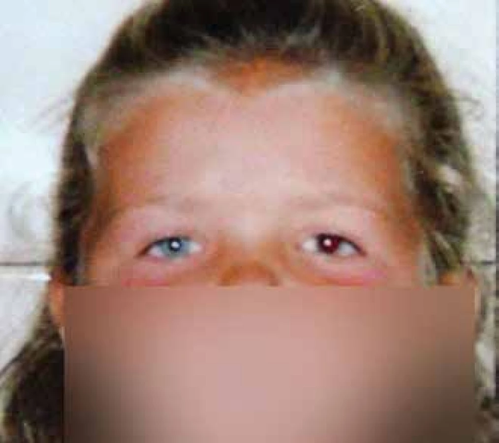
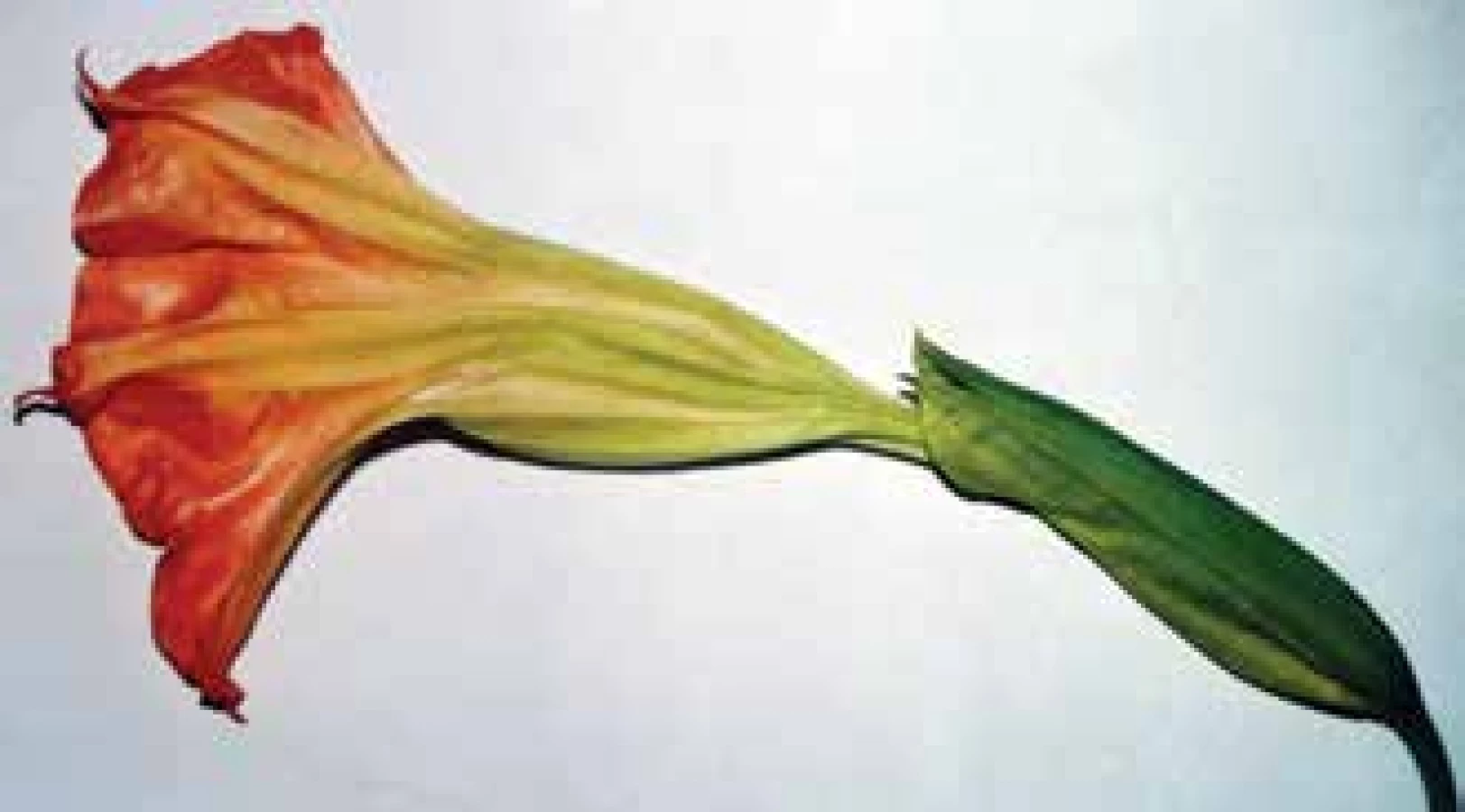
During emergency service, advice of an ophthalmologist was requested in the case of a 6-year-old patient accompanied by her mother due to anisocoria in the sense of an extended pupil of the left eye (Fig. 1). The child was examined by a paediatrician and neurologist, with a negative finding. An examination of the brain by computer tomography indicated by the paediatrician produced a negative result. In the patient's medical history the mother stated that after the child had returned from playing in a garden where children were cycling, she noticed a pronounced dilation of the pupil of the left eye. The mother unequivocally negated trauma (fall from bicycle). According to her statement, the child was slightly agitated, with flushed cheeks. The mother negated other subjective symptoms of the character of double vision, general complaints (nausea, vomiting, dizziness). The child had not come into contact with pharmaceuticals or eye drops. Upon examination, best corrected central visual acuity (BCCVA) in the right eye was 6/6, in the left eye 6/9 with a mydriatic pupil. Pupil width was 3 mm in the right eye, 9 mm in the left eye, in which direct and consensual photo-reaction was present in the right eye and not in the left eye. The other finding in the anterior segment of the eye, as well as on the retina, was bilaterally within the norm. A pharmacological test with the application of 0.1% Pilocarpine (1 ml 1% Pilocarpine diluted in 9 ml of physiological solution) in the left eye was negative – there was no constriction of the pupil, which therefore excluded cholinergic hypersensitivity as the cause of anisocoria. Weak Pilocarpine solution has virtually no influence on the width of a normal pupil, but upon heightened sensitivity of the pupillary sphincter (typical in the case of “pupillotonia”) to cholinergic stimulation, the pupil constricts under its influence (2). After administration of 1.0% Pilocarpine in the left eye, there was also no constriction of the pupil. A test with 1.0 Pilocarpine differentiates a neurogenic cause of mydriasis from mydriasis caused by a cholinergic antagonist. If the muscarinic receptors on the affected side are occupied by an anti-cholinergic agent, 1% Pilocarpine leads to weak or no miosis. By contrast, in the case of neurogenic causes of mydriasis, clear constriction of the pupil takes place upon its application (2). In this case therefore the pharmacological test confirmed mydriasis induced by an anti-cholinergic agent. After a number of targeted questions (what was the child doing, where was it playing, with what did she come into contact, for example plants?) we determined contact with the plant Datura suaveolens, known as “Brazil's white angel's trumpet” (Fig. 2). The child broke off and held in her hand a goblet shaped flower of this plant. The medical history of contact with Datura suaveolens, which contains alkaloids such as L-hyoscyamine, atropine and above all scopolamine, therefore corresponded with the objective finding in the child – juice from the flower probably came into contact with only one eye. The child's agitation and reddening of the cheeks could also have been a symptom of mild general intoxication. Spontaneous correction ad integrum took place without therapy within 24 hours (Fig. 3).

CASE REPORT 2
An ocular consultation examination was requested for a 16-year-old female patient hospitalised with a medical history of aniscoria persisting for one day – mydriatic left pupil (Fig. 4). Since the onset of anisocoria the patient had also subjectively perceived a deterioration especially of near vision in the left eye, and had also experienced increased perspiration in the armpits for approximately 2 weeks. Other than these subjective complaints, the patient negated other symptoms, ocular or otherwise. She did not state any headache, manifestations of spasmophilia or diplopia. The neurological finding was within the norm. Upon an examination of BCCVA for distance vision the patient recorded 6/6 bilaterally, near visual acuity according to Jaeger charts in the right eye 1.0 and in the left eye 0.63 (with addition of +2.0 Ds 1.0). Pupil width is 4-5 mm in the right eye, 7 mm in the left eye, in which direct and consensual photo-reaction is present in the right eye, in the left eye slightly retarded. In dark conditions both pupils dilated, and the asymmetry between the pupils therefore did not essentially change. In this point the pupils behaved like physiological anisocoria (Fig. 5). The active motility of both eyes is correct in all directions. The other finding on the anterior segment of the eye, as well as on the retina, is bilaterally within the norm. As in the case of the first patient, a pharmacological test with administration of 0.1% Pilocarpine in the left eye was negative – no constriction of the pupil occurred (Fig. 6). After application of 1.0% Pilocarpine to the left eye the pupil constricted to 3-4 mm, thereby excluding pharmacological mydriasis generated by an anti-cholinergic agent (Fig. 7). The objective ocular finding corresponded to neurogenic damage to the parasympathetic part of the left-sided oculomotor nerve, therefore ophthalmoplegia interna comes into consideration. Additional MRI of the brain focusing on the left-sided oculomotor nerve and MRI angiography of the brain capillaries were without evident structural changes. With regard to the negative MRI finding, as well as the fact that spontaneous correction of the width of the left pupil as well as near vision occurred in the patient within less than 24 hours (Fig. 8), we concluded this episode of anisocoria as benign episodic unilateral mydriasis syndrome (BEUM).


DISCUSSION
In patients with anisocoria, at the beginning of the examination we always attempt to exclude mechanical causes of unequal pupil width, as well as physiological anisocoria – in this respect older photographs of the patient may be of use. In addition it is necessary to determine which pupil is not physiological. It applies that if anisocoria is more pronounced under light conditions, the wider pupil is abnormal. If the difference in pupil size is more significant in dark conditions, the narrower pupil is abnormal. We also take into account the speed of pupil reactions, or the relative afferent pupillary defect, presence of ptosis or disorders of motility of the eyeballs (12).
Within the wide spectrum of causes of anisocoria, in the majority of patients with isolated unilateral mydriasis this fortunately concerns a benign condition, for example pupillotonia, or a condition following local or general administration of mydriasis-inducing preparations (14). These substances also include alkaloids contained in certain plants, especially Datura stramonium, Atropa belladonna or Hyoscyamus niger. These are highly toxic plants, capable of causing fatal poisoning, especially in children (3). Slight intoxication is manifested in unilateral or bilateral mydriasis (13), after ingestion of infusion or leach from the plant there is a rapid onset of general manifestations. Patients are generally disoriented, suffer from hallucinations or succumb to lethargy (18). Also typical is general agitation, irritation to aggression. Patients also suffer from an increase in body temperature, dryness of the mucous membranes, tachycardia and an increase in systemic blood pressure (1). Pupils are mydriatic, with weakened or absent reaction to light exposure (18).
In the case of benign episodic unilateral mydriasis (BEUM), the triggering mechanism is not precisely known. As a rule this concerns a symptom of a short duration, the identification and objective evaluation of which is complex upon examination (14). According to the available literature, the temporary asymmetry of pupil width may be caused by hyperactivity of the sympathetic component of innervation, or by contrast hypoactivity of the parasympathetic nervous system contributing to the innervation of the pupil. At the same time, there is an assumed relationship between episodes of BEUM and migraine (19), known as ophthalmoplegic migraine (7). Temporary unilateral mydriasis may be a certain form of migraine aura, in which the progression of the pathology itself may be preceded by episodes of change in pupil width for as long as a number of years. Imprecisely defined temporary blurred vision is described relatively frequently in patients with migraine. At the same time, BEUM may be an overlooked symptom inasmuch as the condition is linked only with mild visual complaints in the patient (17).
Within the framework of differential diagnostics, it is also appropriate to consider spastic mydriasis as a source of temporary complaints. However, in child patients this is a less probable cause, since we encounter irritating lesions of the cervical sympathetic nervous system relatively rarely at this age.
CONCLUSION
Anisocoria is defined as unequal pupil width, in which there is a wide spectrum of triggering causes. Acquired and acute anisocoria in particular may be a serious manifestation of certain life threatening conditions. Determining the anatomic cause of origin is of key importance in differential diagnostics. An understanding of the anatomical, physiological and pharmacological factors influencing the pupillomotor system is essential (2). It is also important to adopt a systematic approach in differential diagnosis of the causes of origin of unequal pupil width. Physiological mydriasis and mechanical causes influencing pupil width must be excluded at the start of the examination. After initial determination of which pupil is pathological, pharmacological diagnostic tests serve as an effective method for eliminating medicamentously-induced anisocoria (21). A thoroughly taken anamnesis, frequently with targeted questions concerning contact with certain pharmaceuticals or plants in the preceding period is an indispensible component of the examination of the patient (15). Auto photo-documentation of the patient which is ordinarily available today (“selfie” photographs from mobile telephones) may be very useful in the diagnosis especially of transitional anisocoria, primarily in adolescent and young adult patients, in which this situation occurs relatively often. In general, in the case of isolated unilateral short-term mydriasis without further ocular or general manifestations, in which we are able to exclude an organic cause of origin by means of imaging methods and the pharmacological test is positive, it is necessary also to consider a rare nosological unit – that of BEUM.
The authors of the study declare that no conflict of interest exists in the compilation, theme and subsequent publication of this professional communication, and that it is not supported by any pharmaceuticals company.
Korešpondujúcí autor:
MUDr. Karolína Kapitánová,
Očná nemocnica UVEA MEDIKLINIK
s.r.o., Zelená 1, 036 01 Martin - Priekopa
Received: 19. 1. 2019
Accepted: 4. 4. 2019
Available on-line: 19. 10. 2019
Sources
1. Adegoke, SA., Alo, LA.: Datura stramonium poisoning in children. Niger J Clin Pract, 16; 2013 : 116-8.
2. Antonio-Santos, AA., Santos, RN., Eggenberger, ER.: Pharmacological testing of anisocoria. Expert Opin Pharmacother, 6/12; 2005 : 2007-20.
3. Bednářová, B.: Herbár, alebo od alchemilky po žindavu. Bratislava: Fortuna Libri, 2017. ISBN: 978-8-081-42670-4.
4. Bowling, B. et al.: Kanski's clinical ophthalmology, 8th edition. A systematic approach. Philadelphia: Elsevier - Saunders Ltd., 2015. ISBN: 978-0-702-05572-0.
5. Caglayan Batur, HZ., Colpak, IA., Kansu, T.: A diagnostic challenge: dilated pupil. Curr Opin Ophthalmol, 24; 2013 : 550–557.
6. Doran, M., Karmel, M., Stuart, A. et al.: 4 Neuro conditions not to be missed. American Academy of Ophthalmology, 2012: dostupné na: https://www.aao.org/eyenet/article/4-neuro-conditions-not-to-be-missed.
7. Edelson, RN., Levy, DE.: Unilateral pupillary dilation in young adults. Arch Neurol, 31; 1974 : 12-14.
8. Furdová, A., Babál, P., Kobzová, D.: Orbital optic nerve sheat meningioma. Cesk Slov Oftal, 1/74; 2018 : 23-30.
9. Furdová, A., Horkovičová, K., Justusová, P. et al.: Is it sufficient to repeat LINEAR accelerator stereotactic radiosurgery in choroidal melanoma? Bratisl Med J, 117; 2016 : 456–62.
10. Furdová A., Ružička J., Šramka, M. a kol.: Choroidálny melanóm v štádiu T1 – porovnanie plánu ožiarenia pri stereotaktickej rádiochirurgii a pri protónovej terapii. Cesk Slov Oftal, 4/68; 2012 : 156–161.
11. Furdová, A., Šramka, M., Chorváth, M. et al.: Clinical experience of stereotactic radiosurgery at a linear accelerator for intraocular melanoma: Melanoma Res, 27; 2017 : 463–8.
12. Gross, JR., McClelland, CM., Lee, MS.: An approach to anisocoria. Curr Opin Ophthalmol, 27; 2016 : 486–492.
13. Havelius, U., Asman, P.: Accidental mydriasis from exposure to Angel’s trumpet (Datura suaveolens). Acta Ophthalmol Scand, 80; 2002 : 332–335.
14. Jacobson, DM.: Benign episodic unilateral mydriasis - clinical characteristics. Ophthalmology, 102; 1995 : 1623-1627.
15. Kawasaki, AK.: Diagnostic approach to pupillary abnormalities. Continuum (Minneap Minn), 20/4; 2014 : 1008–1022.
16. Kuchynka, P. a kol.: Oční lékařství, 2., přepracované a doplněné vydání. Praha: Grada, 2016. ISBN: 978-8-024-75079-8.
17. Maggioni, F., Mainardi, F., Malvindi, ML. et al.: The borderland of migraine with aura: episodic unilateral mydriasis. J Headache Pain, 12; 2011 : 105–107.
18. Rakotomavo, F., Andriamasy, C., Rasamoelina, N. et al.: Datura stramonium intoxication in two children. Pediatr Int, 56; 2014 : 14–16.
19. Skeik, N., Jabr, FI.: Migraine with benign episodic unilateral mydriasis. Int J Gen Med, 4; 2011 : 501–503.
20. Vachata, P., Zikmund, L., Kozák, J. et al.: Orbital Tumors. Cesk Slov Neurol N, 78/111(6); 2015 : 617-638.
21. Yeo, DCM., Wijetilleka, S., Sharma, B. et al.: Benign episodic unilateral mydriasis in children: presentation and features in 2 young siblings. Can J Ophthalmol, 2018: v tlači.
Labels
OphthalmologyArticle was published in
Czech and Slovak Ophthalmology

2019 Issue 3
Most read in this issue
- Pupillary abnormalities in childhood – 2 case presentations
- Influence of cornea on intraocular pressure measurement by ICARE PRO and ORA
- Methods of improving the quality of life in patients with stable maculopathy-pilot results of a new study
- Ranibizumab for the treatment of choroidal neovascularization due to cause other than age related macular degeneration