
Dvoufázová tromboembolektomie u akutní viscerální ischemie s atypickým odstupem pravé jaterní tepny, komplikovaná sekundárním střevním infiltrátem
Authors:
M. Škrabal 1,2; J. Chlupáč 1,3; Š. Malý 1; J. Froněk 1,3,4
Authors‘ workplace:
Transplantation Surgery Department, Institute for Clinical and Experimental Medicine, Prague, Czech Republic
1; Surgical Department, Kolín Regional Hospital, a. s., Hospital of the Central Bohemian Region, Czech Republic
2; Department of Anatomy, Second Faculty of Medicine, Charles University, Prague, Czech Republic
3; First Surgical Clinic, First Faculty of Medicine, Charles University, Prague, Czech Republic
4
Published in:
Rozhl. Chir., 2026, roč. 105, č. 6, s. 286-293.
Category:
Case Report
doi:
https://doi.org/10.48095/ccrvch2026286
Overview
Úvod: Akutní mezenteriální ischemie je vzácná, ale závažná cévní komplikace, která je často spojena s poškozením střev. Léčba obvykle zahrnuje kombinaci chirurgických a endovaskulárních zákroků. Přesná diagnostika a včasná intervence jsou klíčové pro prevenci závažných následků, jako je syndrom krátkého střeva, sepse nebo nutnost opakovaných operací. Na základě kazuistiky naše publikace podává odborný přehled o léčbě akutní mezenteriální ischemie s postižením střev se zaměřením na zavedené i moderní terapeutické postupy.
Kazuistika: Prezentujeme případ akutní viscerální ischemie u 57leté pacientky s tyreotoxikózou, způsobené subtotální obstrukcí a. mesenterica superior s infarktem tenkého střeva. Po úspěšné chirurgické rekanalizaci tepny a střevní resekci se navzdory hmatným pulzacím v jaterní krajině objevil nový uzávěr akcesorní pravé jaterní tepny. Následovala druhá revize s obnovou průtoku. Během sledování došlo k rekurenci abdominálních potíží vyžadující ileocekální resekci pro pozdní ischemické změny.
Závěr: Příčiny viscerální ischemie jsou mnohé. Účinná diagnostika a léčba vyžadují interdisciplinární přístup, který zahrnuje otevřené, endovaskulární nebo hybridní techniky, často v kombinaci s resekcí střeva.
Klíčová slova:
mezenteriální ischemie – zobrazovací metody – akcesorní jaterní tepna – střevní resekce – thyreotoxikoza
Introduction
Mesenteric ischemia is a rare but serious vascular condition characterized by acute reduction or occlusion of blood flow to the visceral arteries. It often leads to intestinal and organ ischemia, which can progress to secondary inflammation and sepsis.
The estimated incidence is 6–9 cases per 100,000 people per year, with approximately one case of acute mesenteric ischemia (AMI) occurring in every 2,000 hospital admissions [1,2].
Risk increases with age. If left untreated, AMI carries a mortality rate of over 70%. Common causes of perioperative death include persistent mesenteric ischemia with intra-abdominal sepsis and multi-organ failure, as well as cardiac events or hemorrhage. This acute abdominal emergency often presents with severe abdominal pain that is disproportionate to physical findings and is unresponsive to analgesics [3,4].
The superior mesenteric artery (SMA) is the primary blood supply to the small intestines. It receives collateral flow from the celiac arterial system, via the superior and inferior pancreaticoduodenal arteries, and from the inferior mesenteric artery. Acute visceral ischemia refers to the sudden interruption of blood flow to the abdominal organs. It can result from:
- arterial embolism;
- arterial thrombosis;
- non-occlusive mesenteric ischemia (NOMI);
- mesenteric vein thrombosis.
Etiologically, embolism accounts for 40–50% of cases, arterial thrombosis is 25–30%, NOMI is 20%, and mesenteric vein thrombosis is 5–15% [5]. Embolism results in abrupt occlusion. Mesenteric emboli often originate in the left atrium or ventricle due to atrial fibrillation or ventricular dysfunction by infected cardiac valves (endocarditis) or occasionally from an atherosclerotic aorta. Risk factors include arrhythmias, valvular disease, endocarditis, aortic pathology – aneurysms or atherosclerosis, recent myocardial infarction, ventricular aneurysm, cardiac surgery, and cardiogenic shock [6].
Arterial thrombosis typically involves plaque-induced stenosis at the origin of the SMA. Arterial thrombosis stems from stagnant flow, hypercoagulability due to Factor V Leiden, prothrombin mutation, protein C and S, antithrombin deficiency antiphospholipid syndrome, or from vascular inflammation (mycotic aneurysm). Rare causes of acute visceral ischemia are vasculitis, arterial dissection, fibromuscular dysplasia, retroperitoneal fibrosis, or trauma [7]. NOMI is linked to hypoperfusion states – dehydration, cardiac failure, sepsis, or may be drug-induced [8]. Mesenteric venous thrombosis is associated with prior surgery, pancreatitis, thrombophilia, malignancy, and oral contraceptive use [7].
Mesenteric circulation has high resistance, low oxygen extraction, and limited autoregulation, making it vulnerable to ischemia despite receiving up to 35% of the cardiac output [9]. Collaterals develop in the presence of more than 70% of stenoses. The SMA is involved in 85% of cases [10,11].
The aim of this study is to reveal a surgical management to acute visceral ischemia burdened by atypical hepatic artery branching with subsequent formation of thrombosis. Also, aligned with contemporary medical approaches, we want to draw attention to the upcoming diagnostic tools facilitating the assessment of such an acute disorder.
Case report
A 57-year-old woman was transferred to IKEM vascular and transplant center with worsening abdominal pain, following an initial surgical evaluation. Gastrointestinal symptoms had appeared 5 days before the overall deterioration that ultimately led the patient to the emergency department.
Early in the course, the patient reported non-specific complaints of weakness, abdominal discomfort, diarrhea, and subsequently vomiting. Abdominal CT performed at the referring hospital incidentally revealed thrombosis of the superior mesenteric artery (Fig. 1, 2) prompting transfer to our department, after a previous negative ultrasound and X-ray examinations.
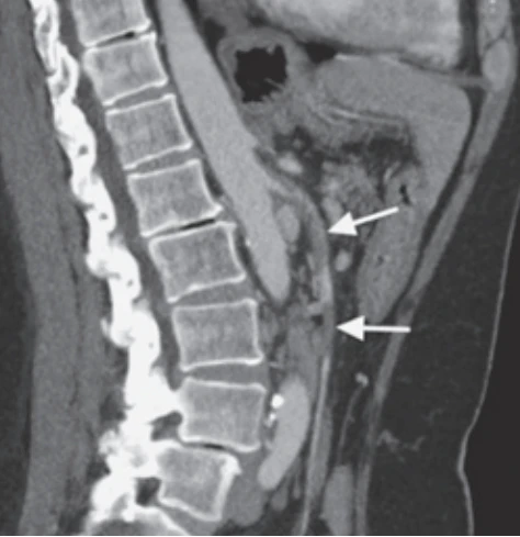
Defekt v náplni a. mesenterica superior s přerušením pasáže kontrastní látky (bílá šipka).
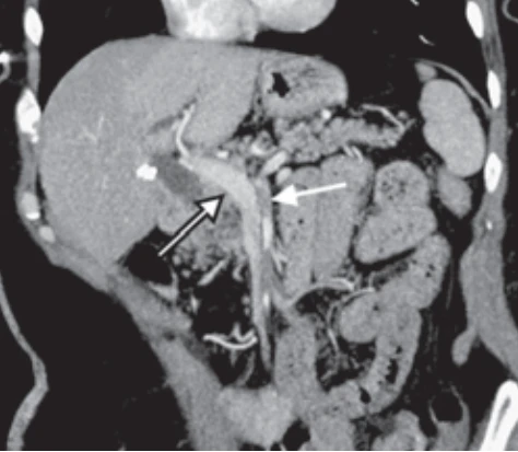
interrupted passage of contrast material (white arrow); portal
vein (black-bordered arrow).
Defekt v náplni a. mesenterica superior s přerušením pasáže
kontrastní látky (bílá šipka); portální žíla (černě ohraničená
šipka).
Upon admission to our center, 2 days after brief improvement, the patient was tachycardic and hypertensive. Abdominal examination revealed a soft abdomen without signs of peritoneal irritation or localized tenderness. The CT scan also uncovered choledocholithiasis (Fig. 3) with dilated bile ducts, likely a post-cholecystectomy complication, and a suspected occlusion of the right hepatic artery.
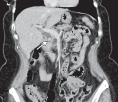
Konkrement v ductus choledochus velikosti 1 cm.
Arterial hypertension, iron-deficiency anemia and untreated thyroid disease with no medication for the last 3 years were the records in the patient’s medical history. She had undergone laparoscopic cholecystectomy years ago. CT imaging showed a subtotal occlusion of the SMA with a significant filling defect (Fig. 4), directing us toward urgent surgical exploration. Although the bowel loops appeared non-distended and adequately perfused on imaging, intestinal ischemia could not be definitively ruled out.
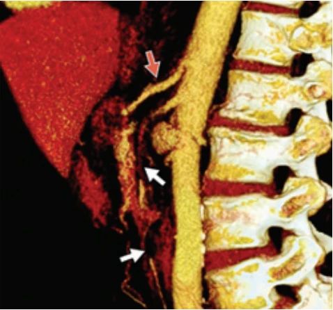
Defekt v náplni AMS v délce 9 cm s téměř subtotální okluzí lumen AMS (bílé šipky). Intaktní truncus coeliacus (červená šipka).
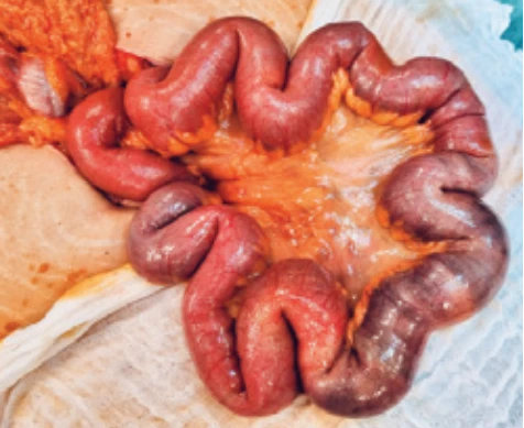
Tenké střevo a ischemické ileum v délce 25 cm.
In addition, the CT scan showed absence of a right hepatic artery – leading us to suspect atypical origin from SMA and thrombosis from the same region. Given the patient’s escalating abdominal pain, tachycardia, rising inflammatory markers, and findings suggestive of early intestinal ischemia at this stage, we proceeded with exploratory laparotomy.
Upon exposing the ileum, we identified two short, early ischemic segments, livid and malodorous, measuring 25 cm in total, located 20–45 cm from the Bauhin’s valve (Fig. 5). The colon was unaffected. The SMA was exposed peripherally from the pancreas, including proximal branches, and dissected over 5 cm (Fig. 6). A purple embolus was removed via arteriotomy, with no signs of a fresh or old fibrous coagulum. Thrombectomy restored backflow in the accessible SMA branches, followed by retrograde thrombectomy from the aorta, achieving strong flow.
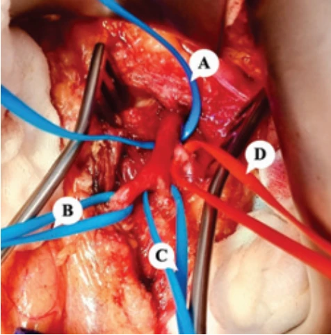
A) superior mesenteric artery (before thromboembolectomy); B) right colic artery;
C) ileal artery branch;
D) jejunal artery branch. Větvení arteria mesenterica superior, vypreparované větve periferně od okraje slinivky břišní. Průvlaky značící:
A) a. mesenterica superior (před trombektomii);
B) a. colica dextra;
C) ileální větev;
D) jejunální větev.
Thereafter, we turned our attention towards the hepatic area because of suspected impairment of the right hepatic artery. Palpation of hepatic arteries revealed a strong central pulsation. Furthermore, we targeted the suspected accessory hepatic artery behind the dilated bile duct; however, it was not found in its typical anatomical location. In light of this, and to prevent unnecessary risk, we avoided further dissection. Due to the dual hepatic blood supply, there was no immediate risk from postponing ultimate hepatic thrombectomy until the second-look procedure.
A 25 cm segment of ischemic ileum was resected using a stapler, with the bowel left temporarily closed until the second-look surgery. A drain was placed in the subhepatic space. The following day, the patient reported significant pain, and lactate levels were mildly elevated. Abdominal ultrasonography showed reduced arterial flow in the right hepatic lobe, while the left lobe had good perfusion.
A re-exploration was performed. Intraoperatively, the liver presented a striking map-like contour, while the intestinal loops appeared vital. We aimed at selective thrombectomy of the accessory right hepatic artery (aRHA), not from the usual coeliac trunk, but branching directly from the SMA – a rare variation seen in just 9–15% of patients [12]. With precision, we released the anomalous aRHA in the hepatoduodenal ligament, incised it and launched a bidirectional thrombectomy to clear the obstructing clot. The thrombosis was found intrahepatically. Although an ostial stenosis was suspected, the restored inflow proved to be sufficient. A 1 cm stone was also retrieved from the common bile duct via transverse incision. Finally, the ileum was reconstructed in an end-to-end fashion.
Within the next course, we explored the cause of the thromboembolism. Transthoracic and transesophageal echocardiography ruled out an intracardiac thrombus despite signs of pulmonary hypertension and right ventricular dilation and a patent foramen ovale. CT angiography showed no pulmonary embolism, only minor pleural effusions and residual infiltrates. Persistent sinus tachycardia (115–120 bpm) prompted Holter monitoring. Meanwhile, the patient remained on therapeutic low molecular weight heparin (LMWH).
Endocrinology consultation revealed a mild tremor, normal thyroid ultrasound, and no ocular signs. Findings (low TSH, elevated anti-TPO, anti-TG and fT3) suggested hyperthyroidism with resting sinus tachycardia. Subclinical Graves-Basedow disease was suspected in the context of autoimmune thyroiditis. Thiamazole was up-titrated to control hormone levels, beta-blockers were continued, and follow-up was arranged. Lipid profile was normal.
On postoperative day 12, despite prior negative ultrasounds, a third surgical revision was required due to a wound infection. After initial silver-charcoal therapy, we applied negative pressure wound therapy (NPWT) with limited debridement. No purulent collections were found. The wound was successfully closed 3 days later over healthy granulated tissue and intact fascia. Cultures grew Enterococcus faecalis and Enterobacter aerogenes (AmpC), treated with piperacillin/tazobactam. The patient was discharged on therapeutic LMWH with follow-up plans including Holter monitoring. Cardioembolic origin in the setting of hyperthyroidism remained a probable cause of the visceral artery occlusion.
Three weeks later, with a non-complicated home course, the patient returned with acute abdominal pain, fever, and elevated inflammatory markers. The home Covid-19 test was negative. Imaging revealed a 3 × 3 cm pericecal infiltrate with gas bubbles and an indistinct appendix (Fig. 7). The SMA was patent; the aRHA showed gracile flow. Considering the septic condition and suspected perforated appendicitis, urgent surgical intervention was indicated.
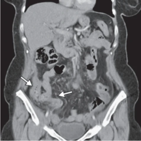
Šipky značící pericékální infiltrát se suspektní perforovanou apendicitidou.
Upon reopening the abdominal cavity, we found the cecum adhered to the distal ileum (Fig. 8). Adhesiolysis was performed, followed by ileocecal resection and reconstruction via an ileo-ascending anastomosis. The procedure terminated with thorough peritoneal lavage.
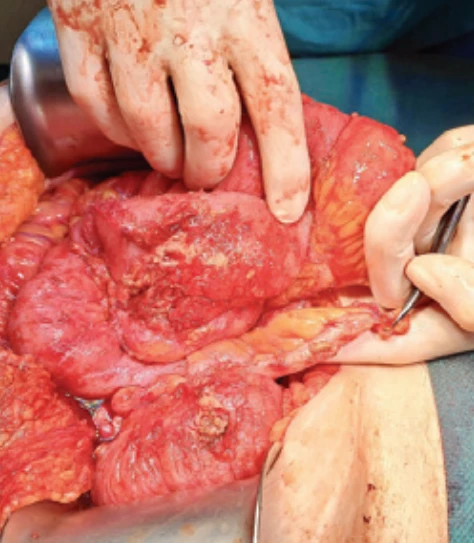
Paracékální infiltrát se suspektní perforovanou appendicitidou.
Three days later, a wound revision became necessary due to purulent effluent. We evacuated the pus, debrided the fascia, and resutured the site, placing a NPWT system (Fig. 9). By the 7th postoperative day, inflammatory markers plateaued, IL-6 surged, and lactate levels rose. In response, we escalated antibiotic therapy (meropenem, ampicillin) and initiated antifungal treatment (fluconazole) after detecting mildly elevated b-D-glucan – an early indicator of invasive fungal infections like Candida or Aspergillus. Cultures identified E. coli, Enterococcus avium, two anaerobes, and Clostridium difficile in the stool. Vancomycin was added to treat the clostridial infection, while Ampicillin was continued until the wound was definitively closed 2 weeks after bowel reconstruction.
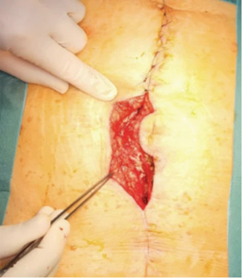
Revize části laparotomie po terapii NPWT před definitivním uzávěrem.
In the following days, b-d-Glucan levels normalized, wound cultures remained negative, and bowel habits stabilized. The patient was discharged on postoperative day 20 with a clean laparotomy wound and resumed oral intake. Final histology confirmed ischemic necrosis of the mucosa and submucosa, with hemorrhagic changes and thrombi in submucosal arteries.
In the evaluation of embolic etiology, CT imaging revealed minor calcified atherosclerotic plaques in the subrenal aorta, with no evidence of atherosclerotic changes in the visceral arteries. Ambulatory Holter monitoring demonstrated no sustained arrhythmias, aside from sporadic ventricular extrasystoles. The patient maintained a sinus rhythm under an increased dose of beta-blockers, with an average heart rate of 91 bpm, decreasing further to 80 bpm. Hemoglobin levels returned to normal, with only mild thyroid hormone elevation. Blood testing for thrombophilia confirmed acquired protein S deficiency. At 13 months post-surgery, the patient is doing well with persistent diarrhea despite unremarkable colonoscopy. The findings point to a multifactorial origin of mesenteric ischemia likely induced by thyrotoxicosis--provoked tachycardia and coagulopathy.
Discussion
In this case study, we present a patient with acute mesenteric ischemia, most likely caused by cardiac embolism in the setting of hyperthyroidism, who was successfully treated with surgical thrombo-embolectomy of the superior mesenteric artery. Revision surgeries were required due to concomitant occlusion of the accessory right hepatic artery, ischemic necrosis of the ileocecal region, and impaired healing of the laparotomy. This case highlights the complex presentation of mesenteric ischemia in the presence of vascular anomalies.
Early clinical signs of acute visceral ischemia are often unclear, as illustrated by our case report. The hallmark symptom is severe abdominal pain with minimal physical findings. Approximately one-third of patients present with nausea, vomiting, diarrhea, or rectal bleeding. A history of postprandial pain, weight loss, and food aversion (sitophobia) suggests underlying chronic mesenteric ischemia.
Mucosal injury is typically the earliest feature of AMI, as it has the highest metabolic demand, and therefore it is most vulnerable to hypoperfusion. At this stage, the damage may still be reversible if recognized and promptly treated. Patients may initially have severe pain despite a relatively sparse physical examination finding.
Intestinal fatty acid-binding protein (I-FABP), released from epithelial cells in the villi, is a promising early biomarker of enterocyte injury and intestinal ischemia. It rises significantly within 2–3 hours of ischemia onset and may predict systemic inflammatory response or sepsis [13,14]. Other valuable markers include ischemia-modified albumin (IMA) and D-lactate that show early elevation with high sensitivity and specificity [14,15]. Transmural necrosis and perforation happen once ischemia extends to the muscular and serosal layers. Complete vascular occlusion can cause irreversible damage within 6 hours, although the bowel may tolerate up to 12 hours of 75% flow reduction [16].
Additional biomarkers linked to gut barrier dysfunction include a-defensins, a-glutathione S-transferase (a-GST), and citrulline [17]. Moreover, microRNAs associated with apoptotic pathways have been explored as a potential early marker of AMI, (miR-122, miR-150), although available evidence remains limited and heterogeneous [18].
Beyond laboratory testing, advanced imaging techniques such as indocyanine green fluorescence (ICG) reveal ischemic regions not visible to the naked eye. Another innovative non-invasive dye-free method – laser speckle contrast imaging (LSCI) – enhances diagnostic precision based on detection of real-time blood flow of red blood cell motion (Fig. 10) [19].
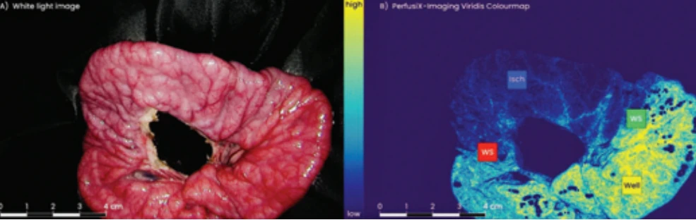
A) White light image as produced by a standard laparoscopic system.
B) Visualization of the perfusion levels during the laser speckle contrast imaging. Isch – ischemic tissue blue, WS – watershed areas green & red, Well – well-perfused tissue yellow
Použití metody LSCI. Tenké střevo na černé roušce. V čase T0 byla ischemie navozená podvazem tepny; centrální mezenteriální ischemie vznikla koagulací přívodné tepny [19]. Prasečí model střevní ischemie (se svolením Hoffman T. J. a Springeru).
A) Snímek v bílém světle pořízený standardním laparoskopickým systémem.
B) Vizualizace úrovně perfuze během laserového kontrastního speckle zobrazování (laserová zrnitost). Isch – ischemická tkáň modrá, WS – oblasti hraniční perfuze červená a zelená, Well – dobře prokrvená tkáň žlutá
Both ICG and LSCI can delineate the extent of mucosal necrosis, helping to avoid unnecessary bowel resection and the risk of short bowel syndrome. By clearly visualizing viable tissue, these tools may reduce the need for second-look operations, especially beneficial in elderly, multi-morbid patients [20,21].
The best approach to mesenteric ischemia remains debated, but imaging and clinical signs determine therapeutic management. Prompt diagnosis relies on early CT angiography and assessment of serum biomarkers, both requiring treatment strategy.
Endovascular treatment is preferred in the era of minimally invasive settings [22], although its limitations are evident – missed bowel ischemia, anatomical challenges, risk of reocclusion, and incomplete revascularization. As reported by Moláček et al., unless imaging findings suggest advanced bowel injury with likely irreversible ischemia, an endovascular-first approach is more commonly indicated in AMI [22]. Once the findings shows advanced ischemia – free air, pneumatosis, portal venous gas or peritonitis, laparotomy is required.
In the present case, the discrepancy between initially non-diagnostic bowel features on CT scan and progressive abdominal deterioration supported ongoing suspicion of acute mesenteric ischemia.
The 2025 ESVS guidelines explicitly note that AMI may be present before definitive radiological signs of bowel ischemia develop. For thrombotic or embolic SMA occlusion, endovascular therapy is recommended as the preferred first-line approach, whereas laparotomy is indicated when peritonitis is present or bowel infarction is strongly suspected [6]. Our rationale for immediate operative management was supported by progressive clinical abdominal findings and concerns of threatened bowel vitality.
The principal endovascular technique described is aspiration thrombectomy: a catheter is introduced via the femoral artery into the SMA to aspirate emboli. Mechanical thrombectomy with devices like AngioJet, Rotarex, and Penumbra may also be used [23]. Occlusions can be recanalized antegrade via femoral/brachial access or retrograde via exposed SMA after laparotomy and then be stented [24]. Intra-arterial thrombolysis with rt-PA has shown success [25]. Atherosclerotic occlusions are treated with balloon angioplasty and stent placement. Surgery options include thromboembolectomy with a Fogarty catheter or mesenteric bypass from the infrarenal/supraceliac aorta or iliac artery.
Synthetic grafts are preferred in uncontaminated cases due to reduced risk of kinking compared to vein grafts [3]. Non-occlusive mesenteric ischemia resulting from severe vasospasm, such as after aortofemoral surgery, may be treated with intra-arterial vasodilators directly to SMA (commonly nitrates, prostaglandins, glucagon, or iloprost) [25].
Although most comparative studies are non-randomized and have not shown an overall superiority of one strategy over the other, current evidence increasingly supports an endovascular-first approach in selected patients, with more favorable short-term outcomes but broadly comparable longer-term results, as stated in the 2025 ESVS guidelines [6].
Conclusion
AMI remains a life-threatening condition with high morbidity and mortality, despite significant advances in medical and surgical care. Advanced imaging modalities and revascularization techniques are the key to early diagnosis, guiding treatment, and organ preservation. CT angiography combined with physical examination serve as the cornerstone of diagnosis.
Although mini-invasive approaches are increasingly favored for selected patients, we advocate for open surgical embolectomy in cases of acute thromboembolic occlusion along with bowel injury, enabling direct intraoperative bowel viability assessment.
In our case, the patient’s escalating abdominal pain, persistent tachycardia, presence of extensive thromboemboli in the SMA and rising inflammatory markers prompted urgent surgical exploration, despite absence of clear ischemic CT signs.
Borderline cases of acute mesenteric ischemia will occur and remain challenging both diagnostically and therapeutically, especially when selecting the treatment approach. Current guidelines gradually endorse an endovascular-first approach when definite bowel infarction is not evident. Novel diagnostic methods may improve risk stratification and help guide therapeutic strategy; however, their integration into acute settings remain challenging and only broader clinical implementation will validate their efficacy.
Dedication
This research was supported by the project National Institute for Research of Metabolic and Cardiovascular Diseases (Programme EXCELES, ID Project No. LX22NPO5104) – Funded by the European Union – Next Generation EU; and by the Ministry of Health of the Czech Republic, grant nr. NW24J-02-00061. All rights reserved.
Conflict of interests
The authors declare that they have no conflict of interest related to the creation of this article, and that this article has not been published in any other journal with access to congress abstracts.
Sources
1. Tamme K, Reintam BA, Laisaar KT et al. Incidence and outcomes of acute mesenteric ischaemia: a systematic review and meta-analysis. BMJ Open 2022; 12 (10): e062846. doi: 10.1136/ bmjopen-2022-062846.
2. Kase K, Reintam Blaser A, Tamme Ket al. Epidemiology of Acute Mesenteric Ischemia: A Population--Based Investigation. World J Surg 2023; 47 (1): 173–181. doi: 10.1007/s00268-022-06805-5.
3. Fuglseth H, Søreide K, Verthus M. Acute mesenteric ischaemia. Br J Surg 2023; 110 (9): 1030–1034. doi: 10.1093/bjs/znad021.
4. Rivoire E, Tresson P, Long A. L’ischémie intestinale: y penser, pour qui et que faire? La Presse Médicale Formation 2024; 5 (4): 260–269. doi: org/10.1016/j.lpmfor.2024.04.002.
5. Molyneux K, Beck-Esmay J, Koyfman Aet al. High risk and low prevalence diseases: Mesenteric ischemia. Am J Emerg Med 2023; 65 : 154–161. doi: 10.1016/j.ajem.2023.01.00.
6. Editor’s Choice – European Society for Vascular Surgery (ESVS) 2025 Clinical Practice Guidelines on the Management of Diseases of the Mesenteric and Renal Arteries and Veins. Eur J Vasc Endovasc Surg 2025; 70 (2): 153–218. doi: 10.1016/j.ejvs.2025.06.010.
7. Bala M, Kashuk J, Moore EE et al. Acute mesenteric ischemia: guidelines of the World Society of Emergency Surgery.World J Emerg Surg 2017 : 12 : 38. doi: 10.1186/s13017-017-0150-5.
8. Bourcier S, Klug J, Nguyen LS. Non--occlusive mesenteric ischemia: diagnostic challenges and perspectives in the era of artificial intelligence. World J Gastroenterol 2021; 27 (26): 4088–4103. doi: 10.3748/wjg.v27.i26.4088.
9. Oldenburg WA, Lau LL, Rodenberg TJet al. Acute mesenteric ischemia: a clinical review. Arch Intern Medicine 2004; 164 (10): 1054–1062. doi: 10.1001/archinte.164.10.1054.
10. Kim AY, Ha HK. Evaluation of suspected mesenteric ischemia. Radiol Clin North Am2003; 41 (2): 327–342. doi: 10.1016/s0033-8389 (02) 00075-1.
11. Salim H, Ozgur O, Erman K et al. Collateral circulation develops in stenosis of the celiac trunk, and superior mesenteric artery. Surg Radiol Anat 2023; 45 (4): 479–486. doi: 10.1007/s00276-023-03104-z.
12. D’Souza D. Replaced right hepatic artery. 2025 [online]. Available from: https: //radiopaedia.org/cases/replaced-right-hepatic-artery.
13. Khadaroo RG, Fortis S, Salim SY et al. I-FABP as biomarker for the early diagnosis of acute mesenteric ischemia and resultant lung injury. PLoS One 2014; 9 (12): e115242. doi: 10.1371/journal.pone.0115242.
14. Montagnana M, Danese D, Lippi G. Biochemical markers of acute intestinal ischemia: possibilities and limitations. Ann Transl Med 2018; 6 (17): 341. doi: 10.21037/atm.2018.07.22.
15. Gunduz A, Turedi S, Mentese A et al. Ischemia-modified albumin in the diagnosis of acute mesenteric ischemia: a preliminary study. Am J Emerg Med 2008; 26 (2): 202–205. doi: 10.1016/j.ajem.2007.04.030.
16. Van Petersen AS, Kolkman JJ, Meerwaldt R et al. Mesenteric stenosis, collaterals, and compensatory blood flow. J Vasc Surg 2014; 60 (1): 111–119. doi: 10.1016/j.jvs.2014.01.063.
17. Treskes N, Person AS, van Zanten AR. Diagnostic accuracy of novel serological biomarkers to detect acute mesenteric ischemia: a systematic review and meta-analysis. Intern Emerg Med 2017; 12 (6): 821–836. doi: 10.1007/s11739-017-1668-y.
18. Wang F, Gu L, Wang Y et al. MicroRNA--122a aggravates intestinal ischemia/reperfusion injury by promoting pyroptosis via targeting EGFR-NLRP3 signaling pathway. Life Sci 2022; 15 : 307. doi: 10.1016/j.lfs.2022.120863.
19. Hoffman JT, Heuvelings DJ, van Zutphen T et al. Real-time quantification of laser speckle contrast imaging during intestinal laparoscopic surgery: successful demonstration in a porcine intestinal ischemia model. Surg Endosc 2024; 38 (9): 5292–5303. doi: 10.1007/s00464-024-11076-3.
20. Nakagawa Y, Kobayashi K, Kuwabara S et al. Use of indocyanine green fluorescence imaging to determine the area of bowel resection in non-occlusive mesenteric ischemia: a case report. Int J Surg Case Rep 2018; 51 : 352–357. doi: 10.1016/j.ijscr.2018.09.024.
21. Pérez-Benítez OA, Pérez-Cantú A, De LD et al. Intestinal ischemia and indocyanine green fluorescence: its use as a therapeutic tool, apropos of a clinical case. Rev Mex Cir Endoscop 2021; 22 (3–4): 150–157. doi: 10.35366/106480.
22. Moláček J, Třeška V, Čertík B et al. Indikace k otevřené chirurgické revaskularizaci viscerálního řečiště v endovaskulární éře – souhrnné sdělení. Rozhl Chir 2018; 97 (11): 487–492.
23. Raupach J, Fejfar T, Hudák A et al. Endovaskulární léčba mezenteriální ischemie. Ces Radiol 2021; 75 (2): 123–133. doi: 10.55095/CesRadiol2021/015.
24. Saadi EK, Oderich G, Medronha E et al. Endovascular recanalization of occluded superior mesenteric artery using retrograde access through the inferior mesenteric artery. J Vasc Surg Cases Innov Tech 2017; 3 (3): 155–158. doi: 10.1016/j.jvscit.2017.04.005.
25. Bjornsson S, Bjorck M, Block T et al. Thrombolysis for acute occlusion of the superior mesenteric artery. J Vasc Surg 2011; 54 (6): 1734–1742. doi: 10.1016/j.jvs.2011.07.054.
Labels
Surgery Orthopaedics Trauma surgeryArticle was published in
Perspectives in Surgery
2026 Issue 6
Most read in this issue
- Monteggiova zlomenina v detskom veku
- Tumory mediastina
- Dvoufázová tromboembolektomie u akutní viscerální ischemie s atypickým odstupem pravé jaterní tepny, komplikovaná sekundárním střevním infiltrátem
- Chirurgie 21.2