
Mají antikoagulační a protidestičkové léky lokální účinky na zvětšení chronického subdurálního hematomu?
Authors:
B. Ertuğrul 1; A. C. Ergün 2; M. Kaplan 1; H. Kömürcü 1; S. Tanyıldızı 3; G. Dağoğlu 3
Authors‘ workplace:
Department of Neurosurgery, Firat University Faculty of Medicine, Elazığ, Türkiye
1; Department of Neurosurgery, Elazığ Fethi Sekin City Hospital, Elazığ, Türkiye
2; Department of Pharmacology and Toxicology, Firat University Faculty of Veterinary Medicine, Elazığ, Türkiye
3
Published in:
Cesk Slov Neurol N 2026; 89(4): 263-266
Category:
Original Paper
doi:
https://doi.org/10.48095/cccsnn2026263
Overview
Úvod: Chronický subdurální hematom je častým neurochirurgickým stavem, který je často spojen s antikoagulační a protidestičkovou terapií. Není jasné, zda tyto látky mají lokální biochemický vliv na zvětšení hematomu. Tato studie zkoumala přítomnost aktivních metabolitů kyseliny acetylsalicylové, warfarinu, klopidogrelu a enoxaparinu v tekutině hematomu a tkáni kapsuly získaných během operace s použitím trepanace. Materiály a metody: Prospektivní observační studie zahrnovala 42 pacientů (25 mužů, 17 žen; průměrný věk 68,5 roku), kteří podstoupili drenáž pomocí trepanace bez přerušení antikoagulační nebo protidestičkové léčby. Vzorky tekutiny hematomu a kapsuly byly odebrány intraoperativně a analyzovány pomocí vysokoúčinné kapalinové chromatografie. Výsledky: Pacienti užívali kyselinu acetylsalicylovou (n = 12), warfarin (n = 8), klopidogrel (n = 6), enoxaparin (n = 7) nebo kombinaci kyseliny acetylsalicylové a klopidogrelu (n = 9). V žádném vzorku tekutiny hematomu ani kapsuly nebyly detekovány žádné aktivní metabolity. Nevyskytly se žádné intraoperační komplikace. Závěr: Antikoagulancia a protidestičkové léky jsou uznávanými systémovými rizikovými faktory pro vznik a progresi chronického subdurálního hematomu. Tato studie však nezjistila žádné důkazy o jejich lokální akumulaci v tkáni hematomu. Výsledky naznačují, že jejich role v patogenezi chronického subdurálního hematomu je zprostředkována spíše systémovými hemostatickými mechanismy než lokálními biochemickými účinky.
Klíčová slova:
Antiplatelet therapy – Anticoagulant therapy – chronic subdural hematoma – hematoma expansion – high-performance liquid chromatography
Introduction
Chronic subdural hematoma (CSDH) is a collection of blood and blood breakdown products between the dura and arachnoid membranes, encapsulated by a pseudomembrane. The condition is frequently encountered in neurosurgical practice. First described in 1857 by Virchow as “hemorrhagic pachymeningitis,” CSDH typically occurs after trauma but may also arise from non-traumatic causes such as arteriovenous malformations or intracranial tumors [1]. Several factors contribute to its development and expansion, including cerebral atrophy, intracranial hypotension, iatrogenic over-drainage of CSF, hematological disorders (e. g., coagulopathies and hemophilia), and the use of anticoagulant or antiplatelet drugs [2].
The pathophysiology underlying the expansion of CSDHs remains a subject of ongoing debate, with multiple etiological factors implicated. In recent years, the widespread use of anticoagulant and antiplatelet agents, driven by the rising prevalence of cardiovascular and cerebrovascular diseases, has contributed to the increasing incidence of CSDH. The ARISE I Consensus Statement (2024), an international multidisciplinary consensus document on the management of CSDH, also recognizes the growing global burden of CSDH and highlights the increasing use of antithrombotic therapy as one of the major contributing factors [3].
The originality of this study lies in its focus on the direct detection of anticoagulant and antiplatelet agents within hematoma material. Although numerous studies have examined the association between these drugs and CSDH expansion, few have investigated the presence of these drugs or their metabolites in hematoma samples using analytical methods. In this study, we measured the levels of acetylsalicylic acid, warfarin, clopidogrel, and enoxaparin active ingredients in hematoma fluid and capsule tissue obtained from patients with CSDH to evaluate whether these agents exert a local effect on hematoma expansion.
Materials and methods
This prospective observational study was conducted at a single tertiary care hospital. Forty-two symptomatic adult patients (≥ 18 years) with a radiologically confirmed diagnosis of CSDH who were receiving anticoagulant or antiplatelet therapy at the time of presentation and underwent emergency burr-hole surgery without discontinuation of these medications were included. Because this study included cases requiring emergency surgery, the exact timing of the last anticoagulant or antiplatelet dose could not be reliably documented, reflecting an inherent limitation of emergency CSDH management.
Inclusion criteria were: (1) CSDH confirmed by imaging, (2) ongoing use of anticoagulant and/or antiplatelet therapy, and (3) emergency burr-hole decompression. Patients with subacute/acute hematoma, individuals with known non-drug-induced coagulopathy, and those with missing data were excluded.
Clinical data, including patient age, sex, comorbidities, Glasgow Coma Scale score at admission, type of medication used, and dosage, were recorded. During surgery, approximately 5–10 mL of hematoma fluid sample and capsule tissue were collected and stored under appropriate conditions for analysis.
Concentrations of the drug active ingredients were determined using high-performance liquid chromatography (HPLC). The HPLC system (Shimadzu, Kyoto, Japan) was equipped with a pump (LC-20AT, controlled by CBM-20A), autosampler (SIL-20A), degasser (DGU-20A), column oven (CTO-20A), and UV-VIS detector (SPD-20A). A C18 column was used for separation, and active ingredient levels of acetylsalicylic acid, warfarin, clopidogrel, and enoxaparin were quantified.
Descriptive statistics were generated using the SPSS v26.0 program (IBM, Armonk, NY, USA).
This study was conducted and reported in accordance with the Strengthening the Reporting of Observational Studies in Epidemiology (STROBE) Statement for cohort studies [4]. A completed STROBE checklist is available only online as supplementary material (Supplement 1).
Results
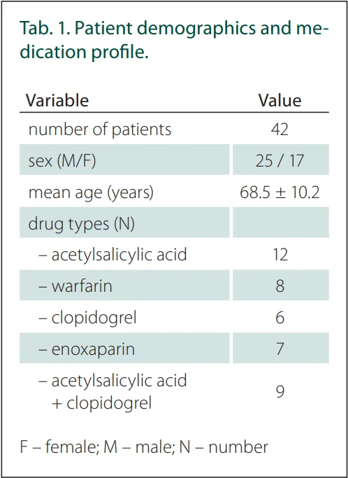
A total of 42 patients (25 males, 17 females) were included in the study. The mean age was 68.5 years. All patients were using at least one anticoagulant/antiplatelet drug before surgery. Patient demographics and medication profile are presented in Tab. 1.
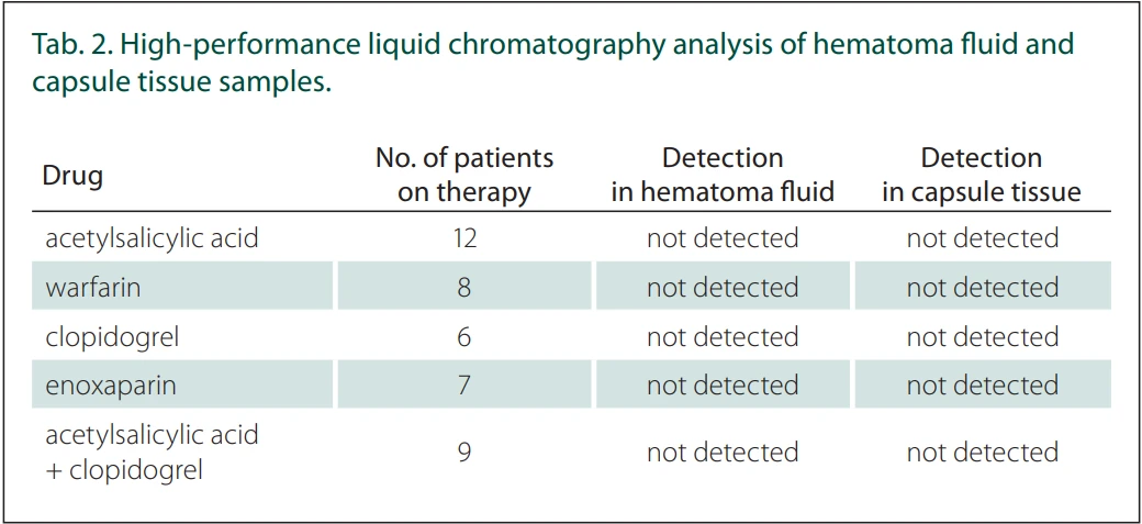
High-performance liquid chromatography analysis revealed no detectable levels of the active ingredients of acetylsalicylic acid, warfarin, clopidogrel, or enoxaparin in either hematoma fluid or capsule tissue samples. All measurements were below the established limits of detection (LOD: 0.01 μg/mL) and limit of quantification (LOQ: 0.03 μg/mL). Samples were stored at −80 °C and subjected to a single freeze-thaw cycle before analysis (Tab. 2).
Discussion
Chronic subdural hematoma is a complex pathology comprising three main morphological components: the outer membrane, the hematoma cavity, and the inner membrane. While the inner membrane contains relatively few vessels, the outer membrane often harbors numerous fragile macrocapillaries that are a source of recurrent, multifocal bleeding [5]. Several factors contribute to CSDH expansion, including recurrent bleeding, exudation from the outer membrane into the interior, orthostatic mechanisms, and rapid expansion secondary to CSF compression [6]. Clinically, advanced age and the use of anticoagulant or antiplatelet agents are recognized as the most significant risk factors for hematoma progression. In large hematomas, increased tension in the bridging veins predisposes to minor rebleeding, which further augments hematoma volume [7].
Excessive fibrinolytic activity has also been identified as an important contributor to CSDH progression. Fibrinolysis compromises the integrity of membranous and vascular structures, thereby promoting hematoma enlargement [8]. Shim et al. reported significantly higher levels of fibrin degradation products in CSDH fluid compared with patient plasma [9], indicating increased fibrinolytic activity in the subdural space. Fibrin degradation products exert both anticoagulant and vasodilatory effects. Although physiological levels of fibrinolysis are necessary for clot resorption, abnormal upregulation of this process can lead to recurrent bleeding from the membranes surrounding the hematoma. Therefore, excessive local fibrinolytic activity plays a critical role in the pathophysiology of CSDH expansion [10].
Previous clinical studies have primarily focused on the association between antithrombotic therapy and the development, recurrence, and perioperative management of CSDH [11,12]. In contrast, previous pathophysiological studies have mainly investigated angiogenesis, vascular proliferation, inflammatory activity, and hyperfibrinolysis within the hematoma cavity [5,9]. However, the direct evaluation of commonly used anticoagulant and antiplatelet agents within hematoma fluid and capsule tissue has received little attention. The present study extends the existing literature by evaluating the local presence of these agents using HPLC, and our findings suggest that their contribution to CSDH progression is more likely mediated through systemic hemostatic alterations rather than direct local accumulation.
Clinically, anticoagulant and antiplatelet drugs are recognized as major risk factors for the development and recurrence of CSDH [13]. However, it remains uncertain whether their effect is mediated exclusively through systemic alterations in hemostasis or also through local accumulation within the hematoma. In this study, we analyzed hematoma fluid and outer membrane samples obtained during emergency burr-hole drainage from patients who continued anticoagulant and/or antiplatelet therapy at the time of surgery. Specifically, we investigated the presence of acetylsalicylic acid, warfarin, clopidogrel, and enoxaparin. None of the active ingredients of these drugs was detected in any of the samples.
Several explanations may account for this negative finding. First, pharmacokinetic properties such as metabolism, tissue distribution, and plasma half-life likely limit the passage of active metabolites into the hematoma cavity. For example, acetylsalicylic acid is rapidly deacetylated in plasma. Clopidogrel is a short-half-life prodrug, and its active metabolite binds extensively to plasma proteins. Enoxaparin, however, cannot cross into intracranial regions due to its molecular size and polarity. Second, the timing of the last drug dose prior to surgery could not be precisely determined. Third, biochemical degradation or dilution within the hematoma matrix may have reduced concentrations below the analytical sensitivity limit despite systemic exposure.
Our findings suggest that the contribution of anticoagulant and antiplatelet agents to the pathophysiology of CSDH occurs primarily through systemic rather than local mechanisms. These agents may promote hematoma formation and expansion by increasing the susceptibility of fragile neocapillaries in the outer membrane to microbleeding. However, they do not appear to act via direct accumulation within the hematoma cavity. This interpretation aligns with previous reports indicating that endogenous mechanisms, such as local hyperfibrinolysis, play a central role in driving hematoma growth [9].
Clinically, the absence of local accumulation of anticoagulant and antiplatelet agents within hematoma tissue suggests that emergency surgical interventions may be safely performed in selected patients, provided that systemic hemostasis is carefully evaluated. In clinical practice, such assessment typically includes platelet count, coagulation parameters (prothrombin time / international normalized ratio and activated partial thromboplastin time), identification of the antithrombotic agent used, and consideration of appropriate reversal strategies when indicated. Nevertheless, larger cohorts and well-designed prospective studies are needed to confirm these findings and to further clarify the role of antithrombotic agents in the pathophysiology of CSDH.
Several methodological considerations should be taken into account when interpreting the findings of this study. Because the work was conducted in an emergency setting, the exact timing of patients’ last anticoagulant or antiplatelet doses could not be reliably determined. This factor is particularly relevant for drugs with short half-lives, as dose timing may influence the detectability of drug or metabolite levels within the hematoma. In addition, due to the practical limitations of obtaining extra blood samples during urgent surgery, concurrent plasma drug or metabolite concentrations could not be measured. This limits the ability to directly correlate systemic exposure with local intrahematomal levels.
Furthermore, the complex biological matrix of hematoma tissue may reduce analytical detection sensitivity. Factors such as high protein content, enzymatic degradation, and local fibrinolytic activity can affect metabolite stability. Although all samples were stored at −80 °C, degradation of certain antithrombotic metabolites during the freeze–thaw process cannot be entirely excluded. Therefore, the absence of detectable drug or metabolite levels in HPLC analysis does not necessarily indicate true absence; concentrations may simply fall below the detection limits of the analytical method.
This study has several limitations. The relatively small sample size and the single-center design may limit the generalizability of the findings. The exact timing of the patients‘ last anticoagulant or antiplatelet doses could not be reliably determined, although all patients were receiving active antithrombotic therapy at the time of admission. This reflects a limitation inherent to emergency clinical practice rather than uncertainty regarding ongoing antithrombotic therapy. In addition, concurrent plasma drug or metabolite levels were not obtained. Due to the complex biological composition of hematoma material, matrix validation for the HPLC method could not be performed, and the possibility of low-level metabolite degradation during processing or storage cannot be entirely ruled out. These methodological limitations should be considered when interpreting the absence of detectable drug or metabolite levels within the hematoma.
In conclusion, this study demonstrated that anticoagulant and antiplatelet agents did not accumulate locally in CSDH material. Their contribution to CSDH pathogenesis appears to occur primarily through systemic hemostatic alterations rather than local pharmacological effects. These findings may have important clinical implications, particularly in guiding the safety of emergency surgical interventions in patients receiving antithrombotic therapy.
Ethics approval
The entire study was conducted in accordance with the Helsinki Declaration of 1975 (as revised in 2004 and 2008). Ethical approval was obtained from the Clinical Research Ethics Committee of Fırat University Faculty of Medicine (Decision No: 2021/08-02; dated June 24, 2021).
Consent to participate
Written informed consent was obtained from all participants for the use of their clinical data and tissue samples for scientific purposes.
Conflict of interest
The authors declare they have no potential conflicts of interest concerning drugs, products, or services used in the study.
Data availability
The datasets generated and/or analyzed during the current study are not publicly available due to patient confidentiality and ethical restrictions, but they are available from the corresponding author on reasonable request and with appropriate institutional approvals.
Author contributions
B.E. – conceptualization, investigation, resources, validation, and writing – review & editing
A.C.E. – data curation, methodology, and visualization
M.K. – Project administration, supervision, and writing – original draft
H.K. – data curation, resources, validation
S.T., G.D. – formal analysis
All authors have read and approved the final version of the manuscript and agree to be accountable for all aspects of the work. All authors meet the ICMJE authorship criteria.
Acknowledgments
The authors thank Enago (www.enago.com) for the English language review.
Sources
1. Markwalder TM. Chronic subdural hematomas: a review. Neurosurgery 1981; 54 : 637–645.
2. Torihashi K, Sadamasa N, Yoshida K. Independent predictors for recurrence of chronic subdural hematoma: a review of 343 consecutive surgical cases. Neurosurgery 2008; 63 : 1125–1129. doi: 10.1227/01.NEU.0000335782.60059.17.
3. Kan P, Fiorella D, Dabus G et al. ARISE I consensus statement on the management of chronic subdural hematoma. Stroke 2024; 55 (5): 1438–1448. doi: 10.1161/STROKEAHA.123.044129.
4. von Elm E, Altman DG, Egger M et al. Strengthening the Reporting of Observational Studies in Epidemiology (STROBE) Statement: guidelines for reporting observational studies. BMJ 2007; 335 (7624): 806–808. doi: 10.1016/j.jclinepi.2007.11.008.
5. Yamashima T, Yamamoto S. How do vessels proliferate in the capsule of a chronic subdural hematoma? Neurosurgery 1984; 15 : 672–678. doi: 10.1227/00006123-198411000-00006.
6. Yadav YR, Parihar V, Namdev H et al. Chronic subdural hematoma. Asian J Neurosurg 2016; 11 (4): 330–342.
7. Asan Z. Growth potential of subdural hematomas under clinical observation: which subdural hematomas tend to grow and why they do. World Neurosurg 2018; 113: e598–e603.
8. Liu W, Bakker NA, Groen RJM. Chronic subdural hematoma: a systematic review and meta-analysis of surgical procedures. J Neurosurg 2014; 121 : 665–673. doi: 10.3171/2014.5.JNS132715.
9. Shim YS, Park CO, Hyun DK et al. What are the causative factors for a slow, progressive enlargement of a chronic subdural hematoma? Yonsei Med J 2007; 48 : 210–217. doi: 10.3349/ymj.2007.48.2.210.
10. Safain MG, Roguski M, Antoniou A et al. A single--center experience with the bedside subdural evacuating port system: a useful alternative for chronic subdural hematoma evacuation. J Neurosurg 2013; 118 : 694–700. doi: 10.3171/2012.11.JNS12689.
11. Poon MTC, Rea C, Kolias AG et al. Influence of antiplatelet and anticoagulant drug use on outcomes after chronic subdural hematoma drainage. J Neurotrauma 2021; 38 (8): 1177–1184. doi: 10.1089/neu.2018.6080.
12. Liu E, Tilbury N, Morin T et al. Antiplatelet and anticoagulation use and outcomes following chronic subdural hematoma drainage: systematic review and meta-analysis. World Neurosurg 2026; 208 : 124873. doi: 10.1016/j.wneu.2026.124873.
13. Bumpetch D, Sitthinamsuwan B, Nunta-Aree S. A prospective study of the effect and safety of atorvastatin on the recurrence of chronic subdural hematoma after burr hole surgery. Asian J Neurosurg 2023; 18 (3): 567–572. doi: 10.1055/s-0043-1771372.
Labels
Paediatric neurology Neurosurgery NeurologyArticle was published in
Czech and Slovak Neurology and Neurosurgery
2026 Issue 4
Most read in this issue
- Changes in the clinical practice of cognitive disorders – testing, treatment, and driving
- Roztroušená skleróza a plánování těhotenství – historie, současnost a úloha registrů
- Urinary biomarkers of oxidative stress in amyotrophic lateral sclerosis – a pilot study
- Do anticoagulant and antiplatelet drugs exert local effects in chronic subdural hematoma expansion?