-
Medical journals
- Career
MEASUREMENT OF THE VALUES OF RADIOFREQUENCY ELECTROMAGNETIC FIELDS AROUND THE HEAD OF ADOLESCENTS
Authors: Hana Habinakova 1; Viera Jakusova 2; Miroslav Kohan 1; Jakub Misek 1; Jan Jakus 1
Authors‘ workplace: Department of Medical Biophysics, Jessenius Faculty of Medicine in Martin, Comenius University in Bratislava, SR 1; Department of Public Health, Jessenius Faculty of Medicine in Martin, Comenius University in Bratislava, SR 2
Published in: Lékař a technika - Clinician and Technology No. 2, 2017, 47, 60-67
Category: Original research
Overview
Cell phones and other communication devices have become the primary source of socialization, especially among adolescents. The aim of the study was to assess the levels of radiated radiofrequency (RF) power (1788.5 MHz, max. 30 V/m) around the head of adolescents. The measurements were performed in 2016 at the Department of Medical Biophysics of Jessenius Faculty of Medicine in Martin. The sample group consisted of 44 adolescents of Viliam Pauliny-Toth, Grammar School in Martin. To measure the performance levels of electromagnetic fields (EMF), we used selective radiation meter NARDA SRM 3006 (9 kHz–6 GHz) with the function of a spectrum analyzer. The average values of power were recorded in eight positions around the head with six minutes exposure length of each of them. Every adolescent filled out a short questionnaire on personal perception of the effects of RF radiation on the body after the exposure. The statistical evaluation showed a significant decrease in the intensity of power on the left side of the adolescent’s head compared to the right side (p < 0.01–0.001), which confirmed different degrees of absorption by the head tissues. The highest level of absorption was measured at temporal area of the head connecting both ears. Short-term exposure to RF radiation did not cause strong adverse health effects in adolescents, however in a few cases tachycardia, drowsiness, headache, fatigue and restlessness appeared. It is necessary to pay more attention to the examination of the relationship between exposure to RF EMF and the potential adverse health reactions mainly in adolescents.
Keywords:
adolescents, cell phone, radiofrequency electromagnetic fields, power measurement, questionnaireIntroduction
Widespread use of radio-communication devices is raising concerns on possible adverse effects of radio-frequency (RF) electromagnetic fields (EMF) on human health. The highest amount of radiation from cell phones is absorbed by the head as most exposed biological tissue. However, depth of the penetration of electromagnetic (EM) energy depends on frequency of EMF, tissue permittivity and conductivity of tissue [1]. Exposure to RF EMF depends on the morphology of biological objects, where induced local fields in the brain may be significantly higher in children than in adults [2, 3]. Simulations of the EMF layout in head phantoms allow using numerical methods to define the equivalent of the real environment and compare the rate of energy absorption in correlation with age [4, 5]. In our study, we wanted to analyse the level of penetration of RF EMF in different areas of the head by experimental procedures. The aim of our study was to measure and evaluate the values of RF EMF power around the head of adolescents during a short term exposition.
Material and Methods
In May and June 2016 we conducted measurements of power P (dBm or mW) of RF EMF at 8 positions around the head in adolescents of Grammar School of Viliam Pauliny-Toth (GVPT) in Martin. Part of the measurements was a brief questionnaire monitoring subjective health complaints of adolescents immediately performed after measurements.
The sample consisted of 44 adolescent - volunteers, from them 23 (52.27%) were women and 21 (47.73%) men. Each adolescent particitated on the measurements just once. The mean age of adolescents was (18.4 ± 0.4) (mean ± SD) years. Only healthy adolescents took part in the measurements, whose blood pressure and body temperature were taken before and after the examination. Subsequently, body mass index (BMI) was calculated. Also a width and lenght of head were taken before examination. Adolescents did not consume coffee, tea, energy drinks or any medication prior to the measurements. All procedures on human subjects were permitted by Ethical Board of Jessenius Faculty of Medicine in Martin (JLFUK) Comenius University in Bratislava and by the laws of Slovak Republic and European Union under permissions covered by APVV project 0189-11 (prof. Jakus). Each adolescent/legal representative provided an informed agreement to participate in the study.
Measurements of RF power in areas around the head of adolescents were conducted at the Department of Medical Biophysics of JLFUK. Adolescents during trial took a supine position with the head placed in a stable cage. The cage was made of styrodur material with the 20 mm walls thickness. The styrodure cage contained eight openings for securing receiving antenna (RA) in the same possition for ensuring repeatabily of the measurement. The power values of RF EMF were measured inside the cage by RA at two levels on a vertical plane in 4 positions on each side of the head (Fig. 1).
Fig. 1: The head of person within the styrodur cage. (With the consent of adolescent) Source: Department of Medical Biophysics of JLF Martin. 
Electric intensity control measurements inside the cage were performed by the Narda NBM550 (Narda, Germany) high frequency broadband meter with three axis E field probe within frequency range 100 kHz–3 GHz, which is certified for near field measurements. This measuring device was also used for regular calibration in prior of each measurement. Thus, the average intensity of the applied electric field never exceeded value of 30 V/m in the place of head exposure inside of styrodure cage. Effective values of all measurements were in line with the safety guidlines for RF EMF parameters set by International Commission on Non-Ionizing Radiation Protection (ICNIRP) and derived regulations [6–9], that equals to 58.15 V/m for given frequency 1788.5 MHz measured at 6-minutes intervals. This frequency is close to the DCS-1800 standard, however it is not used by any mobile operator in Slovak Republic.
The signal generator Agilent N9310A (Agilent, USA) was used as a source of RF EMF wave with 1788.5 MHz frequency. The signal was amplified by 5 W amplifier model 5S1G4 (Amplifier Research, USA) [10] set for constatnt output 2 W. A 10 dB directional coupler was used to verify the output power to transmitting antenna (TA). This measurement was carried out before each measurement by the Selective Radiation Meter NARDA SRM3006 (Narda, Germany) with function of spectrum analyser. As TA was utilized Multi-Band T-Bar (RF Solutions ANT-TBARQB-SMA, UK) [11] antenna optimized for cellular applications on GSM and 3G frequency bands (824–960 MHz, 1710–1990 MHz, 1900–2200 MHz) measuring (104×10×3) mm (l×h×w) with active gain +3dBi (Fig. 2A). RA was PCB (printed circuit
board) triple band (880–960 MHz, 1710–1880 MHz, 1850–1990 MHz) antenna suitable for mobile applications (Yageo Part No. CAN 4313330109191B, Taiwan) measuring 35×6×0.4 mm (l×h×w) with cable and connector for mobile applications (SMA), linear polarisation and maximum power 1 W (Fig. 2B). The power values from RA were recorded in dBm by NARDA SRM3006 (SA). Evaluation frequency band was set up to 1710.1–1880 MHz and RBW (resolution bandwidth) to 10 MHz.Fig. 2: Transmitting (A) and receiving antenna (B). 
TA and RA were placed parallel to main radiation lobes directed opposite each other. The orientation of the antennas is displayed in Fig. 3.
Fig. 3: Relative orientation of antennas. 
Basic calibration was done in prior of the trial, while the output of the amplifier was directly connected to the input of the SA. As a result was set of attenuation coefficients that were inserted into SA as calibration constans for the coaxial wires. There were realized measurements without the head in the cage for verification of exposed power by TA and air attenuation at the distance 220 mm that corresponds the width of the cage. All parameters on the spectrum analyser were at the beginning automatically checked to ensure precise and repeatable measurements. The evaluation of the power (P) was done in mW (P [mW] = 10P[dBm]/10). The block diagram with the measuring equipments is shown on Fig. 4.
Fig. 4: Block diagram of equipment for measurement of power RF EMF around the head. 
Equipments in the block diagram:
G generator - Agilent N9310A
A amplifier -Amplifier Research 5S1G4
TA transmitting antenna - Multi-Band T - BarGSM Quad Band Antenna
H head of the adolescent
RA receiving antenna - Triple Band PCB Antenna
P1–P8 positions of RA
SA spectrum analyser - NARDA SRM 3006
The arrangement of measuring equipments is shown in Fig. 5.
Fig. 5: Measuring of RF EMF around the head of an adolescent. 
Positions P1–P4 were defined on right side of a head (Fig. 6). TA was fixed inside the cage on the right side between positions P1–P4. P1, P2 were located at the top level of the cage, P3, P4 at the lower level. P4 was set up at the right ear of the head as a fix point for each adolescent. Positions P5–P8 were defined to lie to positions P1–P4 on the left side of the styrodur cage: P5 at a same level with P1, P6 with P2, P3 with P7 and P8 with P4. The distance between corresponding pairs was 220 mm. Only the position of the RA was changed, the position of the TA was fixed.
Fig. 6: Scheme of the location of the transmitting antenna (TA) and a distribution of positions P1–P4 on the right side in the styrodur cage. 
Each adolescent immediately after the measurements filled out a short questionnaire on possible subjective health feelings:
1 - fatigue, 2 - headache, 3 - drowsiness, 4 - exhaus-tion, 5 - dizziness, 6 - restlessness, 7 - problems with concentration, 8 - palpitations, 9 - dexterity problems, 10 - tachycardia, 11 - disturbed balance, 12 - burning eyes, 13 - feeling of warmth in the ear canal, 14 - feeling of heat behind the ear, 15 - chest tightness, 16 - sore fingers, 17 - tingling in muscles, 18 - anxiety, 19 - skin problems, 20 - problems with word expression, 21 - knocking sound in the ear, 22 - hyperactivity, 23 - longer reaction time, 24 - noise in the ear, 25 - pain in the whole hand.
Questionnaire included also questions about the model of the cell phone currently in use and questions regarding the age of first cell phone usage. In a survey of subjective discomfort after the exposure to RF radiation we used the method of written non-standardized anonymous questionnaire.
Adolescents were asked to agree or disagree with each statement based on a 5-grade Likert scale (A - agree, B - somewhat agree, C - I can not agree or disagree, D - disagree, E - strongly disagree). Replies were received from all adolescents (n = 44). A total of 1100 data was evaluated in absolute (n) and relative (%) values.
The data was processed by Microsoft Office Excel 2010. For testing of dependence between variables we used GraphPad Instad with determination of the arithmetic mean (x ± SD) and Mann-Whitney paired and unpaired tests. We considered p < 0.01 as significant.
Results
We recorded 352 measurements at positions P1–P8 and 352 control measurements inside the cage without the presence of the adolescent´s head.
BMI in men (n = 21) was (21.52 ± 1.49) kg.m-2 and women (n = 23) was (20.23 ± 2.19) kg.m-2. The average value of the width of foreface in men was (27.19 ± 1.99) cm, in women (24.87 ± 1.84) cm. The average value of the length of foreface in men was (25.1 ± 1.34) cm, in women (21.61 ± 1.19) cm.
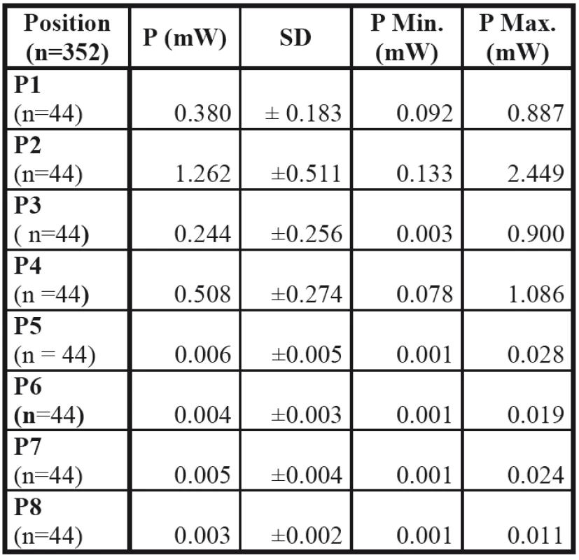
1. Basic values of power P, P Min, P max (in mW) at positions P1–P8. 
The exposure time of adolescents lasted (49.16 ± 0.4) min. The time of individual measurement was (60.17 ± 0.56) min.
Tab. 1 includes average data at positions P1–P8: the mean power values (P ± SD) in mW, the minimum power value (P Min.), the maximum power value (P Max.) around the head within a cage (n = 352 - total number of all measurements, n = 44 - for each P, respectively) in each file.
The difference in absorption of RF EFM by the head at different areas was confirmed by a visualization of EMF power distribution (Fig. 7).
Fig. 7: Visualisation of distribution of EMF power. 
Fig. 8: Comparison of values of power P(mW) of EMF at positions P1-P5, P2-P6, P3-P7, P4-P8. ooo p < 0,01 comparison with P1 – P5 with head; xxx p < 0,01 comparison with P2 – P6 with head; --- p < 0,01 comparison with P3 – P7 with head; +++ p < 0,01 comparison with P4 – P8 with head; *** p < 0,01 comparison without head. 
We revelaed that the highest power passed in the position P5 - red color and the lowest in the P8 - blue color.
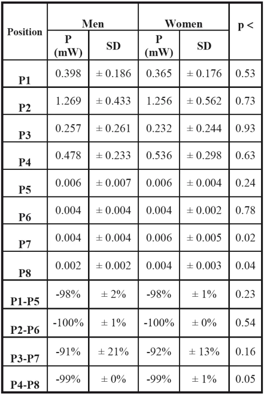
2. Average power values at positions P1–P8 in men and women. 
To compare the power on the right and left side of the head in an adolescent at positions P1 – P5, P2 – P6, P3 – P7, P4 – P8, we determined the percentage differences visualized graphically (Fig. 8).
The average power in the area P2 – P6 was 1.5% lower than in P1 – P5. The average power in the area P3 – P7 was 6.3% higher than in P1 – P5, 7.8% higher than in P2 – P6 and 7.6% higher than in P4 – P8. The average power in the area P4 – P8 was 1.3% lower than in P1 – P5 and 0.2% higher than in P2 – P6.
By evaluation of the power comparison on the right and left side of the head, we found a difference (p < 0.01–0.001) between power on the right and left side of the head at mutually corresponding points (P1 – P5 - frontal area, P2 – P6 - facial area, P3 – P7
- parietal area, P4 – P8 - temporal area). The highest level of RF radiation absorption was measured in the horizontal plane of the head - at temporal areas, connecting both ears at positions P4 – P8.Several significant differences occurred when comparing the genders. On the right side of head, we recorded higher levels of power in men compared with women at positions P1, P2, P3. While comparing power levels in men and women in specific areas, we found differences only at P7, P8 and P4 – P8 (p ≤ 0.05) between the right and the left ear. The increased absorption of RF EMF was observed at this area in women. The comparison of average power values at positions P1–P8 in men and women is indicated in Tab. 2.
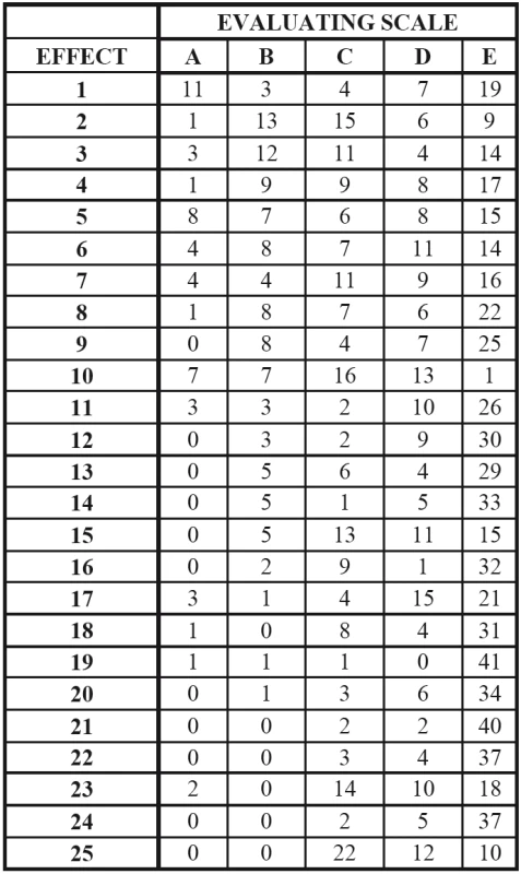
Each adolescent actively used one phone, the average specific absorption rate (SAR) of their cell phones was (0.73 ± 0.23) W/kg. The average age of a first use of cell phone by the adolescents was (9.1 ± 1.5) years. The evaluation of results indicated in the questionnaire (Tab. 3) showed that the most prevalent symptoms of RF EMF were tachycardia (3.14 ± 0.26 on a 1–5 scale), drowsiness (2.91 ± 0.27), headache (2.79 ± 0.26), fatigue (2.55 ± 0.23), restlessness (2.45 ± 0.22). Symptoms with the lowest abundance were skin problems (1.13 ± 0.05), tingling in muscles (1.2 ± 0.08) and problems with word expression (1.22 ± 0.06).
3. Most frequent answers of adolescenst in questionaire (see a text for explanation). 
Symptoms with options A (25%) were reported by the adolescents regarded feelings of fatigue, the options B (29.55%) accounted for headache, the options C (50%) accounted for pain in the hand of adolescents, the options D (34.09%) accounted for chest pain in adolescents, and the options E (93.18%) accounted for tingling in muscles.
Discussion
The results of our study showed that the levels of power of RF EMF with 1788.5 MHz frequency at positions P1 – P4 and P5 – P8 significantly differs at the right and left side of a head. Our findings support a hypothesis that various areas of the head with different bulk of a head tissue may have different absorbency for RF EMF radiation. However, when addressing the monitoring of EMF power and level of absorption one must consider the refraction and interference of EM waves in an environment. We found significant difference in power between P4 – P8 points at temporal areas between the right and left ear both in men and women. This indicates a linear relationship between the absorption and a bulk of head tissue including the brain, skull bones, fat and muscles. However, some differences could be caused also due to head geometry in men and women, with a different volume and density of the hairs, and even by an usage of metalic hair products in women.
Pinosova et al. [1] describes the distribution of energy of EMF of cell phones in the tissue of a human head using 3CAD model SAM Phantom XB 030. The most irradiated parts of the head are within the close proximitty (above the ear, behind the ear) on the side where the cell phone is used, which is consistent with our findings. The spread of SAR and dependance on the frequency of the EMF and the location of the antenna has been shown in other research study [4] on a human head phantom.
From a topography map of EMF distribution is clearly visible that the highest decrease of RF EMF radiation occurred at the position P8, defining area of the left ear. The significant drop in power was also found at positions P2 – P6 (in facial area above the ears). Thus, the area with the highest absorption of the EM radiation is supported by the lowest power measured at position P8. Therefore, it is reasonable to suppose that a magnitude of absorption depends on a size and mass of the brain tissue, throughout the radiation passes.
Biological tissue of a human head forming the layer structure of a skin, fat, skull and brain can be considered as a lossy dielectric material, where the permeability μ = μο and the conductivity is dependent on the properties of the tissue and the frequency of the signal. The depth to which radio waves penetrate the exposure can be used to present the thermal effect of the RF signal within biological tissues of the head at frequency 1800 MHz. The diversity of the exposure within tissues of the head region in children and adults is of particular importance for the interpretation of the biological effects of RF EMF.
The influence of variability and morphology on the RF absorption has been shown in several scientific studies using simulation with models of heads [2,4,12]. Wiart et al. [5] compared the distribution of SAR in the brain using models of heads in the age range 5–15 years and six adult head models developed in various international laboratories. They proved that in 1g of peripheral brain tissue at models of children aged 5–8 years, the percentage of RF absorption is approximately two times higher than in adult models. Due to the thinner pinna, skin and skull in children there is a smaller distance between the peripheral brain tissue and the source of RF EMF and therefore there is higher exposure induction in tissues of children.
Other factors in our work which we believe may contribute to the mechanism of energy absorption include the nature of the radiating antenna, its distance from the interacting peripheral tissue and the reflection of the waves in environment. The effect of the distance between a human head and the internal antenna of a mobile device on SAR values has been studied by Hossain et al. [13]. The results show that by increasing the distance from the head SAR is reduced, however increase in the angle between the head and antenna does not reduce the SAR. Absorption of radiation analysed by SAR values in correlation with the location of the cell phone directly on a side of the head compared with a tilted cell phone (900 MHz, 1800 MHz) with relation to the head (15° and 30°) was investigated by Iqbal-Faruque et al. [14] using simulation technique. There are significant differences in SAR in both of the inclined planes and in relation to the antenna used as a source of EM emissions - higher conductivity results in a higher induced tension and the induced current and thus the greater absorption of EM energy head. SAR values at a 15° angle were higher. The highest absorption of radiation is in the head area, mainly around the frontal lobe of brain, which is the common location of a cell phone in speaking mode. This is consistent with the conclusions of our study. While the brain is one of the conductive parts of the body, this area is more sensitive to radiation.
Distribution of the rate of RF energy into the head is determined by length of exposure and ways of cell phone use – is repeatedly in close proximity ipsilateral to the ear while speaking mode. Our findings confirm the significant rate of RF absorption in the coresponding temporal area of adolescents. Scientific findings of connections between health consequences associated with peripheral auditory system and auditory function and exposure to EMF of cell phones vary [15,16]. A causal relationship between auditory symptoms (tinnitus, audio frequency distortion, warmth or pressure in the ear) and short-term exposure to cell phone was confirmed by Medeiros and Sanchez [17].
There are also technical factors that should be taken into consideration within our target group e.g. the source of signal (natural - from the base transceiver stations or a cell phone or artificial - from a generator, as being used in this study), intensity of radiation, time of exposure, distance from a source, polarisation, modulation, etc. Also the biological factors must be taken into account like unfinished biological growth in adolescents, the possible cumulative effect of RF EMF on autonomic nerve system [18], changes in a sugar metabolism [19], syndrom of EM intolerance, first time use of cell phones in children. E.g. while the use in 2008 was 12.5 years, in 2010 an average age decreased to 10 years and in 2013 even to 8.4 years [20]. Age for a first active use of cell phone in our adolescents was (9.1 ± 1.5) years. Our adolescents used cell phones with a SAR value lower than is limited by the European standard ENV 50166-2 [21]. A prerequisite for the implementation of research projects focusing on young people is a permanent monitoring of their habits in use of mobile devices and the evaluation of EMF characteristics under specific real conditions [22–25].
The adolescent´s adverse health reactions following short time exposure to RF EMF in our study were rare. However, symptoms like tachycardia, drowsiness, headache, fatigue, problems with concentration sometimes appeared. Hence our findings are in a line with reports of several other studies [26–28].
The current level of knowledge on effects of RF EMF radiation on a health of children and adolescents is still not sufficient. Further research should focus mainly on relationships between a long-term exposure of low intensity of natural RF EMF (from the base stations, cell phones and WiFi routers) and the health conditions. Based on the INTERPHONE study RF EM radiation from the cell phone was classified as a possible carcinogen (Group 2B) in humans [29]. Until clear scientific proofs on possible harmful effects of RF EMF on human health, it is necessary to protect the health of children and adolescents [30, 31], by continuous monitoring of the effects of RF EMF sources performed by dosimetric measurements on human volunteers.
Conclusion
The aim of this study was to find power distribution of RF EMF in vicinity of the head of adolescents while they were exposed to a short - term frequency of 1788.5 MHz generated by a signal generator. We also look for subjective health complaints of adolescents following the exposure. We found different absorption of RF energy at different head areas, based on “a size“ principle. The most significant decrease in RF power was observed at a temporal areas between the right and left ear. The subjective adverse health reactions of adolescents on short-term radiofrequency exposure were rare. However, occasionally symptoms like tachycardia, drowsiness, headache, fatigue, problems with concentration had appeared. Our study may support and protect the health of children and adolescents and eliminate the potential health risks.
Acknowledgements
The study was supported by „The Agency for Research and Development based on contract number APVV – 0189-11“ (prof. Jakus).
Mgr. Hana Habiňáková
Institute of Medical Biophysics
Jessenius Faculty of Medicine in Martin
Comenius University in Bratislava
Malá hora 4A, 036 01 Martin
Slovak Republic
E-mail: hhabinakova@gmail.com
Sources
[1] Pinosova, M. et al.: Mobile phones and possible effects onhuman health. 2013. EXTRA [online]. http://www.engineering.sk/
images/stories/2013/04april/extra.pdf (in Slovak).
[2] Christ, A. et al.: Age-dependent tissue-specific exposure of cell phone users. In: Physics in Medicine and Biology. 2010. Physics in Medicine and Biology, 2010, vol. 55, no. 7,
p. 1767–1783.
[3] Morgan, L. et al.: Why children absorb more microwave radiation than adults: The consequences.2014. Journal of Microscopy and Ultrastructure [online]. 2014, vol. 2, no. 4,
p. 197–204. http://www.sciencedirect.com/science/article/pii/
S2213879X14000583
[4] Karanasiou, I. et al.: SAR estimation in human head models related to TETRA, GSM and UMTS exposure using different computational approaches. 2014. WSEAS TRANSACTIONS on BIOLOGY and BIOMEDICINE [online]. 2014, no. 11,
p. 101–110. http://www.wseas.org/multimedia/journals/biology/
2014/a125708-130.pdf
[5] Wiart, J. et al.: Analysis of RF exposure in the head tissues of children and adults. 2008. Physics in Medicine and Biology [online]. 2008, vol. 53, p. 3681–3695.
[6] International Commission on Non-Ionizing Radiation Protection. Guidelines for Limiting Exposure to Time-Varying Electric, Magnetic and Electromagnetic Fields (up to 300 GHz). 1998. Health Physics, 1998, vol. 74, no. 4, p.494–522.
[7] Ministry of Health of the Slovak Republic Codex No. 534/2007 on details and requirements about sources of electromagnetic radiation and limits of exposure to electromagnetic radiation in the environment on the citizens (in Slovak).
[8] Government Regulation. No.217/2008 Codex of Slovak Republic of minimum health and safety requirements to protect workers from the risks related to exposure to electromagnetic fields (in Slovak).
[9] Ministry of Health Codex of the Slovak Republic No. 233/2014 on the details of the assessment of the effects on public health (in Slovak).
[10] http://docs-europe.electrocomponents.com/webdocs/1110/0900
766b811108dd.pdf
[11] https://cdn.sos.sk/productdata/20/ef/2013cf94/can4313330009191b.pdf
[12] Psenakova, Z., Benova, M.: Measurement evaluation of EMF effect by mobile phone on human head.2008. Electrical and Electronic Engineering. 2008, vol.7, no. 1-2, p. 350–353, ISSN 1336-1376.
[13] Hossain, M. I. et al.: Analysis on the Effect of the Distances and Inclination Angles between Human Head and Mobile Phone on SAR. 2015. Progress in Biophysics and Molecular Biology [online]. https://www.ncbi.nlm.nih.gov/labs/articles/25863147/.
[14] Iqbal-Faruque, M. R. et al.: Effects of Mobile Phone Radiation onto Human Head with Variation of Holding Cheek and Tilt Positions.2014. Journal of Applied Research and Technology. 2014, vol. 12, no. 5, p. 871–876.
[15] Bortkiewicz, A. et al.: Changes in tympanic temperature during the exposure to electromagnetic fields emitted by mobile phone. 2012. Int J Occup Med Environ Health, 2012, vol. 25, no. 2, p. 145–150.
[16] Gupta, N. et al.: Effect of Prolonged Use of Mobile Phone on Brainstem Auditory Evoked Potenntials. 2015. J Clin Diagn Res, 2015, vol. 9, no. 5, CC07-9.
[17] Medeiros, L.N., Sanchez: Tinnitus and cell phones: the role of electromagnetic radiofrequency radiation. 2016. Braz J Otorhinolaryngol, 2016, vol. 82, p. 97–104.
[18] Misek, J., Jakus, J., Tonhajzerova, I., Vasicko, T., Veternik, M., Spiguthova, D., Jakusova ,V., Osina, O.: Heart rate variability affected by high frequency electromagnetic field in adolescent students. 2015. BioEM 2015: The Annual Meeting of Bio-electromagnetics Society, European Bioelectromagnetics Association. Londyn: Lawson Health Research Institute, 2015, p. 294–296. ISBN 9781510810440.
[19] Jakus, V., Sapak, M., Kostolanska, J.: Circulating TGF-β1 glycation and oxidation in children with diabetes mellitus type 1. 2012. Experimental Diabetes Research 2012,vol. 2012, p. 1–7.
[20] Buyn, Y. et al.: Mobile Phone Use, Blood Lead Levels, and Attention Deficit Hyperactivity Symptoms in Children: A Lon-gitudinal Study. 2013. PLOS ONE [online]. 2013, vol. 8, no. 3, e59742. http://journals.plos.org/plosone/article?id=10.1371/
journal.pone.0059742.
[21] CENELEC: EMF exposure in the high range (10 kHz–300 GH). 1995. Bruxelles, CENELEC, 1995, 44 p. (in Slovak).
[22] Osina, O., Vasicko, T., Habinakova, H., Spiguthova, D., Osinova, D., Jakusova, V., Jakus, J.: The environmental exposure of young people to electromagnetic fields – personal exposimetric evaluation. 2014. EHE 2014: International Conference of Electromagnetic Fields, Health and Environment, 2014, p. 1–2. ISBN 978-972-8822-28-6.
[23] Spiguthova, D., Habinakova, H., Misek, J., Jakusova, V., Jakus, J.: Measurement of parameters of electromagnetic fields in the use of means of mobile communication in the school environment. 2015. The Clinician and Technology Journal [online]. 2015, vol. 45, no. 4, p. 122–128 (in Slovak).
[24] Habinakova, H. et al.: Adolescents and exposure to radio-frequency of electromagnetic fields from mobile communication devices. 2015. Current issues in public health research and practice [electronic source]. Martin, 2015, p. 51–55. ISBN 978-80-971836-6-0 (in Slovak).
[25] Jakusova ,V., Spiguthova, D., Jakus, J., Osina, O.: High school students' knowledge and their need for education on electromagnetic radiation from mobile phones. 2013. Current issues in public health research and practice. Martin, 2013, p. 127–132. ISBN 978-80-89544-39-4 (in Slovak).
[26] Sangmin, J.: The reciprocal longitudinal relationships between mobile phone addiction and depressive symptoms among Korean adolescents. 2016. Computers in Human Behavior [online]. 2016, vol. 58, p. 179–186. http://www.sciencedirect.
com/science/article/pii/S0747563215303320.
[27] Chiu, C.T. et al.: Mobile phone use and health symptoms in Children. 2015. J Formos Med Assoc., 2015, vol. 114, no.7, p. 598–604.
[28] Zheng, F. et al.: Association between mobile phone use and self-reported well-being in children: a questionnaire-based cross-sectional study in Chongqing, China. 2015. BMJ Open, 2015, doi:10.1136/bmjopen-2014-007302.
[29] International Agency for Research on Cancer. IARC classifies radiofrequency electromagnetic fields as possibly carcinogenic to humans. 2011. [online].
www.iarc.fr/en/mediacentre/pr/2011/pdfs/pr208_E.pdf.
[30] World Health Organization. Maternal, newborn, child and adolescent health, 2015. [online]. http://www.who.int/maternal_
child_adolescent/topics/adolescence/dev/en/.
[31] Belyaev, I. et al.: EUROPAEM EMF Guideline 2016 for the prevention, diagnosis and treatment of EMF-related health problems and illnesses. 2016. Environ Health [online]. 2016, DOI10.1515/reveh-2016-0011. http://www.stralskyddsstiftelsen.se/wp-.
Labels
Biomedicine
Article was published inThe Clinician and Technology Journal

2017 Issue 2-
All articles in this issue
- PERISTALTIC FLOW OF LITHOGENIC BILE IN THE VATERI’S PAPILLA AS NON-NEWTONIAN FLUID IN THE FINITE-LENGTH TUBE: ANALYTICAL AND NUMERICAL RESULTS FOR REFLUX STUDY AND OPTIMIZATION
- /GD-TRACKER/ A SOFTWARE FOR BLOOD-BRAIN BARRIER PERMEABILITY ASSESSMENT
- EXPERIMENTAL ANALYSIS OF THE LUMBAR SPINE KINEMATICS
- RESPIRATORY SOUNDS AS A SOURCE OF INFORMATION IN ASTHMA DIAGNOSIS
- MEASUREMENT OF THE VALUES OF RADIOFREQUENCY ELECTROMAGNETIC FIELDS AROUND THE HEAD OF ADOLESCENTS
- The Clinician and Technology Journal
- Journal archive
- Current issue
- Online only
- About the journal
Most read in this issue- /GD-TRACKER/ A SOFTWARE FOR BLOOD-BRAIN BARRIER PERMEABILITY ASSESSMENT
- PERISTALTIC FLOW OF LITHOGENIC BILE IN THE VATERI’S PAPILLA AS NON-NEWTONIAN FLUID IN THE FINITE-LENGTH TUBE: ANALYTICAL AND NUMERICAL RESULTS FOR REFLUX STUDY AND OPTIMIZATION
- RESPIRATORY SOUNDS AS A SOURCE OF INFORMATION IN ASTHMA DIAGNOSIS
- MEASUREMENT OF THE VALUES OF RADIOFREQUENCY ELECTROMAGNETIC FIELDS AROUND THE HEAD OF ADOLESCENTS
Login#ADS_BOTTOM_SCRIPTS#Forgotten passwordEnter the email address that you registered with. We will send you instructions on how to set a new password.
- Career

